Contents
- Introduction and Scope
- SECTION I --- GENERAL CONSIDERATIONS
- SECTION II --- INDIVIDUAL TUMORS
- II.1 Osteoma
- II.2 Osteoid Osteoma
- II.3 Osteoblastoma
- II.4 Osteochondroma (and Hereditary Multiple Exostoses)
- II.5 Enchondroma (and Enchondromatosis; Periosteal Chondroma)
- II.6 Chondroblastoma
- II.7 Chondromyxoid Fibroma
- II.8 Giant Cell Tumour of Bone (Osteoclastoma)
- II.9 Haemangioma of Bone
- II.10 Desmoplastic Fibroma of Bone
- II.11 Other Benign Lesions (Brief)
- SECTION III --- SYNTHESIS AND EXAM AIDS
- References
- Figure Credits and Licences
Introduction and Scope
This review covers Topic 2 of the orthopaedics syllabus, the benign bone tumours. These are the true neoplasms of bone that as a rule do not metastasise, yet they range from entirely innocent “leave-me-alone” lesions to locally aggressive tumours that destroy bone and recur. It is the companion to Topic 1 (tumour-like lesions) and Topic 3 (malignant tumours), and several differential diagnoses cross those boundaries.
The single most important conceptual shift in the current (2020, 5th edition) WHO Classification of Tumours: Soft Tissue and Bone is that “benign” is no longer a binary. WHO now grades each entity by behaviour: benign, intermediate (locally aggressive), intermediate (rarely metastasising), or malignant. Two tumours traditionally taught as benign now sit in the intermediate tier, osteoblastoma (ICD-O 9200/1, locally aggressive) and giant cell tumour of bone (9250/1, locally aggressive and rarely metastasising), and desmoplastic fibroma (8823/1) is likewise intermediate. WHO 2020 also attaches a defining molecular alteration to most entities, and these are now part of routine diagnosis. This document foregrounds both.
The sources are the standard references in the user’s library: WHO 2020 (classification, ICD-O codes, molecular genetics), Schajowicz, Campanacci, and Dorfman & Czerniak (pathology, clinical, treatment), Greenspan & Beltran (imaging), Wold (pathology atlas), the surgical-oncology texts of Bickels/Wittig/Malawer and Malawer & Sugarbaker (staging, biopsy, operative technique), and the Bulgarian operative atlas of Boychev for the indigenous techniques and terminology. Molecular and therapeutic facts that post-date the older texts are cited to WHO 2020 or current practice.
SECTION I --- GENERAL CONSIDERATIONS
I.1 The Benign-Intermediate-Malignant Spectrum
A “benign” bone tumour is a true neoplasm that grows locally and, with rare and defined exceptions, does not metastasise. WHO 2020 grades behaviour in four tiers, and it is the tier rather than the word “benign” that should drive management:
- Benign: grows locally, does not recur destructively after complete removal, does not metastasise. Most osteomas, osteochondromas, enchondromas, chondroblastomas, chondromyxoid fibromas, and haemangiomas.
- Intermediate, locally aggressive: infiltrates and destroys local bone, recurs after incomplete removal, but does not metastasise. Osteoblastoma, desmoplastic fibroma.
- Intermediate, rarely metastasising: locally aggressive and capable of occasional (usually pulmonary) metastasis that is histologically benign. Giant cell tumour of bone (and, exceptionally, chondroblastoma).
- Malignant: the territory of Topic 3, reached here only through the malignant-transformation risks of osteochondroma and enchondroma (to secondary chondrosarcoma) and the rare sarcomatous change of giant cell tumour.
Two genuinely benign tumours nonetheless behave with surprising aggression, and these are the recurring “trap” lesions of this topic: the giant cell tumour and the locally destructive osteoblastoma and desmoplastic fibroma.
I.2 Classification (WHO 2020)
WHO 2020 groups bone tumours by tissue of differentiation (chondrogenic, osteogenic, fibrogenic, osteoclastic or giant-cell-rich, vascular, notochordal, and others) and within each group by behaviour. The benign and intermediate entities of this topic, with their ICD-O codes and defining molecular alterations, are:
| Tumour | Tissue | WHO behaviour / ICD-O | Defining molecular alteration (WHO 2020) |
|---|---|---|---|
| Osteoma | osteogenic | benign, 9180/0 | none (Gardner → APC; bone islands/osteopoikilosis → LEMD3) |
| Osteoid osteoma | osteogenic | benign, 9191/0 | FOS rearrangement (~91 %) |
| Osteoblastoma | osteogenic | intermediate (locally aggressive), 9200/1 | FOS (~87 %), less often FOSB |
| Osteochondroma | chondrogenic | benign, 9210/0 | EXT1 / EXT2 inactivation |
| Enchondroma | chondrogenic | benign, 9220/0 | IDH1 / IDH2 mutation |
| Chondroblastoma | chondrogenic | benign, 9230/0 | H3-3B (H3F3B) K36M |
| Chondromyxoid fibroma | chondrogenic | benign, 9241/0 | GRM1 rearrangement / upregulation |
| Giant cell tumour | osteoclastic | intermediate (locally aggressive, rarely metastasising), 9250/1 | H3-3A (H3F3A) G34W (~90 %) |
| Haemangioma | vascular | benign, 9120/0 | none (pathogenesis unknown) |
| Desmoplastic fibroma | fibrogenic | intermediate (locally aggressive), 8823/1 | none consistent (exclude FD by absent GNAS, low-grade osteosarcoma by absent MDM2) |
A clean exam point is the gene-pair contrast that WHO diagnosis now turns on. The histone-H3.3 mutations separate two giant-cell-rich cartilage/bone tumours: chondroblastoma carries H3-3B K36M, giant cell tumour carries H3-3A G34W, and immunohistochemistry for the mutant proteins distinguishes them.[14]
I.3 Diagnostic Approach
The diagnosis of a benign bone tumour is the deliberate triangulation of clinical data, imaging, and pathology. For benign tumours, age, site, and the radiographic pattern usually narrow the field before any biopsy.
- Age and site are powerful. Chondroblastoma is the epiphyseal lesion of the immature skeleton; giant cell tumour is the epiphyseal lesion of the mature skeleton; osteoid osteoma and osteoblastoma favour the femur and the posterior spinal elements; osteochondroma and enchondroma arise from displaced or arrested cartilage.
- Read the radiograph systematically. Assess margin and zone of transition (Lodwick grade), cortical integrity, periosteal reaction, and, decisively for this topic, the matrix. A “rings-and-arcs”, stippled, or popcorn matrix means cartilage (enchondroma, chondroblastoma); a dense ground-glass or osteoid matrix means bone-forming tissue; a purely lytic lesion with no matrix at the end of a long bone in an adult suggests giant cell tumour.
- Some lesions are diagnosed on imaging alone and need no biopsy: the classic osteochondroma (cortical and medullary continuity), the incidental enostosis, the corduroy vertebral haemangioma, and the latent non-ossifying fibroma.
- When the radiograph is aggressive, it is not necessarily malignant. Giant cell tumour, aneurysmal bone cyst, osteoblastoma, and osteomyelitis all mimic malignancy.
I.4 Principles of Staging and Treatment
Benign tumours are staged biologically by the Enneking system: stage 1 (latent), stage 2 (active), and stage 3 (aggressive), graded from the reactive rim seen on imaging. The stage, more than the histological label, dictates treatment.
- Stage 1 (latent): observe; intervene only for symptoms or fracture risk.
- Stage 2 (active): intralesional curettage with a high-speed burr, usually curative.
- Stage 3 (aggressive): extended intralesional curettage with a local adjuvant (phenol, liquid nitrogen/cryosurgery, argon-beam, or electrocautery; PMMA cement adds an exothermic and structural effect), or wide en-bloc resection.
The operative work-horse for the cystic and giant-cell lesions is extended intralesional curettage. It comprises a wide cortical window, thorough hand curettage, high-speed burring of the cavity walls, a local adjuvant to sterilise the residual margin, and reconstruction with bone graft, a calcium-phosphate substitute, or PMMA, with internal fixation where stability demands it. Wide resection is reserved for expendable bones, for tumours that have destroyed a joint, and for the rare entity that needs it (adamantinoma; secondary chondrosarcoma; sarcomatous transformation). Biopsy follows staging, never precedes it, and obeys the standard rules: a longitudinal incision in line with the definitive resection, through one compartment, with meticulous haemostasis, and excision of the tract at surgery. A poorly-placed biopsy is a documented cause of unnecessary amputation. # SECTION II - THE INDIVIDUAL TUMOURS
SECTION II --- INDIVIDUAL TUMORS
II.1 Osteoma
Synonyms / WHO. Ivory exostosis; the medullary form is the enostosis / “bone island.” WHO 2020: benign osteogenic tumour, ICD-O 9180/0.[15]
Definition. A benign tumour of dense, well-differentiated lamellar (cortical-type) bone arising on a bone surface; when it lies within the medulla it is a bone island. Many regard it as a hamartoma rather than a true neoplasm.
Epidemiology. Adults, typically in the 4th to 5th decades, with equal sex distribution. Common as an incidental finding.
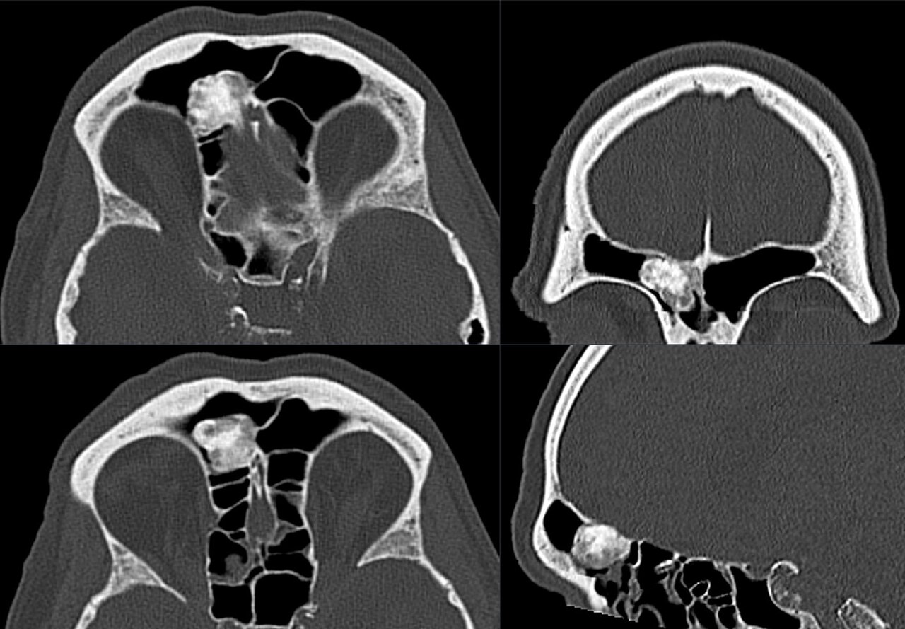
Sites. Almost exclusively the membranous bones: the outer table of the skull and the paranasal sinuses (frontal and ethmoid). Bone islands occur in any cancellous bone.
Clinical features. Usually asymptomatic and slow-growing. Sinus osteomas may obstruct an ostium (sinusitis, mucocele) or deform the orbit. Multiple osteomas should prompt a search for Gardner syndrome (autosomal-dominant APC mutation: premalignant colonic polyposis, plus osteomas, epidermoid cysts, and desmoid tumours).
Imaging. A dense, structureless, sharply marginated radiopaque mass attached to the cortex. The bone island is a homogeneously dense focus with a “brush-border” of thorny radiations blending into the trabeculae. It is cold or only mildly warm on bone scan, the single feature distinguishing it from an osteoblastic metastasis.[16]
Pathology. Dense mature lamellar bone resembling compact cortex, with sparse cellular activity and no cartilage cap (distinguishing it from osteochondroma and parosteal osteosarcoma). Sinus and other facial osteomas, by contrast, are built of immature bone with active surface modelling and are histologically akin to osteoblastoma.[17]
Differential diagnosis. Parosteal osteosarcoma is the lesion to exclude for a surface osteoma. An osteoma has exquisitely smooth borders, uniform sclerosis, and no cleft from the cortex. A giant bone island that grows or is hot on scan must be distinguished from sclerotic metastasis.
Treatment. None if asymptomatic. Symptomatic or cosmetically troublesome lesions are excised. A surface osteoma of a long bone is removed with its underlying cortex because parosteal osteosarcoma cannot be excluded clinically. Does not recur after excision.
Exam pearls. Ivory mass of skull/sinus; bone island = medullary osteoma, COLD on bone scan; multiple osteomas → Gardner syndrome (APC).
II.2 Osteoid Osteoma
Synonyms / WHO. Schajowicz’s “circumscribed osteoblastoma.” WHO 2020: benign osteogenic tumour, ICD-O 9191/0.[18]
Definition. A small (usually <1.5-2 cm), self-limited benign bone-forming tumour consisting of a vascular nidus of woven bone and osteoid rimmed by osteoblasts, surrounded by a zone of reactive sclerosis that is not part of the tumour.
Epidemiology. About 10 to 12 % of benign bone tumours, with a peak in the second decade (most patients 5-25 years) and a male predominance ~2:1.
Pathogenesis. A benign osteoblastic tumour. WHO 2020 defines the osteoid-osteoma/osteoblastoma family by FOS rearrangement (and occasionally FOSB). The classic night pain is driven by prostaglandins (PGE₂) and COX-2 within the nidus, which is the basis both for the dramatic NSAID response and for the inflammatory/synovial changes.[19]
Sites. Long bones, especially the femur (the femoral neck is the single commonest site) and tibia. About 10 to 15 % are spinal, almost always in the posterior arch, where they cause painful scoliosis.
Clinical features. The cardinal symptom is pain, worse at night, relieved within ~20-25 minutes by aspirin or NSAIDs. Relief is obtained in roughly three-quarters of patients rather than all, the pain is classically aggravated by alcohol, and unmyelinated nerve fibres within the nidus are thought to be its substrate; an intra-articular nidus (e.g. at the hip) causes a reactive synovitis with little surrounding sclerosis.[20] A posterior-element spinal lesion causes a painful scoliosis (concave toward the lesion, no rotation); a juxta-physeal lesion in a child can accelerate growth; an intra-articular lesion causes synovitis mimicking arthritis.
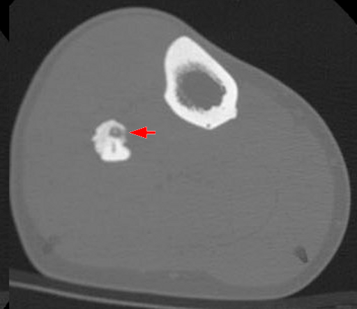
Imaging. A small round lucent nidus (<1.5 cm) with a central sclerotic dot, surrounded by dense reactive sclerosis. Bone scintigraphy is invariably hot, often with a “double-density” sign. Thin-section CT is the key test to localise the nidus; MRI tends to overcall the surrounding oedema and can mimic a more aggressive lesion.[21]
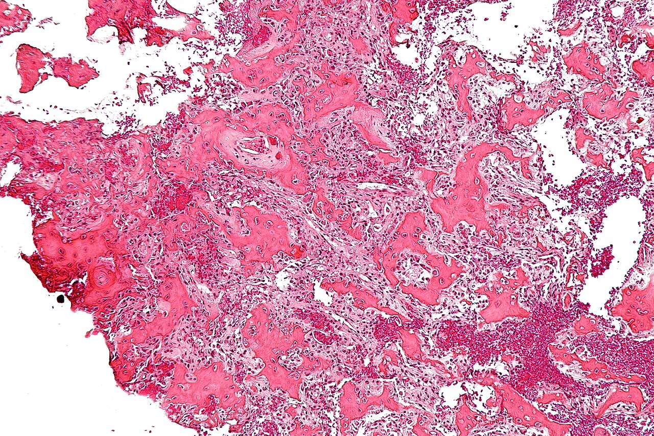
Pathology. A reddish, gritty nidus of interlacing woven-bone/osteoid trabeculae rimmed by plump osteoblasts in a richly vascular stroma, sharply demarcated from the surrounding reactive bone. No cartilage.
Differential diagnosis. Osteoblastoma (larger, less reactive sclerosis); Brodie abscess (serpentine tract, no nidus); stress fracture (lucent line perpendicular to the cortex); intracortical osteosarcoma.
Treatment. CT-guided percutaneous radiofrequency ablation is now the standard of care (success ~80-90 %), having largely replaced en-bloc excision; complete removal of the nidus is curative whatever the technique. Prolonged NSAIDs can give lasting relief, and some lesions regress spontaneously.
Exam pearls. Young patient, night pain relieved by aspirin; nidus <1.5 cm, hot on bone scan, CT to find it; spinal lesion → painful scoliosis; treat by radiofrequency ablation; FOS rearrangement (WHO).
II.3 Osteoblastoma
Synonyms / WHO. “Giant osteoid osteoma.” WHO 2020 reclassifies it as intermediate, locally aggressive: ICD-O 9200/1.[22]
Definition. A bone-forming tumour histologically similar to osteoid osteoma but larger (>2 cm), with greater growth potential and less surrounding reactive sclerosis; locally aggressive.
Epidemiology. About five times rarer than osteoid osteoma, with a peak in the second-third decades and a male predominance ~2:1.
Pathogenesis. Shares the FOS/FOSB rearrangement of osteoid osteoma; the two are one molecular family separated by size and behaviour.
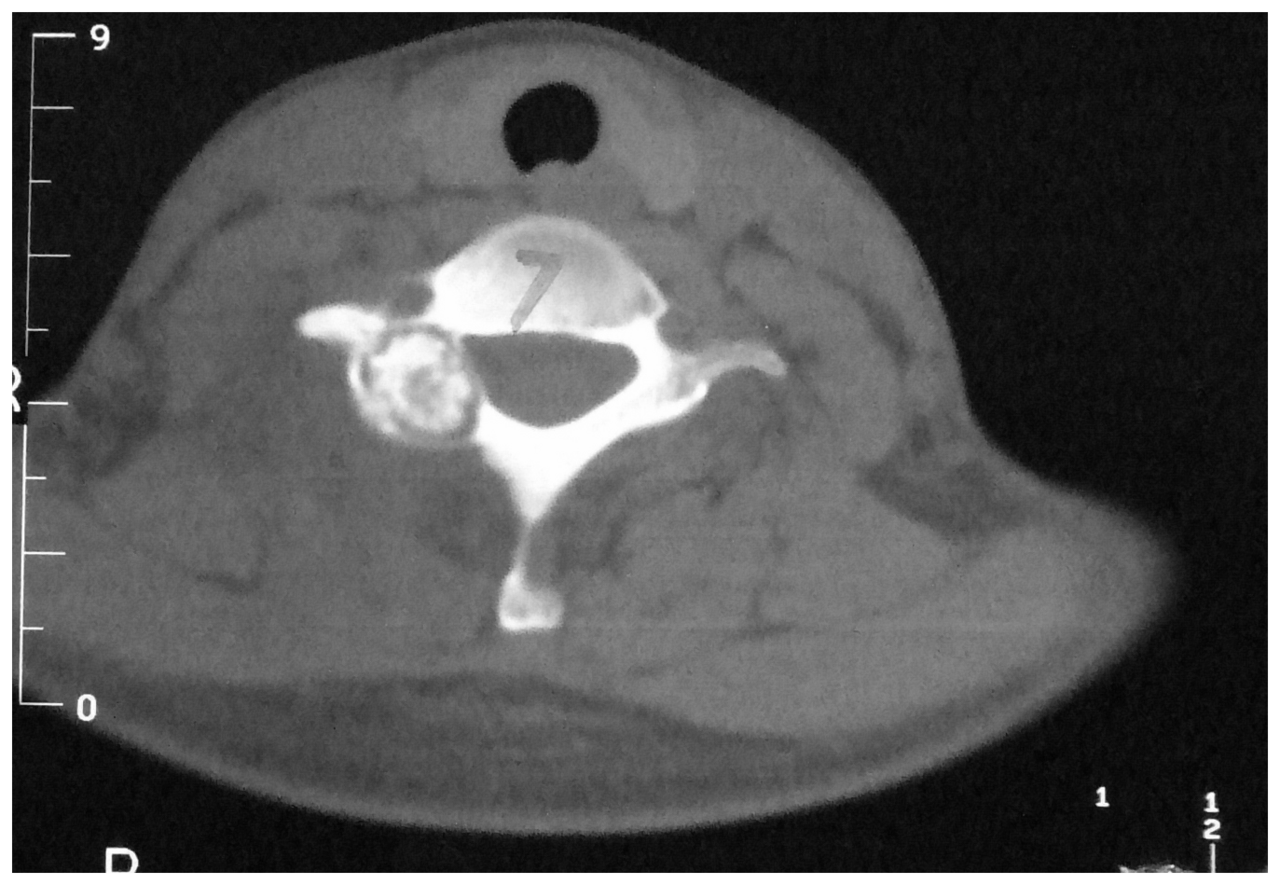
Sites. The only benign bone tumour with a predilection for the vertebral column (>40 % of cases, in the posterior elements); otherwise long-bone metaphysis/diaphysis, pelvis, and craniofacial bones.
Clinical features. Pain, but less reliably relieved by aspirin than osteoid osteoma; bony swelling; spinal lesions cause muscle spasm, scoliosis, and occasionally cord or root compression.
Imaging. An expansile, predominantly lytic lesion containing osseous mineralisation, 2 to 10 cm, often with a “blow-out” aneurysmal-bone-cyst-like appearance in the spine. The margin is demarcated by a rind of sclerosis less dense than in osteoid osteoma, and the lesion is markedly hot on bone scan. Its appearance can suggest a malignant tumour radiographically.
Pathology. Like osteoid osteoma, it shows woven-bone/osteoid trabeculae rimmed by plump osteoblasts in a vascular stroma, but larger and with a sharp, non-permeative interface with host bone. The critical malignant differential is osteosarcoma, distinguished by a permeative margin, atypical mitoses, lace-like osteoid embedding individual cells, necrosis, and malignant cartilage. The epithelioid (aggressive) variant contains large epithelioid osteoblasts and is locally aggressive but does not metastasise.[23] Mirra cautions that most lesions labelled “aggressive osteoblastoma,” and most reports of an osteoblastoma “transforming,” are in reality low-grade osteoblastoma-like osteosarcomas misclassified from the outset, separated by permeation and entrapment of host lamellar bone rather than by cytology.[24]
Treatment. Stage-dependent: intralesional curettage with adjuvant for most lesions, marginal or wide resection for stage-3 disease and accessible sites; preoperative embolisation for vascular spinal/pelvic lesions. Recurrence up to ~25 % after incomplete removal; does not metastasise.
Exam pearls. “Giant osteoid osteoma” >2 cm, spine posterior elements, expansile, pain less aspirin-responsive; osteosarcoma is the make-or-break DDx (permeative margin, atypical mitoses); WHO 2020 now grades it intermediate (9200/1); FOS/FOSB.
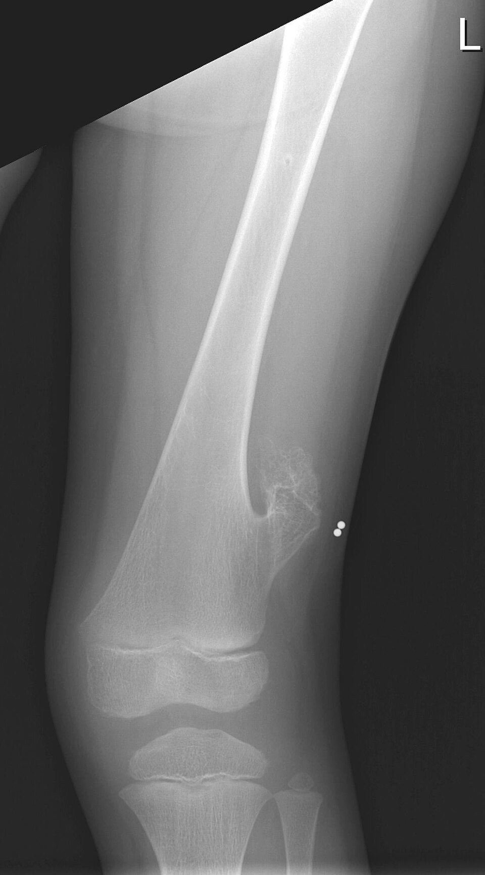
II.4 Osteochondroma (and Hereditary Multiple Exostoses)
Synonyms / WHO. Osteocartilaginous exostosis. WHO 2020: benign chondrogenic tumour, ICD-O 9210/0.[25]
Definition. A bony projection capped by cartilage, arising on the surface of a bone, whose cortex and medullary cavity are continuous with those of the host bone. This continuity is the pathognomonic feature.
Epidemiology. The commonest benign bone tumour (~35 % of benign tumours), usually noticed before age 20, with a male predominance ~1.5-2:1.
Pathogenesis. Arises from a displaced fragment of physeal cartilage that grows by endochondral ossification through its own growth plate, points away from the nearest joint, and stops growing at skeletal maturity. WHO 2020 attributes it to inactivation of EXT1/EXT2 (exostosin glycosyltransferases that synthesise heparan sulfate); in solitary lesions the inactivation is somatic and confined to the cartilage cap.
Sites. Metaphyses of long bones, especially around the knee (distal femur, proximal tibia) and the proximal humerus; also the pelvis and scapula. It does not occur in membranous bones or, normally, the epiphyses.
Clinical features. A hard, fixed, painless mass of long duration. Complications include mechanical impingement on a nerve or vessel (rarely a popliteal pseudoaneurysm), an overlying inflamed bursa (exostosis bursata), snapping scapula, and fracture of a pedunculated stalk. The one that matters is malignant transformation.
Imaging. Cortical and medullary continuity with the host bone is diagnostic (best confirmed on CT); the lesion is pedunculated (pointing away from the joint) or sessile. MRI measures the cartilage cap (high T2 signal), the key to surveillance.
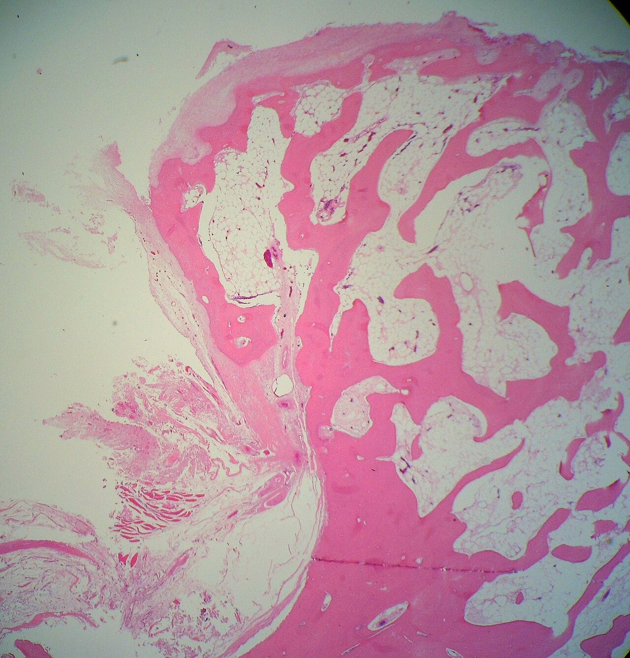
Pathology. A cartilage cap (normally <1 cm, thinning with age) overlying a bony stalk with marrow continuous with the host; the cap resembles a disorganised growth plate maturing to endochondral bone, without cytological atypia.
Malignant transformation. To secondary peripheral chondrosarcoma, with a risk of ~1 % for a solitary lesion and substantially higher (cited ~5-20 %) in hereditary multiple exostoses. Suspect it when an osteochondroma grows after skeletal maturity, becomes painful, or develops a cartilage cap >1.5-2 cm in an adult; bone scan is unreliable for this distinction.[26] Bullough frames the lesion as a herniation of physeal cartilage through the periosteal bone cuff that ossifies and stops growing at physeal closure, so a bosselated cap that thickens again after maturity is the alarm sign.[27]
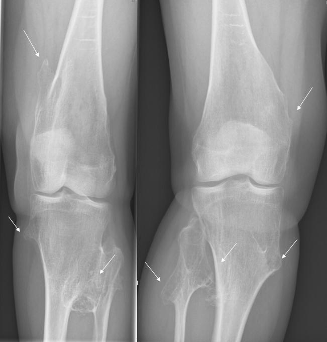
Hereditary multiple exostoses (HME). Autosomal dominant, caused by germline EXT1/EXT2 mutations, with incomplete penetrance in females; multiple symmetrical exostoses with metaphyseal undermodelling, limb-length discrepancy, and a pseudo-Madelung forearm deformity (short ulna, bowed radius, dislocated radial head). The chondrosarcoma risk is much higher than for solitary disease, mandating lifelong surveillance of pelvic and shoulder-girdle lesions.
Treatment. Observe the asymptomatic lesion. Excise a symptomatic one at its base, removing the entire cartilage cap and perichondrium (the only parts capable of growth). Suspected malignant change requires wide en-bloc resection after biopsy.
Exam pearls. Pathognomonic = cortical + medullary continuity; points away from the joint; grows by its own physis, stops at maturity; EXT1/EXT2; new pain/growth/cap >1.5-2 cm in an adult → secondary chondrosarcoma; HME = AD, higher malignancy risk, pseudo-Madelung.
II.5 Enchondroma (and Enchondromatosis; Periosteal Chondroma)
Synonyms / WHO. Central chondroma. WHO 2020: benign chondrogenic tumour, ICD-O 9220/0.[28]
Definition. A benign intramedullary hyaline-cartilage neoplasm lacking the features of chondrosarcoma.
Epidemiology. The second commonest benign bone tumour and the commonest tumour of the hand; any age, peak in the 2nd to 4th decades; no sex predilection.
Pathogenesis. Arises from rests of physeal cartilage that fail to ossify. WHO 2020 attributes it to somatic IDH1/IDH2 mutations, the same mutation found in central chondrosarcoma, so it confirms cartilaginous lineage but does not by itself separate benign from malignant.
Sites. The short tubular bones of the hand (>50 %; proximal phalanx, metacarpal), then the femur, humerus, and ribs. A cartilage tumour of the pelvis, scapula, or a long bone is far more likely to be malignant than one of the hand.
Clinical features. Usually asymptomatic, found incidentally or after a pathological fracture (especially in the hand). Rest pain without a fracture, persisting for months, is the warning sign of malignant change.
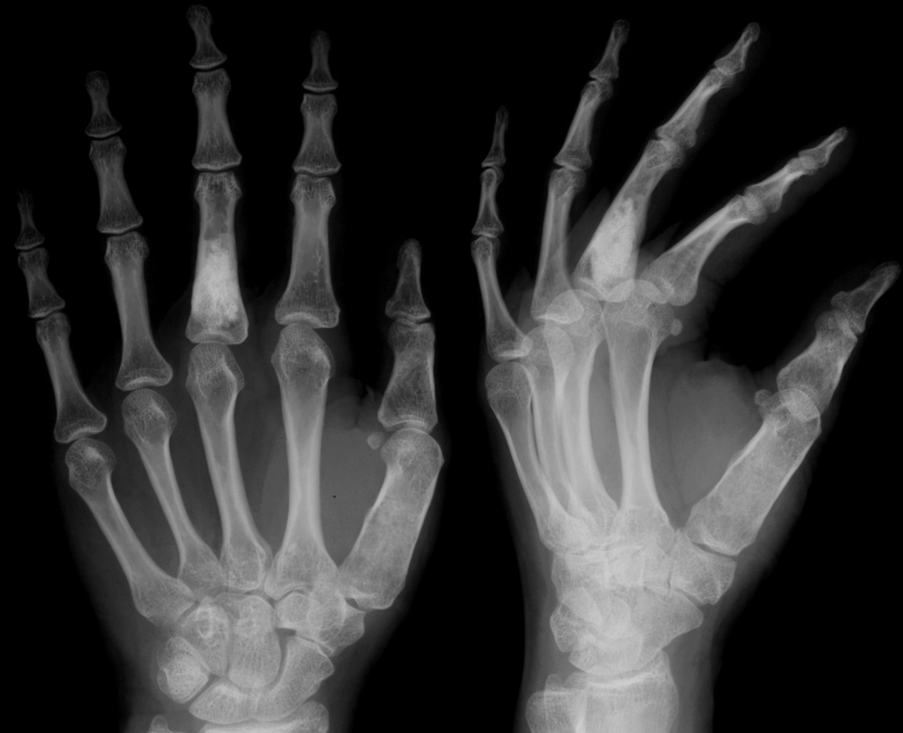
Imaging. A lytic, well-defined medullary lesion with rings-and-arcs, stippled, or “popcorn” chondroid calcification and shallow endosteal scalloping; phalangeal lesions are often purely lytic and expansile. CT/MRI cannot reliably separate a benign enchondroma from a low-grade chondrosarcoma.
Pathology. Lobules of hypocellular, bland hyaline cartilage with small uniform nuclei, often rimmed by reactive bone. Important site-specific caveat: cartilage tumours of the hand/foot, periosteal chondromas, and Ollier lesions are normally more cellular with mild atypia, and this is benign at those sites, whereas the identical picture in a long bone or the pelvis is worrying.
Malignant transformation / the chondrosarcoma problem. Transformation of a solitary enchondroma is rare and essentially never occurs in the hand, but does occur in long and flat bones. The radiological red flags for low-grade chondrosarcoma (now termed atypical cartilaginous tumour in the appendicular skeleton) are deep endosteal scalloping (>2/3 of the cortex), focal cortical thickening or destruction, a soft-tissue mass, size >4-5 cm, and rest pain.[29] Mirra adds two rules. Malignant risk is roughly proportional to the volume of dysplastic cartilage (negligible for a small hand lesion, but 20 to 50 % over a lifetime in extensive Ollier/Maffucci disease). And because about a quarter of chondrosarcomas are too well differentiated to show atypia, low-power architecture, namely host-bone entrapment, permeation, and a soft-tissue mass, outweighs cytology.[30]
Periosteal (juxtacortical) chondroma. A surface cartilage tumour causing saucer-shaped cortical erosion with a sclerotic base and a periosteal buttress, without medullary continuity (distinguishing it from osteochondroma); more cellular histology that is benign at this site; treated by en-bloc excision.
Enchondromatosis - Ollier and Maffucci. Ollier disease = multiple, predominantly unilateral, non-hereditary enchondromas causing limb shortening and deformity, with longitudinal “columns” of cartilage from the physis. Maffucci syndrome = enchondromatosis plus soft-tissue haemangiomas (with phleboliths). Both are caused by somatic mosaic IDH1/IDH2 mutations and carry a much higher malignant-transformation risk (Ollier ~30-40 %; Maffucci the highest of all, approaching certainty over a lifetime), including non-skeletal malignancies (gliomas, ovarian tumours, and in Maffucci visceral angiosarcoma).[31]
Treatment. Observe the asymptomatic, non-weakening lesion. Curettage and grafting for the symptomatic, structurally weakening, or fracture-prone hand enchondroma (curative); wide resection where low-grade chondrosarcoma cannot be excluded. Enchondromatosis is managed functionally, with lifelong surveillance for malignant change.
Exam pearls. Commonest hand tumour; lytic + rings-and-arcs; hypocellular bland cartilage; IDH1/IDH2; hand lesions almost never malignant, but rest pain / deep scalloping / size >4 cm → atypical cartilaginous tumour; Ollier (unilateral, non-hereditary) and Maffucci (+ haemangiomas, highest malignancy) = mosaic IDH.
II.6 Chondroblastoma
Synonyms / WHO. Codman tumour. WHO 2020: benign chondrogenic tumour, ICD-O 9230/0.[32]
Definition. A benign cartilage tumour of immature chondroblasts arising in an epiphysis or apophysis of the skeletally immature.
Epidemiology. Rare (<1 % of bone tumours), with a peak in the second decade in the immature skeleton and a male predominance ~2-3:1.
Pathogenesis. WHO 2020 defines it by the H3-3B (H3F3B) K36M histone mutation (the contrast partner to giant cell tumour’s H3-3A G34W); chondroblasts are S100-positive.
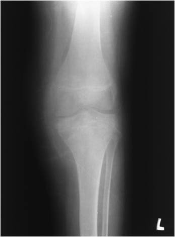
Sites. The epiphyses of the distal femur, proximal tibia, and proximal humerus, and apophyses such as the greater trochanter and greater tuberosity; also the talus and calcaneus. It characteristically crosses the open physis into the metaphysis.
Clinical features. Pain referred to the adjacent joint, with effusion and limited motion (the epiphyseal location irritates the joint), a feature absent in the metaphyseal chondromyxoid fibroma.
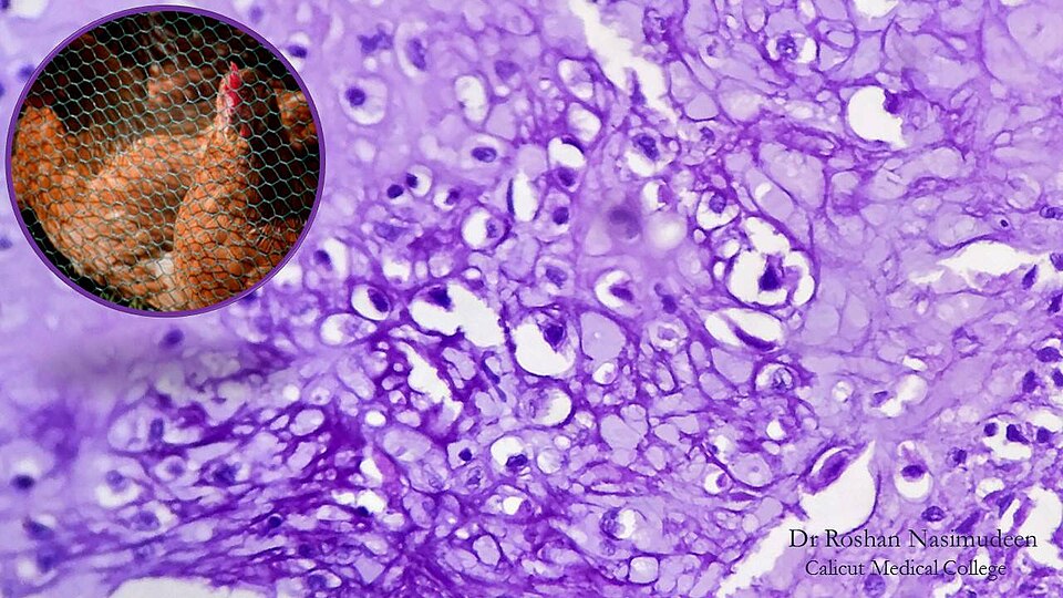
Imaging. A well-defined, eccentric epiphyseal lytic lesion with a thin sclerotic rim and punctate “chicken-wire” matrix calcification, often crossing the physis; MRI typically shows striking peritumoral marrow and soft-tissue oedema and a joint effusion.
Pathology. Sheets of chondroblasts with grooved (“coffee-bean”) nuclei and well-defined cell borders (a “mosaic” pattern), osteoclast-type giant cells, islands of chondroid matrix, and delicate “chicken-wire” pericellular calcification; S100-positive. The S100 marks the chondroblasts but not the giant cells (unlike giant cell tumour); the chicken-wire calcification is pericellular deposition on the reticulin sheath, seen on CT in about half of cases; and a secondary aneurysmal-bone-cyst component supervenes in 10 to 15 %.[33]
Differential diagnosis. Giant cell tumour (skeletally mature, no chondroid matrix), Langerhans cell histiocytosis (epiphyseal equivalents), and, in older patients, clear-cell chondrosarcoma, the malignant epiphyseal mimic.
Treatment. Curettage and bone grafting (with care not to bridge the physis in a child), curative in >90 %; radiofrequency ablation for small lesions; en-bloc resection for aggressive disease. Chondroblastoma can rarely produce histologically benign pulmonary implants. Recurrence ~10 %.
Exam pearls. Codman tumour = epiphyseal cartilage tumour of the immature skeleton; chicken-wire calcification + grooved-nucleus chondroblasts + giant cells, S100+; H3-3B K36M; can give benign lung implants; clear-cell chondrosarcoma is the malignant mimic.
II.7 Chondromyxoid Fibroma
Synonyms / WHO. Fibromyxoid chondroma. WHO 2020: benign chondrogenic tumour, ICD-O 9241/0.[34]
Definition. The rarest benign cartilage tumour, a lobulated lesion of chondroid, myxoid, and fibrous tissue arising in a metaphysis.
Epidemiology. Rare; 2nd to 3rd decades; slight male predominance.
Pathogenesis. WHO 2020 defines it by GRM1 (glutamate receptor) gene rearrangement and upregulation through promoter swapping, a highly specific marker.
Sites. The proximal tibial metaphysis (the single commonest site), elsewhere around the knee, the small bones of the foot, and the pelvis.
Clinical features. Mild, long-standing pain and swelling; no joint effusion (it is metaphyseal, not epiphyseal).
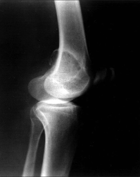
Imaging. An eccentric, metaphyseal, lobulated/scalloped lytic lesion with a sclerotic rim that “bites out of the cortex”; matrix calcification is usually absent radiographically (a contrast with chondroblastoma); high T2 signal on MRI.
Pathology. Lobules with a pale, hypocellular myxoid-chondroid centre and a dark, hypercellular periphery of spindle/stellate cells with osteoclast-type giant cells; about a third show bizarre, hyperchromatic (“pseudomalignant”) nuclei that mimic chondrosarcoma, but atypical mitoses are absent, since the atypia is degenerative. S100/SOX9-positive. This near-total absence of mitoses is the decisive point against chondrosarcoma, and intralesional calcification is radiographically absent in over 98 % of cases, which is why Bullough favours en-bloc excision over simple curettage.[35]
Differential diagnosis. Chondrosarcoma (the dangerous trap, both ways: use young age, sharp lobulated margins, and the absence of mitoses/necrosis); aneurysmal bone cyst; non-ossifying fibroma; chondroblastoma.
Treatment. Thorough curettage and grafting, or marginal/en-bloc resection for expendable bones; recurrence after curettage is relatively high (cited 12.5-25 %, occasionally higher).
Exam pearls. Metaphyseal lobulated lytic lesion that bites the cortex, no calcification; lobules with a pale myxoid centre and hypercellular periphery; bizarre nuclei WITHOUT mitoses (great chondrosarcoma mimic); GRM1.
II.8 Giant Cell Tumour of Bone (Osteoclastoma)
Synonyms / WHO. Osteoclastoma. WHO 2020: intermediate (locally aggressive and rarely metastasising), ICD-O 9250/1.[36]
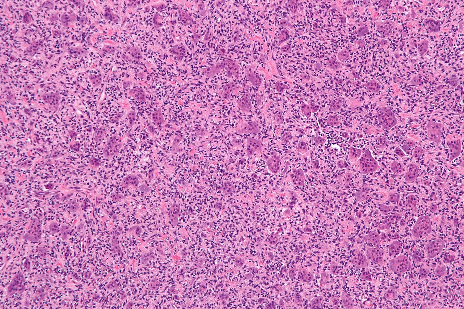
Definition. A locally aggressive neoplasm of neoplastic mononuclear stromal cells with admixed macrophages and uniformly distributed osteoclast-type giant cells, arising at the end of a long bone in a skeletally mature patient.
Epidemiology. It accounts for 4-5 % of primary and roughly 20 % of benign bone tumours. The peak lies in the third and fourth decades, almost always after physeal closure. There is a slight female predominance, a notable exception among bone tumours, and a higher incidence in China.
Pathogenesis. The mononuclear stromal cell is the neoplastic, proliferating element. It expresses RANKL, which recruits and fuses monocytes into the reactive osteoclast-like giant cells (the rationale for denosumab). WHO 2020 defines GCT by the H3-3A (H3F3A) G34W mutation (~90 %), the contrast partner to chondroblastoma’s H3-3B K36M.
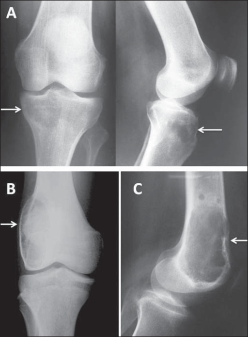
Sites. It favours the epi-metaphysis extending to subchondral bone, with ~50-60 % around the knee (distal femur, proximal tibia), then the distal radius, the proximal humerus, and the sacrum (the commonest axial site). Multicentric GCT is rare (<1 %) and should prompt exclusion of hyperparathyroidism.
Clinical features. Pain, swelling, joint symptoms, and pathological fracture (5-10 %); sacral and spinal lesions cause neurological deficit.
Imaging. An eccentric, purely lytic, geographic lesion at the articular end of a long bone, extending to subchondral bone, with a non-sclerotic margin and no matrix mineralisation (Lodwick IB/IC). A soap-bubble or trabeculated look is uncommon. MRI shows soft-tissue extent and any secondary aneurysmal-bone-cyst fluid levels. Behaviour is staged (Enneking/Campanacci 1-3) clinically and radiologically, but grading does not predict behaviour.
Pathology. Sheets of uniform oval or plump mononuclear stromal cells with evenly distributed osteoclast-type giant cells whose nuclei are identical to the stromal-cell nuclei, the single most useful diagnostic feature. There is little collagen and no tumour matrix; mitoses (typical, not atypical) are frequent; secondary aneurysmal-bone-cyst change and a fibrohistiocytic (xanthomatous) reaction are common.[37]
Differential diagnosis. Brown tumour of hyperparathyroidism (always check calcium, phosphate, and PTH), especially if the lesion is multicentric or atypically sited; giant-cell reparative granuloma; aneurysmal bone cyst; chondroblastoma; and giant-cell-rich osteosarcoma, the malignant mimic, with anaplasia and atypical mitoses.
Treatment. Extended intralesional curettage with a high-speed burr, a local adjuvant (phenol, cryosurgery, or argon-beam), and filling with PMMA cement ± bone graft is the mainstay (recurrence ~5-10 % with adjuvant, higher without). Wide resection is used for extensively destroyed bone, pathological fracture, expendable bones, or destroyed joints (recurrence <5 %). Selective arterial embolisation suits the sacrum and pelvis. Denosumab (anti-RANKL) is the modern targeted therapy for unresectable, recurrent, or metastatic disease and as a neoadjuvant to downstage. Radiotherapy is reserved for inoperable lesions because of the risk of post-radiation sarcoma.
Behaviour, metastases, transformation. “Benign” pulmonary metastases occur in roughly 2-7 % (WHO 2020 reports 3-7 %; older series cite ~1-2 %). They are histologically benign, are often curable by resection, and arise mainly from aggressive or recurrent tumours. Malignant transformation is <5 %, overwhelmingly after radiotherapy. Recurrence is the dominant clinical problem, usually within three years.
Exam pearls. Eccentric, purely lytic, subchondral lesion at the end of a long bone in a skeletally mature adult, ~60 % around the knee; stromal cell is neoplastic, giant cells reactive via RANKL; giant-cell nuclei = stromal-cell nuclei; H3-3A G34W (vs chondroblastoma H3-3B K36M); curettage + burr + adjuvant + cement, denosumab for unresectable; always check PTH; ~2-7 % benign lung mets.
II.9 Haemangioma of Bone
Synonyms / WHO. Angioma. WHO 2020: benign vascular tumour, ICD-O 9120/0 (pathogenesis unknown; molecular pathology not clinically relevant).[38]
Definition. A benign lesion of capillary, cavernous, or venous vascular channels, often regarded as a malformation rather than a true neoplasm.
Epidemiology. It is very common at autopsy (in the vertebral bodies of ~10 % of adults) but rarely symptomatic, predominantly in adults, with a slight female predominance.
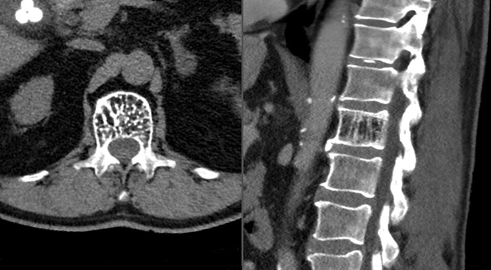
Sites. The vertebral body is the commonest site, then the skull; long bones are rare.
Clinical features. Most are asymptomatic, incidental findings. A rare aggressive thoracic vertebral haemangioma causes cord compression through epidural extension or pathological collapse. Lesions can bleed profusely at biopsy or surgery.
Imaging. In the vertebra, the radiograph shows coarse vertical “corduroy / jail-bar” striations and axial CT a “polka-dot” pattern (cross-sections of thickened trabeculae); MRI shows high signal on both T1 and T2 (fat and slow-flow vessels). In the skull, a lytic lesion shows a “sunburst / spoke-wheel” of radiating spicules.
Pathology. Thin-walled vascular channels (cavernous in the skull, capillary in long bones, mixed in the vertebra) lined by flat, bland endothelium, the feature that distinguishes it from low-grade angiosarcoma.
Treatment. Observe the asymptomatic lesion. For a symptomatic or aggressive vertebral lesion, use radiotherapy, vertebroplasty, transarterial embolisation, or surgical decompression (with embolisation to control bleeding). Resection or curettage suits accessible symptomatic lesions elsewhere.
Brief related vascular lesions. Lymphangioma (dilated lymphatic channels); cystic angiomatosis (disseminated lytic lesions with visceral, especially splenic, involvement); Gorham-Stout disease (massive osteolysis with vascular and lymphatic proliferation; see Topic 1); and the glomus tumour of the terminal phalanx (a small, exquisitely painful lytic lesion cured by excision).
Exam pearls. Vertebral body #1 site; corduroy/jail-bar (radiograph) → polka-dot (CT) → bright on T1 AND T2 (MRI); flat bland endothelium; mostly asymptomatic; aggressive thoracic lesion → cord compression.
II.10 Desmoplastic Fibroma of Bone
Synonyms / WHO. Desmoid tumour of bone. WHO 2020: intermediate, locally aggressive, ICD-O 8823/1.[39]
Definition. The intraosseous counterpart of soft-tissue fibromatosis (desmoid): a rare, locally aggressive tumour of bland fibroblasts in abundant collagen, with no metastatic capacity.
Epidemiology. Extremely rare (<0.1 % of bone tumours); mostly young adults (<30 years); near-equal sex distribution.
Sites. The mandible, long-bone metaphyses, and pelvis.
Clinical features. Pain, swelling, or pathological fracture, often long-standing because of slow growth.
Imaging. A purely lytic, expansile, trabeculated/“bubbly” lesion that may breach the cortex into soft tissue, mimicking malignancy; there is no acute periosteal reaction except at a fracture. MRI signal is variable, classically low on both T1 and T2 from dense collagen, but high on T2 in more cellular lesions.
Pathology. The most collagen-rich, most hypocellular benign fibrous lesion of bone: sparse bland spindle (myofibroblastic) cells in abundant collagen, with an infiltrative, permeative growth into marrow and Haversian canals.
Differential diagnosis. Low-grade/well-differentiated fibrosarcoma is the key and most difficult differential (more cellular, atypical, mitotically active); fibrous dysplasia (absent metaplastic woven bone, since desmoplastic fibroma engulfs lamellar bone) and non-ossifying fibroma (no giant or xanthoma cells).
Treatment. Wide or marginal en-bloc resection, not curettage, which recurs in ~40-50 %. It is the benign bone tumour that behaves most like a low-grade fibrosarcoma, yet it does not metastasise. Mirra separates it from low-grade fibrosarcoma by its extreme nuclear monotony and absence of atypical mitoses; the metastatic potential is nil, versus 5-20 % for low-grade fibrosarcoma.[40]
Exam pearls. Bone counterpart of soft-tissue desmoid; most collagen-rich/hypocellular fibrous lesion; locally aggressive, recurs after curettage, never metastasises → treat by wide excision; WHO 2020 intermediate (8823/1).
II.11 Other Benign Lesions (Brief)
Intraosseous lipoma. A rare benign tumour of mature fat, classically in the calcaneus and the intertrochanteric femur; often asymptomatic; a lytic lesion with a thin sclerotic rim and a central calcified nidus that follows fat on CT/MRI (diagnostic). Milgram stages 1-3 run from viable fat to necrosis with calcification to involution. Treatment is observation, or curettage if symptomatic.
Benign notochordal cell tumour. A benign intraosseous proliferation of notochordal cells in the axial skeleton (clivus, vertebrae, sacrococcyx), usually incidental and sclerotic on imaging; the benign counterpart and differential of chordoma.
Neurilemmoma (schwannoma) of bone. A rare benign nerve-sheath tumour, most often of the mandible or sacrum; a sharply marginated lytic lesion with a sclerotic rim; histology shows Verocay bodies; marginal excision is curative.
SECTION III --- SYNTHESIS AND EXAM AIDS
III.1 Pattern-based Differential Diagnosis
A few patterns organise the benign tumours for the exam:
- The epiphyseal lesion: chondroblastoma in the immature skeleton; giant cell tumour in the mature skeleton. (Add subchondral geode, intraosseous ganglion, and clear-cell chondrosarcoma.)
- The calcified (cartilage) matrix - “rings and arcs”: enchondroma, chondroblastoma (chicken-wire), and osteochondroma’s cap. Absence of calcification in a lobulated metaphyseal lesion favours chondromyxoid fibroma.
- The dense/ground-glass or osteoid matrix: osteoma, osteoid osteoma, osteoblastoma.
- The “aggressive benign” tumours that destroy bone and recur: giant cell tumour, osteoblastoma, desmoplastic fibroma, and aneurysmal bone cyst.
- The surface lesion: osteochondroma (medullary continuity), periosteal chondroma and parosteal osteoma (no continuity), and their malignant mimic parosteal osteosarcoma.
III.2 Quick Reference - Age, Site, and Signature
| Tumour | Typical age | Classic site | Signature |
|---|---|---|---|
| Osteoma | adult | skull, paranasal sinus | dense ivory mass; bone island cold on scan |
| Osteoid osteoma | 2nd decade | femoral neck, tibia, spine | nidus <1.5 cm, hot scan, night pain → aspirin |
| Osteoblastoma | 2nd-3rd decade | spine posterior elements | >2 cm, expansile; intermediate (9200/1) |
| Osteochondroma | <20 y | metaphysis, around knee | cortical + medullary continuity; EXT1/2 |
| Enchondroma | 2nd-4th decade | hand phalanx/metacarpal | rings-and-arcs; IDH1/2 |
| Chondroblastoma | 2nd decade (immature) | epiphysis (knee, prox. humerus) | chicken-wire Ca²⁺; H3-3B K36M |
| Chondromyxoid fibroma | 2nd-3rd decade | proximal tibia metaphysis | eccentric lobulated, no Ca²⁺; GRM1 |
| Giant cell tumour | 20-45 y (mature) | epi-metaphysis, around knee | lytic to subchondral, no matrix; H3-3A G34W |
| Haemangioma | adult | vertebral body | corduroy/polka-dot; bright T1 & T2 |
| Desmoplastic fibroma | <30 y | mandible, metaphysis, pelvis | bubbly lytic; behaves like low-grade fibrosarcoma |
III.3 Malignant-Transformation Risks
| Lesion | Becomes | Risk |
|---|---|---|
| Solitary osteochondroma | secondary peripheral chondrosarcoma | ~1 % |
| Hereditary multiple exostoses | secondary peripheral chondrosarcoma | ~5-20 % |
| Solitary enchondroma | chondrosarcoma (long/flat bones; not hand) | low |
| Ollier disease | chondrosarcoma (and non-skeletal) | ~30-40 % |
| Maffucci syndrome | chondrosarcoma + visceral/angiosarcoma | highest of all |
| Giant cell tumour | sarcoma (usually post-radiation) | <5 % |
| Osteoblastoma / desmoplastic fibroma | locally aggressive, do not metastasise | - |
III.4 When to Observe, Biopsy, or Operate
Observe (no biopsy): classic osteochondroma, incidental enostosis, asymptomatic vertebral haemangioma, calcaneal intraosseous lipoma, small asymptomatic enchondroma of the hand. Biopsy/confirm tissue for any lesion with alarm features or where low-grade malignancy cannot be excluded, and stage before biopsy, with the tract planned for excision. Operate: curettage ± adjuvant for active benign lesions (chondroblastoma, chondromyxoid fibroma, symptomatic enchondroma, giant cell tumour); radiofrequency ablation for osteoid osteoma; wide resection for desmoplastic fibroma, for giant cell tumour that has destroyed a joint, and for any suspected secondary chondrosarcoma; treat the cause (parathyroidectomy) before calling a giant-cell lesion a tumour.
III.5 The Bulgarian Operative Tradition (Boychev) and Terminology
Boychev’s Хирургическа ортопедия contributes operative techniques and the vocabulary expected in the Bulgarian examination.
Boychev osteoplastic resection of the knee (костно-пластична резекция по Бойчев). This is used for a large benign periarticular tumour of the distal femur or proximal tibia, a giant cell tumour (остеокластом) or osteochondroma, and for slowly-growing low-grade malignancies. The tumour-bearing bone end is divided transversely well clear of the lesion and removed “ablastically” (no-touch). The defect is bridged by a local autograft rotated 180° (taken from the anterior tibia when the distal femur is resected, or the anterior femur when the proximal tibia is resected) and held between medial and lateral plates whose screws engage both plates, in effect an osteoplastic arthrodesis. Plaster immobilisation continues for more than six months until union.
Marginal excision of a benign tumour (отстраняване на доброкачествени тумори). The tumour is freed with its capsule down to its bony base. The periosteum is incised around the base, and the lesion is removed with a broad gouge chisel together with the periosteum and a shell of healthy bone, an en-bloc marginal excision.
Resection and allograft reconstruction for distal-femoral osteoclastoma. For a giant cell tumour that has thinned but not breached the cortex, curettage and plombage (кюртиране и пломбиране) may suffice. For extensive destruction, the bone end is resected and replaced with a frozen allograft (алотрансплантат) fixed by plate or Küntscher nail (the graft drilled to aid revascularisation), or by an alloplastic arthrodesis of the knee as an alternative to amputation.
III.6 High-Yield Revision Summary
- Osteoma - ivory skull/sinus mass; bone island cold on scan; multiple → Gardner (APC).
- Osteoid osteoma - night pain relieved by aspirin; nidus <1.5 cm, hot scan, CT to find; spine → painful scoliosis; radiofrequency ablation; FOS.
- Osteoblastoma - >2 cm, spine posterior elements; WHO intermediate (9200/1); osteosarcoma is the DDx; FOS/FOSB.
- Osteochondroma - cortical + medullary continuity, points away from joint, stops at maturity; EXT1/2; cap >1.5-2 cm in an adult → secondary chondrosarcoma.
- Enchondroma - commonest hand tumour, rings-and-arcs, hypocellular cartilage; IDH1/2; rest pain/deep scalloping → atypical cartilaginous tumour; Ollier/Maffucci = mosaic IDH, high malignancy.
- Chondroblastoma - epiphysis of the immature skeleton, chicken-wire calcification, grooved chondroblasts; H3-3B K36M; benign lung implants.
- Chondromyxoid fibroma - metaphyseal lobulated lytic lesion, no calcification; bizarre nuclei without mitoses; GRM1.
- Giant cell tumour - mature skeleton, end of long bone, lytic to subchondral, no matrix; stromal cell neoplastic, RANKL; H3-3A G34W; check PTH; denosumab; ~2-7 % benign lung mets.
- Haemangioma - vertebral body, corduroy/polka-dot, bright on T1 and T2.
- Desmoplastic fibroma - bubbly lytic, behaves like low-grade fibrosarcoma, wide excision, no metastasis.
References
- WHO Classification of Tumours Editorial Board. Soft Tissue and Bone Tumours. WHO Classification of Tumours, 5th ed. Lyon: IARC; 2020.
- Schajowicz F. Tumors and Tumorlike Lesions of Bone: Pathology, Radiology and Treatment. 2nd ed. Berlin: Springer-Verlag; 1994.
- Campanacci M. Bone and Soft Tissue Tumors. 2nd ed. Vienna/New York: Springer; 1999.
- Dorfman HD, Czerniak B. Dorfman and Czerniak’s Bone Tumors. 2nd ed. Philadelphia: Elsevier; 2016.
- Greenspan A, Beltran J. Orthopedic Imaging: A Practical Approach. 6th ed. Philadelphia: Wolters Kluwer; 2015.
- Wold LE, Unni KK, Sim FH, Adler C-P, Sundaram M, Inwards CY. Atlas of Orthopedic Pathology. 3rd ed. Philadelphia: Saunders/Elsevier; 2008.
- Bickels J, Wittig JC, Malawer MM, et al. Operative Techniques in Orthopaedic Surgical Oncology. 2nd ed. Philadelphia: Wolters Kluwer; 2015.
- Malawer MM, Sugarbaker PH, eds. Musculoskeletal Cancer Surgery. Dordrecht: Kluwer Academic; 2001.
- Behjati S, Tarpey PS, Presneau N, et al. Distinct H3F3A and H3F3B driver mutations define chondroblastoma and giant cell tumour of bone. Nat Genet. 2013;45(12):1479-1482.
- Amary MF, Bacsi K, Maggiani F, et al. IDH1 and IDH2 mutations are frequent events in central chondrosarcoma and central and periosteal chondromas but not in other mesenchymal tumours. J Pathol. 2011;224(3):334-343.
- Бойчев Б. (Boychev B.) Хирургическа ортопедия (Surgical Orthopaedics). Sofia.
- Mirra JM, Picci P, Gold RH. Bone Tumors: Clinical, Radiologic, and Pathologic Correlations. Philadelphia: Lea & Febiger; 1989.
- Bullough PG. Orthopaedic Pathology. 4th ed. Edinburgh: Mosby; 2004.
Note on currency: the molecular classification (FOS/FOSB, EXT1/EXT2, IDH1/IDH2, H3F3B K36M, H3F3A G34W, GRM1) and ICD-O behaviour codes are taken from the WHO Classification of Tumours: Soft Tissue and Bone Tumours, 5th ed. (2020); denosumab for giant cell tumour and radiofrequency ablation for osteoid osteoma reflect current practice.
- WHO Classification of Tumours: Soft Tissue and Bone Tumours, 5th ed. (IARC, 2020): chondroblastoma p.370 (H3-3B K36M); giant cell tumour of bone p.452 (H3-3A G34W in ≥95 %, ~90 % G34W). The originating study is Behjati S, et al. Distinct H3F3A and H3F3B driver mutations define chondroblastoma and giant cell tumour of bone. Nat Genet 2013;45:1479-82.
- WHO 2020 p.401-403: osteoma 9180/0; “a benign tumour arising on the surface of bone and composed primarily of lamellar/cortical-type bone”; multiple osteomas suggest Gardner syndrome (APC), multiple bone islands suggest osteopoikilosis (LEMD3).
- Bone island cold/mildly-warm on scintigraphy vs hot osteoid osteoma and hot osteoblastic metastasis: Schajowicz p.47; Campanacci p.387; Greenspan p.1462, Table 17.2.
- Bullough, Orthopaedic Pathology, 4th ed., pp.385-387.
- WHO 2020 p.404-405: osteoid osteoma 9191/0; “a benign bone-forming tumour characterized by small size (<2 cm) and limited growth potential”; FOS rearrangement in 91 %, detectable by anti-FOS immunohistochemistry; 10-12 % of primary bone tumours, M:F 2:1.
- Prostaglandin/COX-2 mechanism of pain: Campanacci p.409; Dorfman p.157-159; Makley & Dunn, Lancet 1982 (cited WHO 2020).
- Mirra, Bone Tumors (1989), pp.248-250.
- CT is the modality of choice for the nidus; MRI overcalls peritumoral oedema: Greenspan p.1453; Campanacci p.404-405; Dorfman p.159.
- WHO 2020 p.407-408: osteoblastoma 9200/1, “a locally aggressive bone-forming tumour, morphologically similar to osteoid osteoma but … generally >2 cm”; FOS rearrangement in 87 %, FOSB in a smaller fraction; <1 % of primary bone tumours, peak 2nd-3rd decades, M:F 2:1.
- Permeative margin is the key discriminator of osteosarcoma from osteoblastoma: Campanacci p.430; Dorfman p.186; epithelioid/aggressive osteoblastoma (Dorfman & Weiss 1984) - Dorfman p.193, Schajowicz p.80.
- Mirra, Bone Tumors (1989), p.422.
- WHO 2020 p.366: osteochondroma 9210/0; “a benign cartilaginous neoplasm … a bony projection covered by a cartilage cap … containing a marrow cavity continuous with that of the underlying bone”; caused by biallelic inactivation of EXT1 or EXT2; ~8 % of all and ~35 % of benign bone tumours; ~15 % have multiple lesions.
- Solitary malignant transformation ~1 % (Schajowicz p.188; Campanacci p.205; Greenspan p.1539); HME ~5-20 % (Campanacci p.215; Schajowicz p.188; Greenspan p.1556). Cap >1.5-2 cm in an adult is suspicious but there is no absolute cut-off (Schajowicz p.184).
- Bullough, Orthopaedic Pathology, 4th ed., pp.416-418.
- WHO 2020 p.363-364: enchondroma 9220/0; “a benign hyaline cartilaginous neoplasm that arises within the medullary cavity”; caused by IDH1/IDH2 mutations (52 % of sporadic enchondromas, ~90 % in enchondromatosis); usually 3rd-4th decades, sexes equal.
- Enchondroma vs atypical cartilaginous tumour/low-grade chondrosarcoma: Greenspan p.1527-1528; Wold p.241; the IDH mutation does not discriminate (WHO 2020 p.364). Hand cartilage tumours are almost never malignant: Campanacci p.227.
- Mirra, Bone Tumors (1989), pp.496-500.
- Ollier/Maffucci = somatic mosaic IDH1/IDH2 (WHO 2020; Amary et al., Nat Genet 2011). Malignant risk: Ollier ~30-40 % (Campanacci p.254); Maffucci highest, with visceral and non-skeletal malignancies (Campanacci p.254; Schajowicz p.156).
- WHO 2020 p.369-370: chondroblastoma 9230/0; “a benign tumour … with a predilection for epiphyseal or apophyseal regions, composed of chondroblastic cells and islands of eosinophilic chondroid matrix”; defined by H3-3B (H3F3B) K36M; <1 % of bone tumours, 10-25 years, M:F 2:1.
- Mirra, Bone Tumors (1989), pp.629-639; Bullough, Orthopaedic Pathology, 4th ed., p.426.
- WHO 2020 p.372-373: chondromyxoid fibroma 9241/0; “a benign lobulated cartilaginous neoplasm with a zonal architecture composed of chondroid, myxoid, and myofibroblastic areas”; driven by GRM1 rearrangement/promoter-swap upregulation; rare, most often 2nd-3rd decades, male-predominant.
- Mirra, Bone Tumors (1989), pp.644-654; Bullough, Orthopaedic Pathology, 4th ed., p.429.
- WHO 2020 p.450-452: giant cell tumour of bone 9250/1; “a locally aggressive and rarely metastasizing neoplasm composed of neoplastic mononuclear stromal cells … admixed with macrophages and osteoclast-like giant cells. A small subset of cases are malignant”; ≥95 % harbour H3-3A (H3F3A) mutations, ~90 % G34W; 4-5 % of all and ~20 % of benign bone tumours; peak 20-45 years; slight female predominance; lung metastases in 3-7 %; malignant GCT <10 %.
- The giant-cell nuclei matching the stromal-cell nuclei, with even giant-cell distribution and no reactive matrix, distinguishes GCT from the clustered giant cells of reparative granuloma/brown tumour: Dorfman p.705, p.716; Schajowicz p.301-302; Campanacci p.128.
- WHO 2020 p.436-437: haemangioma 9120/0; “a benign tumour … composed of vascular channels of small or large calibre”; pathogenesis “unknown”, molecular pathology “not clinically relevant”; found at autopsy in vertebrae of ~10 % of adults, symptomatic in <1 %.
- WHO 2020 p.432-433: desmoplastic fibroma 8823/1; “an extremely rare locally aggressive bone tumour composed of bland spindle cells set in abundant collagen, with histology reminiscent of desmoid-type fibromatosis”; no consistent defining alteration - diagnosis of exclusion (absent GNAS excludes fibrous dysplasia; absent MDM2 amplification excludes low-grade central osteosarcoma); <0.1 % of bone tumours.
- Mirra, Bone Tumors (1989), pp.776-777.
Figure Credits and Licences
All figures are radiographs, CT images, clinical images, or photomicrographs reproduced from openly-licensed sources; each remains under its original licence, attributed below.
- Fig 1 Osteoma, frontal sinus CT - Hellerhoff, CC BY-SA 4.0, Wikimedia Commons. https://commons.wikimedia.org/wiki/File:Osteom_im_Sinus_frontalis_rechts_84M_-_CT_-_001.jpg
- Fig 2 Osteoid osteoma, fibula CT - Hellerhoff, CC BY-SA 3.0, Wikimedia Commons. https://commons.wikimedia.org/wiki/File:Osteoidosteom_Fibula_CT_KF.png
- Fig 3 Osteoid osteoma histology - Nephron, CC BY-SA 3.0, Wikimedia Commons. https://commons.wikimedia.org/wiki/File:Osteoid_osteoma_-_intermed_mag.jpg
- Fig 4 Osteoblastoma, C7 CT - Li Z, Zhao Y, et al., PLOS ONE 2013, CC BY-SA 4.0, Wikimedia Commons. https://commons.wikimedia.org/wiki/File:Osteoblastoma_C7_TC.png
- Fig 5 Osteochondroma, distal femur - Lucien Monfils, CC BY-SA 3.0, Wikimedia Commons. https://commons.wikimedia.org/wiki/File:EXOSTOSE.jpg
- Fig 6 Osteochondroma histology (cartilage cap) - Sarahkayb, CC BY-SA 4.0, Wikimedia Commons. https://commons.wikimedia.org/wiki/File:Bone_Osteochondroma_LP_PA.JPG
- Fig 7 Hereditary multiple exostoses - Bovée JVMG, Orphanet J Rare Dis 2008;3:3, CC BY-SA 3.0 NL, Wikimedia Commons. https://commons.wikimedia.org/wiki/File:MO_radiograph.jpg
- Fig 8 Enchondromatosis (Ollier), hands - Hellerhoff, CC BY-SA 3.0, Wikimedia Commons. https://commons.wikimedia.org/wiki/File:Morbus_Ollier_Finger_Roentgen.png
- Fig 9 Chondroblastoma, proximal tibia - Kundu ZS, et al., J Orthop Surg Res 2010;5:47, CC BY, via PMC. https://www.ncbi.nlm.nih.gov/pmc/articles/PMC2921358/
- Fig 10 Chondroblastoma histology (chicken-wire) - R. Nasimudeen, CC BY-SA 4.0, Wikimedia Commons. https://commons.wikimedia.org/wiki/File:Chicken_wire_calcification_chondroblastoma.jpg
- Fig 11 Chondromyxoid fibroma, distal femur - Kumar N, et al., J Cytol 2010;27(3):96, CC BY, via PMC. https://www.ncbi.nlm.nih.gov/pmc/articles/PMC2983082/
- Fig 12 Giant cell tumour, knee - Costa FM, et al., Radiol Bras 2016;49(3):182, CC BY, via PMC. https://www.ncbi.nlm.nih.gov/pmc/articles/PMC4938449/
- Fig 13 Giant cell tumour histology - Nephron, CC BY-SA 3.0, Wikimedia Commons. https://commons.wikimedia.org/wiki/File:Giant_cell_tumour_of_bone_-_intermed_mag.jpg
- Fig 14 Vertebral haemangioma, CT - Hellerhoff, CC BY-SA 3.0, Wikimedia


