Contents
- Scope and orientation
- Part I - General concepts of adult osteonecrosis
- Part II - Osteonecrosis of the femoral head (and Chandler disease)
- Part III - Kienböck disease (lunatomalacia)
- Part IV - Other adult avascular-necrosis sites
- Bulgarian terminology glossary
- Figure credits and licences
Scope and orientation
This topic covers the avascular necroses of the adult skeleton. The centrepiece is osteonecrosis of the femoral head (ONFH), for which Chandler disease is the old eponym for the idiopathic adult form, together with Kienböck disease (lunatomalacia), the osteonecrosis of the lunate named in the syllabus. The other adult sites (knee, talus, scaphoid, humeral head) complete the picture.
A few unifying ideas carry the topic:
- One disease, many sites. Whatever the trigger, bone dies when its blood supply fails. It is repaired by creeping substitution and may then fracture beneath the joint surface (the crescent sign) and collapse. The femoral head, lunate, talar body and scaphoid proximal pole all share a tenuous, often single, end-arterial supply.
- The crescent sign is the hinge of management. Before subchondral collapse the joint can be preserved; after collapse the options narrow toward replacement. The staging systems all turn on this boundary.
- MRI is the early test. Radiographs are normal for weeks; MRI shows the reactive band long before collapse.
- Two controversies recur: whether spontaneous osteonecrosis of the knee is really a subchondral insufficiency fracture rather than primary necrosis, and whether ulnar negative variance causes Kienböck disease or merely drives its progression.
A Bulgarian terminology glossary, a viva self-test appendix, and image credits follow the clinical sections.
Part I - General concepts of adult osteonecrosis
Osteonecrosis (ON) is death of bone and marrow from loss of blood supply; it is a condition, not a single disease, and the synonyms avascular, aseptic and ischaemic necrosis are used interchangeably.[1] By convention, avascular necrosis is applied to epiphyseal/subarticular involvement and bone infarct to metaphyseal/diaphyseal involvement.[2] The femoral head is the commonest site (an end-arterial supply with little collateral), followed by the humeral head, femoral condyles, talus and scaphoid.[3]
The final common pathway is focal intravascular thrombosis of terminal vessels and/or raised intraosseous pressure compressing the subchondral microvasculature.[4] Histologically, marrow fat necrosis is recognisable about five days after the insult and empty osteocyte lacunae are the hallmark. Repair proceeds by creeping substitution (new bone laid on dead trabeculae), but osteoclastic resorption at the reparative front weakens the subchondral bone, producing the subchondral fracture (crescent sign) and eventual collapse.[5] Once the necrotic, empty-lacunae zone exceeds about 1 cm² the lesion is no longer reversible.[6]
Part II - Osteonecrosis of the femoral head (and Chandler disease)
II.1 Definition and Chandler disease
Osteonecrosis of the femoral head is death of the subchondral bone of the head from interruption of its blood supply. Chandler disease is the eponym for idiopathic avascular necrosis of the adult femoral head. In 1948 Chandler described the vascular basis of the condition in a paper entitled “Coronary Disease of the Hip,” drawing the analogy that, just as coronary ischaemia kills heart muscle, ischaemia of the femoral-head vessels kills subchondral bone.[7] The first detailed description of bilateral aseptic necrosis of the femoral heads was Freund’s (1936); Trueta and Harrison mapped the adult vascular anatomy in 1953.[8]
II.2 Epidemiology
- >20,000 new cases per year in the USA; ON accounts for about 10% of all primary total hip replacements, rising to roughly half of THAs in Korea and Taiwan.[9]
- Typically the 3rd-5th decade; the great majority are under 50.[10]
- Bilateral in a high proportion (commonly taught as ~50%, with series reporting 40-80% and higher), which justifies imaging the often-silent contralateral hip.[11]
II.3 Aetiology and risk factors
The two commonest atraumatic causes are corticosteroid and alcohol, which together account for about 80% of non-traumatic cases.[12]
- Corticosteroid: there is a dose relationship, with risk rising above a cumulative dose of ~2 g of prednisone equivalent; in the SARS cohort the rate was 0.6% below 3 g versus 13% above 3 g. The mechanism includes marrow-fat hypertrophy raising intraosseous pressure, fat embolism, and down-regulated angiogenesis.[13]
- Alcohol: a clear dose-response (relative odds rising to ~14 at the highest intake).[14]
- Idiopathic (= Chandler disease), post-traumatic (femoral-neck fracture, hip dislocation; see II.9), and a longer list: sickle-cell disease, Gaucher disease, dysbarism/Caisson disease, SLE, radiation, chemotherapy, organ transplantation, renal failure, coagulopathy, pregnancy.[15] Subtle coagulopathies (thrombophilia, hypofibrinolysis) are found in up to 70% of ON patients when sought.[16]
II.4 Pathophysiology and vascular anatomy
The pathophysiology follows Part I. The relevant vascular point is that the adult head is supplied mainly by the deep branch of the medial femoral circumflex artery, which becomes the posterosuperior (lateral epiphyseal) retinacular vessels. These run extra-osseous but intracapsular and enter the head a few millimetres from the articular margin, with the obturator externus protecting the artery. The lateral circumflex and the artery of the ligamentum teres contribute little in the adult, so head viability depends on the medial circumflex.[17]
II.5 Clinical presentation
The usual complaint is groin pain (occasionally trochanteric or buttock pain), deep and worse on weight-bearing and at night, with a limp and painful, restricted internal rotation. Acute worsening signals subchondral collapse. Many patients are asymptomatic, particularly in the contralateral hip, and there is little correlation between radiographic stage and symptoms (Ficat’s “silent hip,” stage 0).[18]
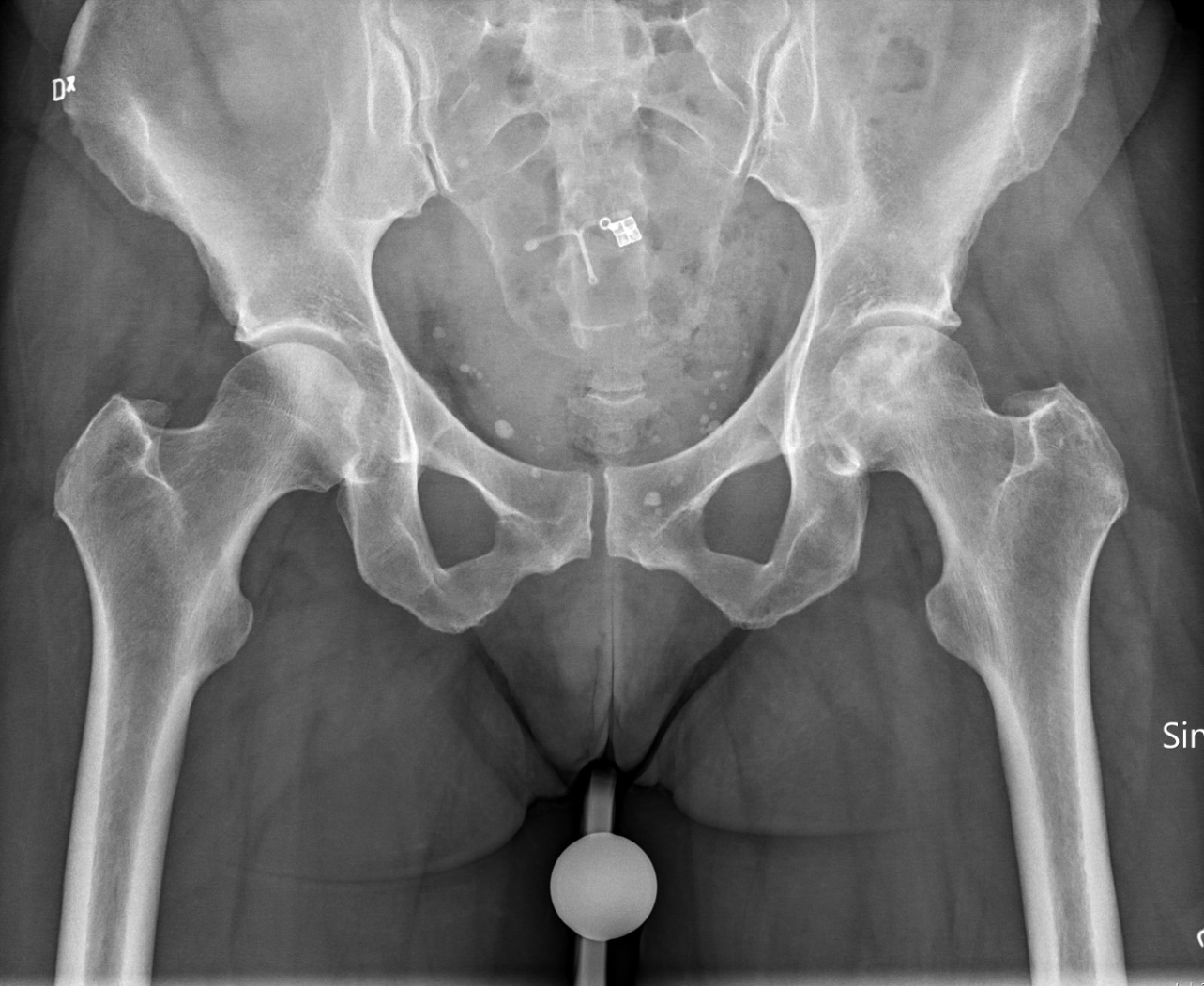
Antero-posterior pelvis showing bilateral idiopathic osteonecrosis of the femoral heads: both heads have mottled subchondral sclerosis and lucency. (Mikael Häggström MD, Wikimedia Commons; CC0.)
Antero-posterior pelvis showing bilateral idiopathic osteonecrosis of the femoral heads: both heads have mottled subchondral sclerosis and lucency. (Mikael Häggström MD, Wikimedia Commons; CC0.)
II.6 Staging and classification
Several systems coexist; all pivot on the crescent sign = subchondral fracture = the pre-collapse → collapse boundary, best seen on the frog-leg lateral.[19]
- Ficat-Arlet (0/I-IV): 0 = silent hip (normal radiograph, no symptoms); I = pain, normal radiograph; II = sclerosis/cysts with a preserved spherical head; III = crescent sign / sequestrum ± segmental flattening, joint space preserved; IV = collapse with secondary osteoarthritis. Its weakness is that it does not quantify lesion size.[20]
- ARCO (0-4 in the 1994 simplification): 0 = normal, histology only; 1 = MRI/scan positive, radiograph normal; 2 = sclerosis/cysts, no subchondral fracture; 3 = subchondral fracture; 4 = osteoarthritis. ARCO also records lesion size and location, and the 2002 Nijmegen revision split stage 3 into early-3 (no collapse) versus late-3 (definite collapse), since joint-preserving results are far better before collapse.[21]
- Steinberg / University of Pennsylvania (0-VI): the first to make MRI and quantitative lesion size integral, subdividing each stage A <15% / B 15-30% / C >30% of the head; stage III = crescent without flattening, IV = flattening, V = joint-space narrowing/acetabular change, VI = advanced degeneration.[22]
- JIC (Japanese Investigation Committee): classifies by location of the lesion on the mid-coronal view, which predicts collapse: type A (medial third) collapses <10%, type B (medial two-thirds) ~40%, type C1 (more than two-thirds, not to the acetabular edge) ~80%, type C2 (extending to the lateral edge) ≥90%. The C-type (lateral) lesion is the high-risk lesion.[23]
- Kerboul combined necrotic angle (sum of the necrotic arc on AP + lateral): <200° is favourable for joint-preserving surgery; the MRI-modified Kerboul (Ha) uses ≤190° low-risk, ≥240° high-risk.[24]
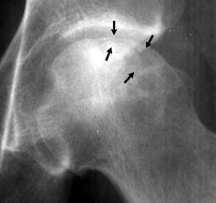
Antero-posterior hip with the subchondral crescent sign (arrows) - a lucent subchondral fracture line beneath the articular surface, marking the pre-collapse-to-collapse boundary. (Mikael Häggström, after OrthopaedicsOne, Wikimedia Commons; CC BY-SA 3.0.)
Antero-posterior hip with the subchondral crescent sign (arrows) - a lucent subchondral fracture line beneath the articular surface, marking the pre-collapse-to-collapse boundary. (Mikael Häggström, after OrthopaedicsOne, Wikimedia Commons; CC BY-SA 3.0.)
II.7 Imaging
Radiographs are normal early, then show mottled sclerosis/lucency, the crescent sign, head flattening and finally secondary osteoarthritis.[25] MRI is the most sensitive and specific test (around 97-99%): the band sign, a serpiginous low-signal rim on T1 demarcating necrotic from viable bone, is the first detectable finding, and the double-line sign on T2 (a high-signal inner line with a low-signal outer rim) is seen in up to 80% and regarded by many as pathognomonic.[26] A bone scan shows the “cold-in-hot” doughnut pattern but misses over 20% of lesions; CT best demonstrates a subtle subchondral fracture.[27] The key differentials are transient osteoporosis of the hip / bone-marrow oedema syndrome (diffuse oedema crossing into the neck, self-limiting, no demarcating band) and subchondral insufficiency fracture (a fracture line without the outer reactive band).[28]
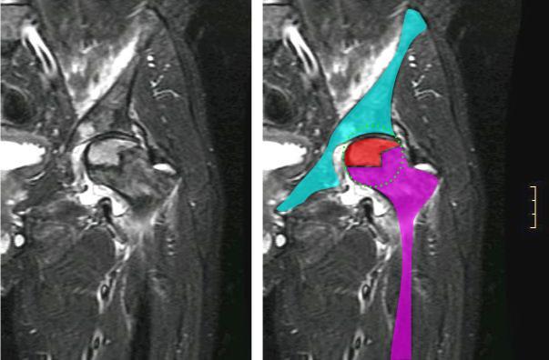
Coronal hip MRI of left femoral-head osteonecrosis: the left panel is the plain image; the right panel colour-codes the demarcated necrotic segment (red) within the head. (Jmarchn, Wikimedia Commons; CC BY-SA 3.0.)
Coronal hip MRI of left femoral-head osteonecrosis: the left panel is the plain image; the right panel colour-codes the demarcated necrotic segment (red) within the head. (Jmarchn, Wikimedia Commons; CC BY-SA 3.0.)
II.8 Natural history and prognosis
Untreated, a symptomatic hip progresses to collapse and arthroplasty in up to ~80%, most within four years.[29] Small, medial, non-weight-bearing lesions may stay asymptomatic for life. The predictors of collapse are the stage (pre- vs post-collapse), lesion size (Kerboul >200°, Steinberg C), head depression (>2 mm being poor), lesion location (the C-type/lateral lesion) and surrounding bone-marrow oedema.[30]
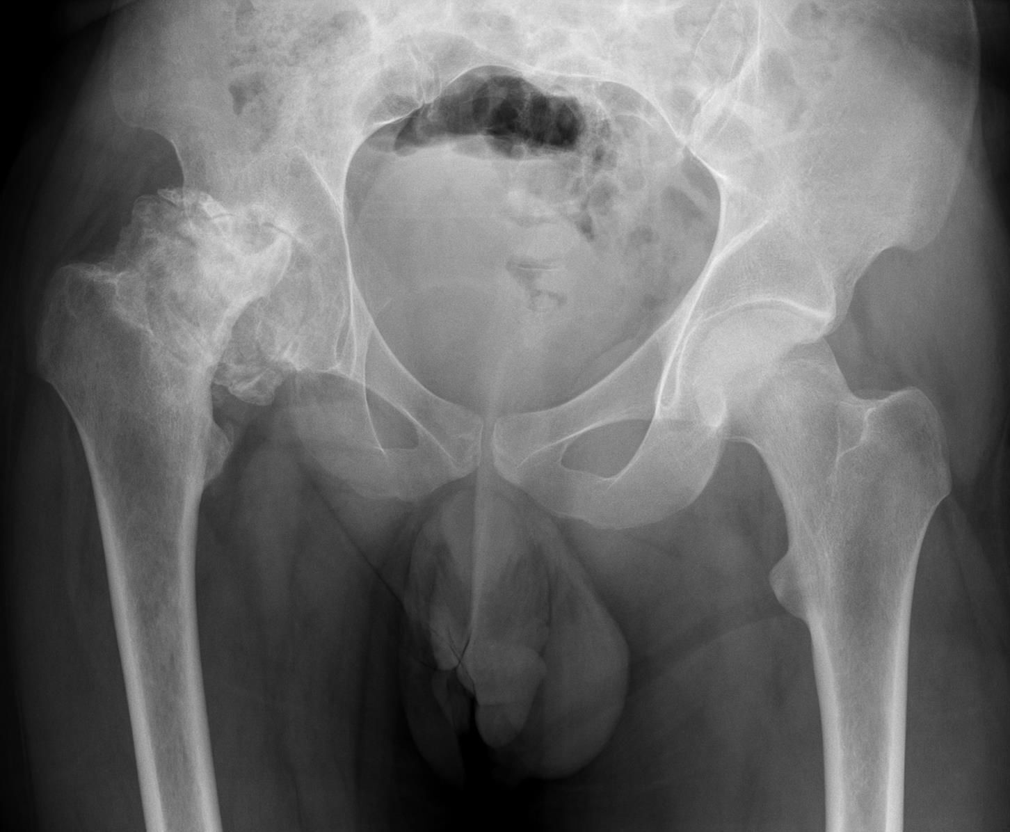
Antero-posterior pelvis in advanced steroid-induced osteonecrosis: the right femoral head (image left) shows collapse, fragmentation and sclerosis. (Hellerhoff, Wikimedia Commons; CC BY-SA 3.0.)
Antero-posterior pelvis in advanced steroid-induced osteonecrosis: the right femoral head (image left) shows collapse, fragmentation and sclerosis. (Hellerhoff, Wikimedia Commons; CC BY-SA 3.0.)
II.9 Management
The aim is early, joint-preserving treatment before collapse, with arthroplasty reserved for end-stage disease; stage and lesion size drive the choice.[31]
- Non-operative. Protected weight-bearing alone is unsatisfactory (pooled satisfactory results only ~22%); pharmacologic agents (bisphosphonates, statins, anticoagulants, iloprost) remain investigational.[32]
- Core decompression (± bone graft, BMP or autologous bone-marrow/MSC; or multiple small drillings) is the mainstay pre-collapse: satisfactory in ~63% versus ~23% for non-operative care, best in Ficat I-II with Kerboul <200°.[33]
- Bone grafting: non-vascularised (Phemister, light-bulb, trapdoor; ~77-80%) and vascularised free fibular graft (Urbaniak ~91% stage II, 77% stage III survivorship) for larger pre-collapse or early-collapse lesions.[34]
- Osteotomy: the Sugioka transtrochanteric anterior rotational osteotomy rotates the necrotic anterosuperior segment off the weight-bearing dome (best in young patients with a viable posterior arc), and varus/valgus intertrochanteric osteotomy; results are best in Asian series.[35]
- Arthroplasty for end-stage (Ficat III-IV): modern total hip arthroplasty with ceramic-on-ceramic or ceramic/metal-on-highly-cross-linked-polyethylene bearings now approaches the results for osteoarthritis (≈90%+ at 10 years), with metal-on-metal avoided in the young; resurfacing and hemiresurfacing are bone-conserving options in selected young patients, while bipolar hemiarthroplasty and tantalum rods are not recommended.[36]
Post-traumatic ONFH deserves separate mention: it follows interruption of the medial circumflex artery after a femoral-neck fracture (~12-23%; higher with subcapital level, displacement, malreduction and delay), a femoral-head fracture (up to ~40%) or a traumatic hip dislocation (~26%; worse if reduction is delayed beyond 6-12 hours). The only effective measure is prevention, by prompt anatomic reduction and capsular decompression.[37]
Part III - Kienböck disease (lunatomalacia)
III.1 Definition and history
Kienböck disease is osteonecrosis of the lunate (German lunatomalacia, “softening of the moon-bone”). Robert Kienböck, a Viennese radiologist, described it in 1910, attributing it to compromised blood supply under stress rather than to a single injury; Peste’s earlier (1843) report was of a fractured, dislocated lunate, not true lunatomalacia.[38] The lunate is the keystone of the proximal carpal row, so its collapse disturbs the whole carpus.[39]
III.2 Epidemiology
Classically the disease of the dominant hand of a male manual worker aged 20-40, usually unilateral, with bilateral disease rare.[40] More recent general-hospital series challenge the stereotype, reporting a mean age in the mid-30s with a near-equal sex ratio, infantile and juvenile cases (which carry a much better prognosis through spontaneous revascularisation), and a separate elderly, female-predominant group that often does well non-operatively.[41]
III.3 Vascular anatomy of the lunate
The lunate is supplied by palmar and dorsal nutrient vessels; about 80% of lunates receive both, but ~20% depend on a single (palmar) supply.[42] Gelberman described three intraosseous patterns, Y (59%), I (31%) and X (10%), and the lunates “at risk” are those with a single vessel or no internal anastomosis. The proximal subchondral pole is a watershed (terminal) zone, and the subarticular venous plexus is vulnerable to a subchondral fracture, so raised intraosseous pressure (venous outflow obstruction) is also implicated.[43]
III.4 Aetiology and pathogenesis
The cause is multifactorial and unresolved: the standard text presents a mechanical/structural theory alongside a biologic/vascular one.[44] The classic mechanical factors all increase load on the lunate: ulnar negative variance (Hultén found it in 78% of cases), an apex-shaped Antuña-Zapico type-I lunate, a small or radially-inclined lunate, a thin proximal subchondral plate (~0.1 mm, prone to a stress fracture), and a single vascular supply.[45]
The ulnar-variance controversy. Hultén’s view that a short ulna shifts load onto the radial half of the lunate has been challenged: several series found neutral or positive variance, “pseudolengthening” of the radius can falsify the measurement, and one meta-analysis (Chung) found no correlation while another (Ståhl) did but without satisfying causation criteria. The consensus position is that ulnar negative variance probably promotes progression of collapse rather than initiating the disease.[46]
III.5 Classification
- Lichtman (the standard, modifying Ståhl): I normal radiograph, MRI-positive; II lunate sclerosis without collapse; IIIA lunate collapse with carpal height and alignment preserved (normal radioscaphoid angle); IIIB lunate collapse with fixed scaphoid rotatory flexion (radioscaphoid angle >60°, proximal capitate migration, scaphoid “ring sign”); IV lunate collapse with radiocarpal/midcarpal arthritis. The IIIA/IIIB distinction rests on the fixed scaphoid flexion (a radioscaphoid angle >60°, chosen because it does not involve the diseased lunate). Later refinements add a stage 0 and a stage IIIC (coronal fracture/fragmentation).[47]
- Bain & Begg arthroscopic classification grades the number of non-functional articular surfaces (0-4) among the proximal lunate, the lunate facet of the radius, the distal lunate and the proximal capitate; it guides whether to unload/revascularise (where surfaces remain functional) or to salvage.[48]
- Schmitt/Nakamura gadolinium-MRI staging grades viability: pattern A (oedema, viable, good prognosis), B (partial necrosis), C (complete necrosis, no enhancement, poor prognosis).[49]
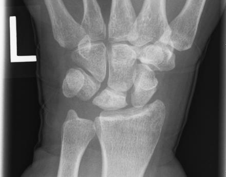
Postero-anterior wrist radiograph in Kienböck disease: the lunate is sclerotic (denser) relative to the adjacent carpal bones. (User Muzichick, Wikimedia Commons; CC BY-SA 4.0.)
Postero-anterior wrist radiograph in Kienböck disease: the lunate is sclerotic (denser) relative to the adjacent carpal bones. (User Muzichick, Wikimedia Commons; CC BY-SA 4.0.)
III.6 Clinical features and imaging
The presentation is insidious dorsal/central wrist pain aggravated by extension, with local swelling and tenderness over the lunate, reduced grip strength and a reduced range of motion. Diagnosis is often delayed by 1-2 years.[50] On the standard PA radiograph (forearm neutral) one measures ulnar variance (normal ≤ 2 mm), the radioscaphoid angle (normal 30-60°) and the carpal height ratio. MRI is the only modality positive in stage I (diffuse low T1 signal, with T2/gadolinium indicating viability), and CT best shows fragmentation and coronal fractures, often upstaging the disease.[51]
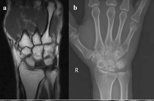
Kienböck disease: (a) coronal wrist MRI with diffuse low signal throughout the lunate; (b) the matching PA radiograph. (Xie et al., via Wikimedia Commons; CC BY-SA 4.0.)
Kienböck disease: (a) coronal wrist MRI with diffuse low signal throughout the lunate; (b) the matching PA radiograph. (Xie et al., via Wikimedia Commons; CC BY-SA 4.0.)
III.7 Treatment by stage
There is no consensus on the best procedure; the decision rests on stage, ulnar variance, the state of the articular cartilage and the patient’s age.[52]
- Stage 0-I (and all children): immobilisation/observation, treating any cause; children frequently revascularise.[53]
- Stages II-IIIA (no fixed carpal collapse, functional cartilage):
- Joint-levelling: radial shortening osteotomy is the preferred procedure for the ulnar-negative wrist (shorten ~2-3 mm; more than 4 mm worsens outcomes); for the ulnar-neutral or positive wrist use a radial wedge or capitate-shortening osteotomy (which does not alter ulnar variance).[54]
- Revascularisation: vascularised bone grafts (the 4+5 extensor-compartmental artery graft is the Mayo/Green’s preference; also 2,3-ICSRA, pisiform transfer, and the free medial-femoral-condyle flap), ideally with an intact cartilage shell, often combined with temporary unloading.[55]
- Core decompression of the lunate or distal radius/metaphysis, and arthroscopic “forage,” for early disease with intact surfaces.[56]
- Stage IIIB (fixed carpal collapse): intercarpal fusion to unload the lunate, that is a scaphotrapeziotrapezoid (STT) or scaphocapitate fusion (redirecting load through the scaphoid and controlling its rotation), or a radioscapholunate fusion if the midcarpal joint is preserved; the fragmented lunate is generally retained.[57]
- Stage IIIC/IV (fragmentation or pancarpal arthritis): salvage by proximal-row carpectomy (best in patients over 40 with an intact capitate head and lunate fossa), total wrist arthrodesis, total wrist arthroplasty, or wrist denervation for pain relief while preserving motion.[58]
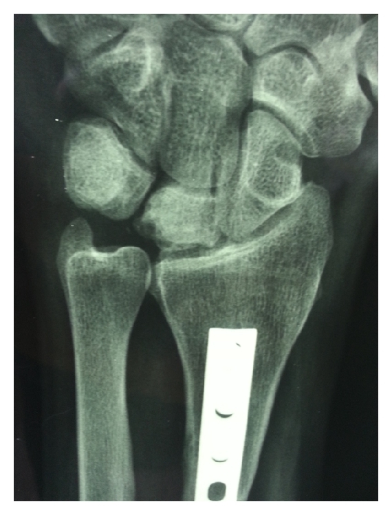
Postero-anterior wrist after a radial shortening osteotomy (distal-radius plate) for Lichtman IIIA Kienböck disease. (Dehghani et al., via Wikimedia Commons; CC BY 4.0.)
Postero-anterior wrist after a radial shortening osteotomy (distal-radius plate) for Lichtman IIIA Kienböck disease. (Dehghani et al., via Wikimedia Commons; CC BY 4.0.)
Part IV - Other adult avascular-necrosis sites
IV.1 Osteonecrosis of the knee
The name covers two principal forms, plus a recognised third variant:[59]
- Spontaneous osteonecrosis of the knee (SPONK / Ahlbäck disease), described by Ahlbäck (1968): typically a woman over 55-60, with the sudden onset of severe medial knee pain, usually a single lesion of the medial femoral condyle, and no risk factors. The modern view is that it represents a subchondral insufficiency (fragility) fracture in osteoporotic bone rather than primary necrosis. Radiographs are normal early (MRI shows a subchondral line with oedema), then show a subchondral lucency with a sclerotic halo and finally flattening; prognosis follows lesion size (favourable below ~3.5 cm² or a width ratio under 0.45; poor above ~5 cm² or a ratio over 0.5). Small lesions are managed conservatively, larger ones by unloading, core decompression, valgus osteotomy or unicompartmental/total knee arthroplasty.[60]
- Secondary osteonecrosis of the knee: a younger patient (under ~45) on corticosteroid/alcohol or with SLE, sickle-cell or Gaucher disease, with multiple, often bilateral, multifocal lesions and usually coexisting femoral-head ON. It responds poorly to non-operative care; core decompression is the favoured procedure in the young.[61]
- Post-arthroscopic osteonecrosis: appearing after meniscectomy/chondroplasty, in middle-aged or older patients, by an uncertain mechanism (whether truly caused by the procedure or a pre-existing lesion missed on early imaging).[62]
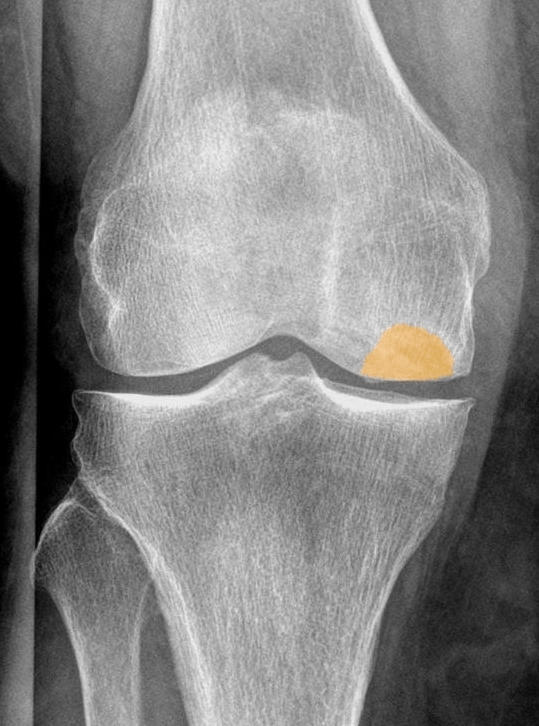
Antero-posterior knee in spontaneous osteonecrosis (Ahlbäck disease) of a 78-year-old woman: the subchondral lucent/flattened lesion of the medial femoral condyle is highlighted (orange overlay on a real radiograph). (Hellerhoff, Wikimedia Commons; CC BY-SA 4.0.)
Antero-posterior knee in spontaneous osteonecrosis (Ahlbäck disease) of a 78-year-old woman: the subchondral lucent/flattened lesion of the medial femoral condyle is highlighted (orange overlay on a real radiograph). (Hellerhoff, Wikimedia Commons; CC BY-SA 4.0.)
IV.2 Osteonecrosis of the talus
The talus is at risk because most of its surface is covered by articular cartilage with no muscular attachments, so it is supplied through few cartilage-free areas; the lateral two-thirds of the body depends on the artery of the tarsal canal (from the posterior tibial), the medial third on the deltoid branch.[63] Most talar ON is post-traumatic, following a talar neck fracture, body fracture or dislocation, with the risk graded by the Hawkins classification of talar-neck fractures: type I (undisplaced) ~0%, type II (subtalar dislocation) 0-50%, type III (body dislocated from ankle and subtalar joints) 75-100%, and type IV (Canale-Kelly addition, plus talonavicular dislocation) the worst.[64]
The Hawkins sign is a good prognostic sign: a subchondral lucent band in the talar dome at about 6-8 weeks (best seen on the AP view) reflects disuse osteopenia of vascularised bone, so its presence argues against osteonecrosis. In Canale and Kelly’s series, assessed at 12 weeks, only ~4% of patients with the sign developed osteonecrosis, versus ~77% of those without it. A persistently dense dome suggests necrosis.[65] MRI is the most sensitive test. Management is debated: a fracture heals despite necrosis, after which the concern is late segmental collapse (creeping substitution takes up to 36 months). Protected weight-bearing is reasonable, and collapse with arthrosis is salvaged by arthrodesis (tibiotalar, tibiotalocalcaneal, or the Blair fusion).[66]
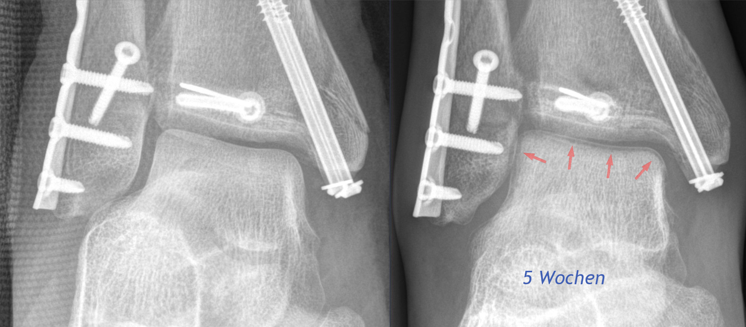
Hawkins sign: AP ankle radiographs after fixation, with arrows (right panel, “5 weeks”) marking the subchondral lucent band in the talar dome - evidence of preserved talar vascularity and a good prognostic sign. (Hellerhoff, Wikimedia Commons; CC BY-SA 4.0.)
Hawkins sign: AP ankle radiographs after fixation, with arrows (right panel, “5 weeks”) marking the subchondral lucent band in the talar dome
- evidence of preserved talar vascularity and a good prognostic sign. (Hellerhoff, Wikimedia Commons; CC BY-SA 4.0.)
IV.3 Osteonecrosis of the scaphoid
The scaphoid’s proximal pole has a retrograde, tenuous blood supply entering distally, so it is prone to necrosis after a fracture or nonunion; proximal-pole fractures are inherently unstable and best fixed internally.[67] In a documented avascular nonunion (no punctate bleeding at surgery), a conventional volar wedge graft unites in only ~30%, so vascularised bone grafting is preferred (union ~88% versus ~47% with a non-vascularised graft; the free medial-femoral-condyle graft outperforms the distal-radius pedicle for the avascular pole). Collapse with arthritis (SNAC wrist) is salvaged by scaphoid excision with midcarpal fusion or proximal-row carpectomy.[68]
Preiser disease is idiopathic (atraumatic) osteonecrosis of the scaphoid, described by Preiser in 1910 and compared by him to Kienböck disease; it is linked variably to corticosteroids, repetitive trauma, connective-tissue disease, or no identifiable cause. The radiograph shows a sclerotic proximal pole with later fragmentation. Treatment (revascularisation, with salvage fusion/carpectomy as a fallback) is not standardised, and a conservative approach is reasonable in the absence of severe symptoms.[69]
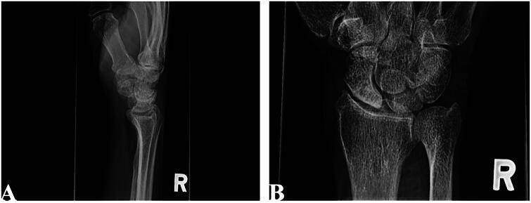
Preiser disease (idiopathic scaphoid osteonecrosis): lateral (A) and PA (B) wrist radiographs showing a dense, sclerotic proximal pole of the scaphoid. (Topham, Wehrli & Kadar, Case Rep Plast Surg Hand Surg 2024; CC BY-NC - non-commercial.)
Preiser disease (idiopathic scaphoid osteonecrosis): lateral (A) and PA (B) wrist radiographs showing a dense, sclerotic proximal pole of the scaphoid. (Topham, Wehrli & Kadar, Case Rep Plast Surg Hand Surg 2024; CC BY-NC - non-commercial.)
IV.4 Osteonecrosis of the humeral head
The humeral head is the second commonest site of atraumatic osteonecrosis after the femoral head. Causes are post-traumatic (displaced proximal-humerus fractures, especially anatomic-neck and four-part patterns, which disrupt the anterior humeral circumflex artery and its terminal arcuate branch) and the same atraumatic factors as elsewhere (corticosteroid, alcohol, sickle-cell). It is staged by the Cruess classification, a modification of Ficat-Arlet (I normal radiograph/MRI-positive; II sclerosis/cysts, sphericity preserved; III crescent sign; IV flattening/collapse; V secondary glenoid arthritis). Management mirrors the femoral head: conservative or core decompression before collapse, and arthroplasty (hemiarthroplasty/resurfacing if the glenoid is spared, total shoulder once the glenoid is involved) afterwards. (The Cruess staging and humeral-head specifics are standard references, not drawn from the page-cited extracts used above.)
Bulgarian terminology glossary
For consistency with the Bulgarian state-examination vocabulary (and the operative terminology of Boychev, Хирургическа ортопедия):
| English | Bulgarian |
|---|---|
| Avascular / aseptic necrosis (osteonecrosis) | Аваскуларна (асептична) некроза (остеонекроза) |
| Osteonecrosis of the femoral head | Аваскуларна некроза на бедрената глава |
| Chandler disease | Болест на Chandler (идиопатична некроза на бедрената глава) |
| Crescent sign (subchondral fracture) | Симптом на полумесеца (субхондрална фрактура) |
| Core decompression | Core декомпресия (декомпресивно пробиване) |
| Femoral / rotational osteotomy | Бедрена / ротационна остеотомия (по Sugioka) |
| Total hip arthroplasty | Тотално ендопротезиране на тазобедрената става |
| Kienböck disease / lunatomalacia | Болест на Kienböck / лунатомалация |
| Lunate | Полулунна кост (os lunatum) |
| Ulnar variance (negative) | Улнарен вариант (отрицателен) |
| Radial shortening osteotomy | Скъсяваща остеотомия на радиуса |
| Vascularised bone graft | Васкуларизиран костен трансплантат |
| Proximal row carpectomy | Резекция на проксималния карпален ред |
| Spontaneous osteonecrosis of the knee (Ahlbäck) | Спонтанна остеонекроза на коляното (болест на Ahlbäck) |
| Talus / Hawkins sign | Талус (скочна кост) / симптом на Hawkins |
| Preiser disease (scaphoid necrosis) | Болест на Preiser (некроза на ладиевидната кост) |
| Magnetic resonance imaging | Ядрено-магнитен резонанс (ЯМР) |
Figure credits and licences
All images were independently opened and visually verified to depict the stated entity before use; licences were confirmed against the Wikimedia Commons API or the NCBI PMC Open-Access service.
- Femoral-head ON - bilateral AP pelvis -
hip_avn_pelvis_bilateral_Haggstrom_CC0.jpg. Mikael Häggström MD. CC0. - Femoral-head ON - crescent sign (annotated) -
hip_avn_crescent_sign_annotated_Haggstrom_CCBYSA3.jpg. Mikael Häggström, after OrthopaedicsOne. CC BY-SA 3.0. - Femoral-head ON - MRI (demarcated segment) -
hip_avn_MRI_left_AIDS_Jmarchn_CCBYSA3.jpg. Jmarchn, Wikimedia Commons. CC BY-SA 3.0. - Femoral-head ON - collapse (steroid) -
hip_avn_AP_cortison_Hellerhoff_CCBYSA3.jpg. Hellerhoff, Wikimedia Commons. CC BY-SA 3.0. - Kienböck - PA wrist radiograph -
kienbock_PA_topview_Muzichick_CCBYSA4.jpg. User Muzichick, Wikimedia Commons. CC BY-SA 4.0. - Kienböck - wrist MRI + radiograph -
kienbock_MRI_Xie_CCBYSA4.png. Xie et al., via Wikimedia Commons. CC BY-SA 4.0. - Kienböck - radial shortening osteotomy -
kienbock_postop_osteotomy_Dehghani_CCBY4.jpg. Dehghani M, et al., via Wikimedia Commons. CC BY 4.0. - SPONK / Ahlbäck - AP knee (annotated) -
sponk_AP_78W_annotated_Hellerhoff_CCBYSA4.jpg. Hellerhoff, Wikimedia Commons. CC BY-SA 4.0. - Talus - Hawkins sign (annotated) -
talus_hawkins_sign_annotated_Hellerhoff_CCBYSA4.jpg. Hellerhoff, Wikimedia Commons. CC BY-SA 4.0. - Preiser disease - scaphoid radiograph -
preiser_scaphoid_radiograph_fig1_PMC11225627_CCBYNC.jpg. Topham J, Wehrli L, Kadar A, Case Reports in Plastic Surgery & Hand Surgery 2024;11(1):2374550, Fig. 1. CC BY-NC - non-commercial use only.
Licensing note for distribution: image 10 (Preiser) is CC BY-NC
- acceptable for a personal, non-commercial study document, but it must be removed or replaced if this material is ever used commercially. The CC BY-SA images require that any redistributed derivative carry the same share-alike licence; the CC0 image is public-domain.
References
-
Osteonecrosis (Koo, Mont & Jones), p.21.
-
Surgery of the Hip (Berry), p.432.
-
Osteonecrosis, p.21; Greenspan, Orthopedic Imaging, pp.176-177.
-
Surgery of the Hip (Berry), p.475.
-
Surgery of the Hip (Berry), pp.475-476; Greenspan, p.176.
-
Osteonecrosis, p.210.
-
Osteonecrosis, p.21.
-
Osteonecrosis, p.21.
-
Osteonecrosis, p.22; The Adult Hip (Callaghan), p.882.
-
Osteonecrosis, p.209; The Adult Hip, p.882.
-
Surgery of the Hip (Berry), p.432; The Adult Hip, p.884.
-
Osteonecrosis, p.23; The Adult Hip, p.882.
-
The Adult Hip, p.882; Surgery of the Hip (Berry), pp.474-475.
-
Surgery of the Hip (Berry), p.474.
-
Osteonecrosis, p.23; Greenspan, pp.173-175.
-
Osteonecrosis, p.22.
-
Osteonecrosis (traumatic-AVN chapter), pp.108-109; Campbell’s, p.1759.
-
Surgery of the Hip (Berry), p.477; Osteonecrosis, pp.195, 197.
-
Greenspan, pp.176, 180; The Adult Hip, p.887.
-
Osteonecrosis, pp.194-195.
-
Osteonecrosis, pp.209-212.
-
Osteonecrosis, pp.196-200.
-
Osteonecrosis, pp.202-204.
-
Osteonecrosis, pp.205-208; The Adult Hip, p.887.
-
Greenspan, pp.176-179.
-
Surgery of the Hip (Berry), p.433; Greenspan, p.177.
-
The Adult Hip, p.886; Greenspan, p.176.
-
Surgery of the Hip (Berry), pp.432-433; Osteonecrosis, pp.218-220.
-
Osteonecrosis, p.26; Surgery of the Hip (Berry), p.482.
-
The Adult Hip, pp.886-888; Osteonecrosis, p.204.
-
The Adult Hip, p.890; Surgery of the Hip (Berry), p.481.
-
The Adult Hip, pp.890-891; Surgery of the Hip (Berry), p.482.
-
Osteonecrosis, pp.265-268; The Adult Hip, p.892.
-
Osteonecrosis, pp.291-303; The Adult Hip, p.893.
-
Osteonecrosis, p.307; The Adult Hip, pp.894-895.
-
Osteonecrosis, pp.28-29, 342-349; The Adult Hip, pp.896-899.
-
Osteonecrosis (traumatic-AVN chapter), pp.110-120; Greenspan, p.176.
-
Lichtman & Bain, Kienböck’s Disease, pp.24-25, 30, 81, 94.
-
Lichtman & Bain, p.34.
-
Lichtman & Bain, pp.94, 115.
-
Lichtman & Bain, pp.99-100, 119-123.
-
Lichtman & Bain, pp.45-46.
-
Lichtman & Bain, pp.46-48, 89-95.
-
Lichtman & Bain, pp.83-84.
-
Lichtman & Bain, pp.55-56, 84-85, 108, 137.
-
Lichtman & Bain, pp.94, 108-112.
-
Lichtman & Bain, pp.115-118, 128-132, 150.
-
Lichtman & Bain, pp.171-174.
-
Lichtman & Bain, pp.149-150.
-
Lichtman & Bain, pp.114-115, 127.
-
Lichtman & Bain, pp.116, 128-143.
-
Lichtman & Bain, pp.140, 188.
-
Lichtman & Bain, pp.119-122.
-
Lichtman & Bain, pp.188-199, 206-210.
-
Lichtman & Bain, pp.196, 211-217; Green’s Operative Hand Surgery, carpal-VBG chapter.
-
Lichtman & Bain, pp.176-177, 185-186.
-
Lichtman & Bain, pp.243-257.
-
Lichtman & Bain, pp.119, 237-242, 258.
-
Insall & Scott, Surgery of the Knee, pp.459, 477, 481.
-
Insall & Scott, pp.459-478, 486-492.
-
Insall & Scott, pp.459, 481, 492-496.
-
Insall & Scott, pp.472, 481, 491.
-
Mann’s Surgery of the Foot & Ankle, pp.2102-2106.
-
Mann’s, pp.2109-2114.
-
Mann’s, pp.2114, 2132.
-
Mann’s, pp.2115, 2132-2133, 2143-2149.
-
Green’s Operative Hand Surgery, scaphoid chapter.
-
Green’s Operative Hand Surgery, scaphoid chapter.
-
Green’s Operative Hand Surgery, scaphoid chapter; Greenspan, p.573.