Contents
- Introduction and scope
- Part I - Rheumatoid arthritis: pathogenesis
- Part II - Rheumatoid arthritis: clinical features and the characteristic deformities
- Part III - Rheumatoid arthritis: extra-articular disease and the cervical spine
- Part IV - Rheumatoid arthritis: diagnosis and classification
- Part V - Rheumatoid arthritis: medical management
- Part VI - Surgical management of the rheumatoid hand, wrist and foot
- Part VII - The seronegative spondyloarthropathies
- Part VIII - Crystal arthropathies
- Part IX - Juvenile idiopathic arthritis
- Bulgarian terminology (Боев / Boychev tradition) - glossary
- Image attributions
- References
Introduction and scope
The inflammatory arthropathies are the systemic diseases an orthopaedic surgeon meets at the joint: rheumatoid arthritis (the prototype), the seronegative spondyloarthropathies, the crystal arthropathies, and juvenile idiopathic arthritis. For rheumatoid arthritis the examiner expects a working command of its pathogenesis (the synovial pannus, rheumatoid factor and anti-citrullinated antibodies), the characteristic hand, wrist and forefoot deformities, the danger of cervical instability, the 2010 classification criteria, the modern treat-to-target medical strategy and the surgical reconstruction of the rheumatoid hand and foot. Beyond that, the defining features of the spondyloarthropathies (ankylosing spondylitis, psoriatic, reactive and enteropathic arthritis, all linked to HLA-B27 and enthesitis), the crystal diseases (gout and calcium pyrophosphate deposition, told apart under polarised light), and juvenile idiopathic arthritis.
Two ideas run through the topic. First, rheumatoid arthritis is a systemic disease with a surgical footprint: the synovitis destroys the soft-tissue restraints in a predictable sequence (synovitis → attenuation → subluxation → tendon rupture and fixed deformity), and modern disease-modifying drugs have changed its course, with rheumatoid hand surgery falling by over 80% since the biologic era. Second, diagnosis turns on pattern and serology. A symmetric small-joint polyarthritis with anti-CCP antibodies is rheumatoid; an asymmetric lower-limb arthritis with enthesitis, sacroiliitis and HLA-B27 is a spondyloarthropathy; and an acute monoarthritis of the first toe is gout until the polarised microscope says otherwise.
Part I - Rheumatoid arthritis: pathogenesis
Rheumatoid arthritis (RA) is a chronic systemic autoimmune inflammatory polyarthritis, affecting roughly 0.5-1% of the population, two to three times more often in women, with peak incidence in the sixth decade (the fifties).[1] Susceptibility is both genetic and environmental. The strongest genetic factor is the HLA-DRB1 “shared epitope” (a conserved amino-acid motif in the antigen-binding groove), with PTPN22 a second contributor; the dominant environmental factor is smoking, which induces citrullination in the lung.[2] Citrullination, the conversion of arginine to citrulline by peptidylarginine deiminases, generates the neo-antigens recognised by anti-citrullinated protein antibodies (ACPA, measured as anti-CCP), and this autoimmunity precedes clinical arthritis by years.[3]
The target organ is the synovium. The normally one-to-two-cell-deep lining becomes hyperplastic (four to ten cells deep), with macrophage-like (type A) and aggressive fibroblast-like (type B) synoviocytes, and forms an invasive pannus that erodes cartilage and bone.[4] The cytokine network of TNF-α, IL-6 and IL-1 perpetuates the inflammation, and RANKL-driven osteoclasts produce the marginal bone erosions.[5] Serologically, rheumatoid factor (an antibody to the Fc of IgG) is present in about 75% but is not specific, whereas anti-CCP/ACPA is found in 80-90% of established disease, is highly specific (>95%), and predicts more aggressive, erosive disease.[6]
Part II - Rheumatoid arthritis: clinical features and the characteristic deformities
RA presents as an insidious, symmetric, additive polyarthritis of the small joints (the metacarpophalangeal [MCP], proximal interphalangeal [PIP], wrist and metatarsophalangeal [MTP] joints) with morning stiffness lasting more than an hour, and it characteristically spares the distal interphalangeal (DIP) joints and the thoracolumbar spine.[7] The radiographic hallmarks are periarticular osteopenia and marginal erosions at the “bare area” where synovium meets bone.[8]
The destruction of the soft-tissue restraints produces the classic deformities:
- the “zig-zag” hand: radial deviation of the wrist with ulnar drift and volar subluxation of the MCP joints;[9]
- the caput ulnae syndrome: dorsal prominence of the unstable ulnar head, depressible like a “piano key”;[10]
- the swan-neck deformity (DIP flexion with PIP hyperextension) and the boutonnière deformity (PIP flexion with DIP hyperextension);[11]
- the rheumatoid (Z) thumb;[12]
- in advanced disease, arthritis mutilans (“opera-glass hand”) with telescoped digits.[13]
RA is the commonest cause of hand tendon ruptures. The Vaughan-Jackson lesion is attritional rupture of the digital extensors at the dorsally prominent ulnar head (the little-finger extensor first), and the Mannerfelt lesion is rupture of flexor pollicis longus on a scaphoid spur within the carpal tunnel.[14] The rheumatoid forefoot is involved in up to 90%: MTP synovitis leads to dorsal subluxation and dislocation of the toes, claw-toe deformities and hallux valgus, with the metatarsal heads driven plantarward so the patient feels they are “walking on marbles,” while the hindfoot collapses into a planovalgus deformity.[15]
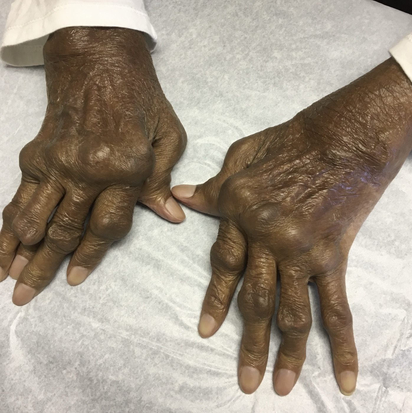
Rheumatoid hand deformities. Dorsal view of both hands: MCP swelling, ulnar deviation of the digits and multiple finger deformities of long-standing rheumatoid arthritis. Image: D. Gannavarapu Narasimha Murthy, CC0, via Wikimedia Commons.
Rheumatoid hand deformities. Dorsal view of both hands: MCP swelling, ulnar deviation of the digits and multiple finger deformities of long-standing rheumatoid arthritis. Image: D. Gannavarapu Narasimha Murthy, CC0, via Wikimedia Commons.
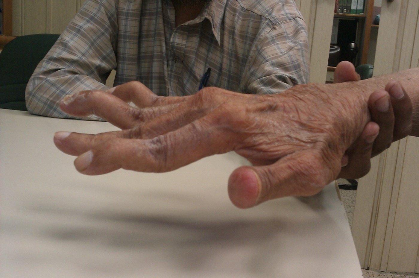
Swan-neck deformity (PIP hyperextension with DIP flexion) in a hand affected by rheumatoid arthritis. Image: Phoenix119, CC BY-SA 3.0, via Wikimedia Commons.
Swan-neck deformity (PIP hyperextension with DIP flexion) in a hand affected by rheumatoid arthritis. Image: Phoenix119, CC BY-SA 3.0, via Wikimedia Commons.
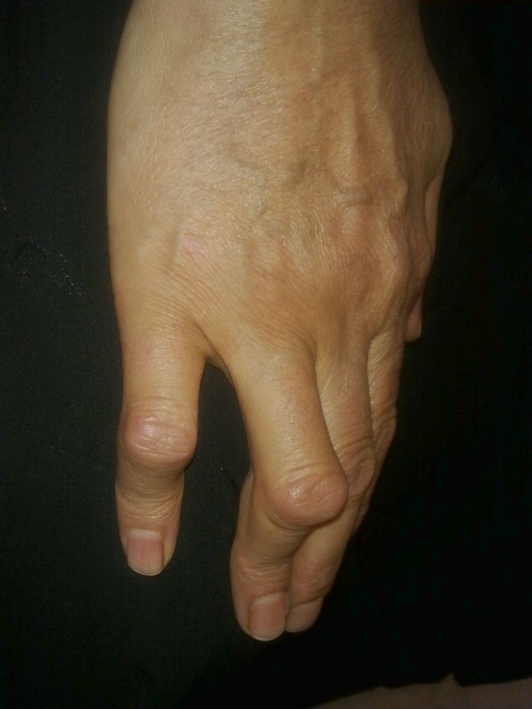
Boutonnière deformity (PIP flexion with DIP hyperextension). Image: Alborz Fallah, CC BY-SA 3.0, via Wikimedia Commons.
Boutonnière deformity (PIP flexion with DIP hyperextension). Image: Alborz Fallah, CC BY-SA 3.0, via Wikimedia Commons.
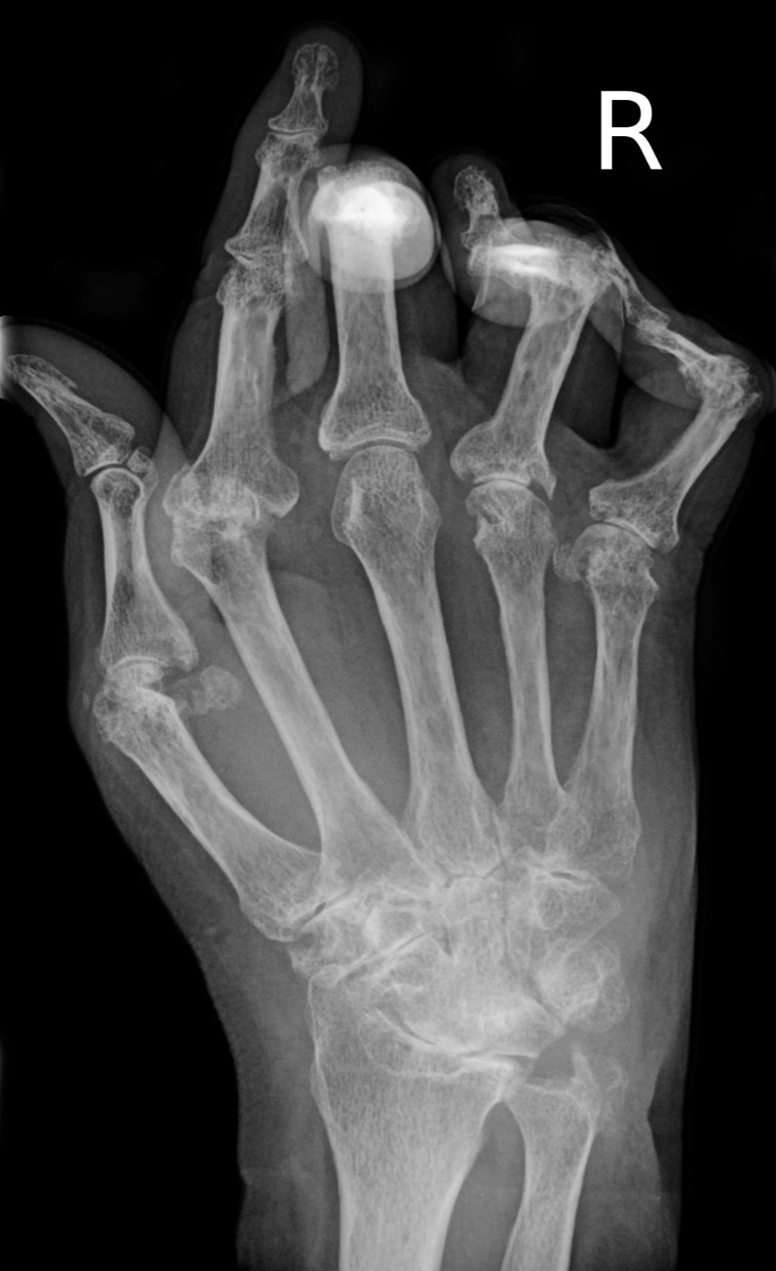
Advanced rheumatoid arthritis of the hand (AP radiograph). Gross metacarpophalangeal destruction, marginal erosions, joint-space loss and periarticular osteopenia. Image: Bernd Brägelmann, CC BY 3.0, via Wikimedia Commons.
Advanced rheumatoid arthritis of the hand (AP radiograph). Gross metacarpophalangeal destruction, marginal erosions, joint-space loss and periarticular osteopenia. Image: Bernd Brägelmann, CC BY 3.0, via Wikimedia Commons.
Part III - Rheumatoid arthritis: extra-articular disease and the cervical spine
Extra-articular features occur in up to half of patients and carry a worse prognosis.[16] They include rheumatoid nodules (subcutaneous, over pressure areas, seen in seropositive disease and, when biopsied, essentially pathognomonic, though methotrexate can paradoxically increase them); rheumatoid vasculitis; pulmonary disease (interstitial lung disease, with the usual-interstitial-pneumonia pattern, pulmonary nodules, and pleuritis with characteristically low glucose and low pH); pericarditis (the commonest cardiac lesion, found at up to half of autopsies); ocular disease (keratoconjunctivitis sicca / secondary Sjögren’s in about 17%, and scleritis); Felty syndrome (the triad of RA, splenomegaly and neutropenia); anaemia; secondary amyloidosis; and accelerated cardiovascular disease, which is the leading cause of the excess mortality.[17]
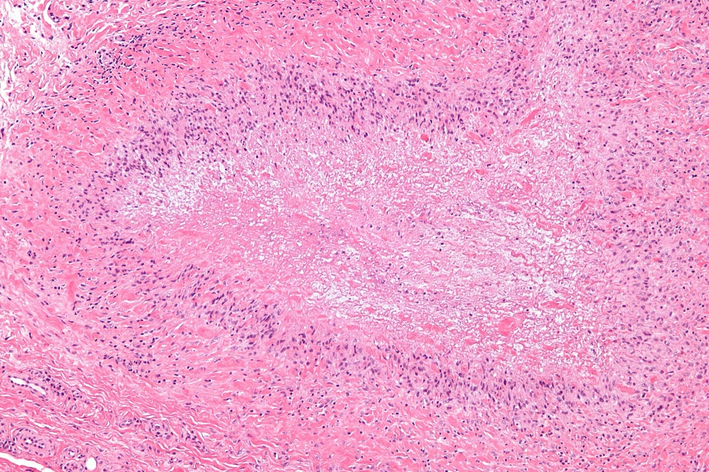
Rheumatoid nodule (histology). Central fibrinoid necrosis surrounded by palisading histiocytes - the characteristic palisading granuloma. Image: Nephron, CC BY-SA 3.0, via Wikimedia Commons.
Rheumatoid nodule (histology). Central fibrinoid necrosis surrounded by palisading histiocytes - the characteristic palisading granuloma. Image: Nephron, CC BY-SA 3.0, via Wikimedia Commons.
For the orthopaedic surgeon the cervical spine is the highest-stakes site: it is involved in up to 80% of RA, most importantly as atlantoaxial (C1-C2) subluxation, then cranial settling and subaxial subluxation.[18] The earliest symptom is pain radiating to the occiput, and progression to cord compression carries roughly a 50% one-year mortality. Because cervical manipulation during intubation can be catastrophic, cervical-spine assessment is mandatory before any general anaesthetic.[19]
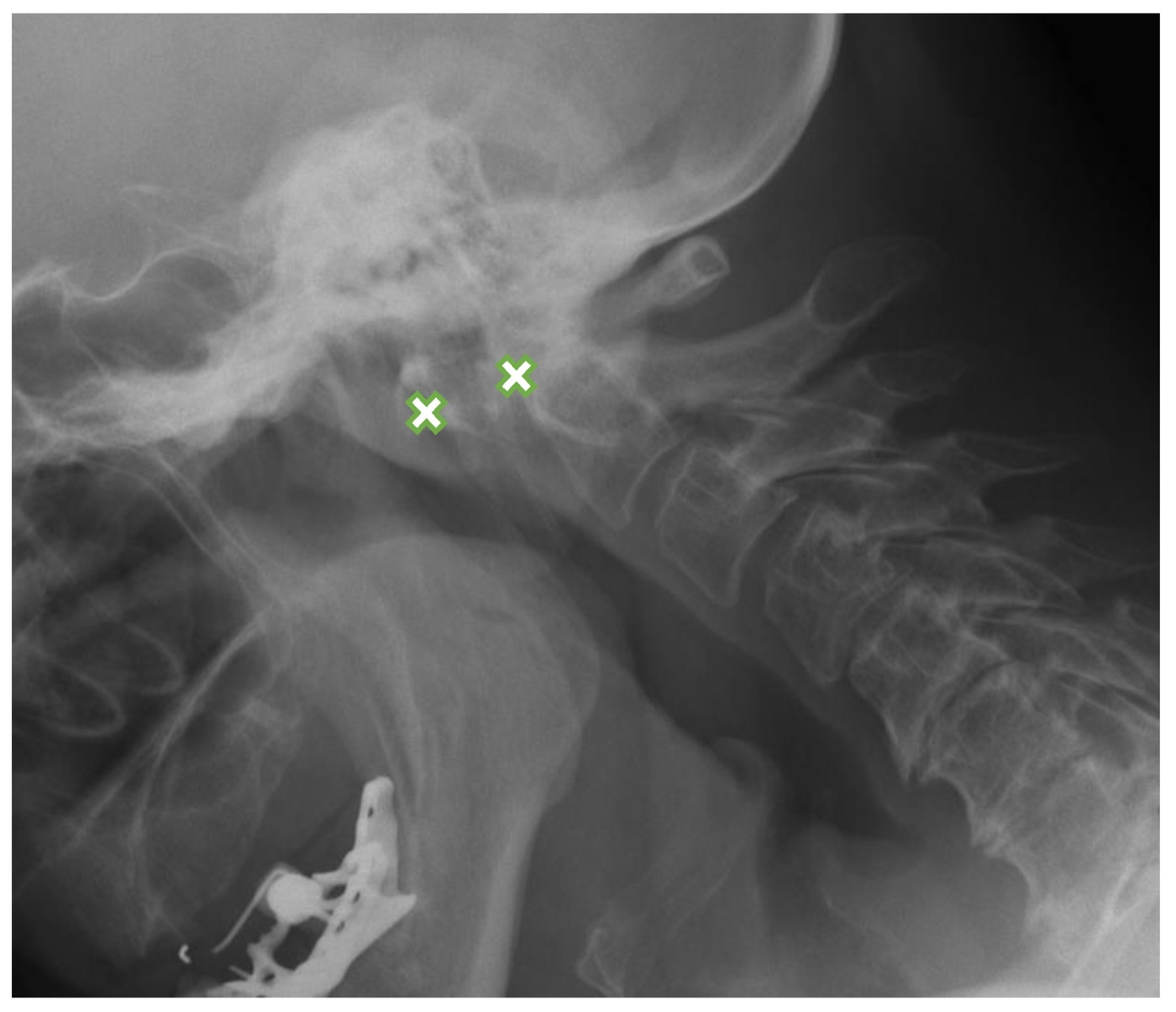
Atlantoaxial subluxation in rheumatoid arthritis. Flexion lateral cervical radiograph; the anterior atlanto-dental interval (between the crosses) measures 8.8 mm, indicating C1-C2 instability. Kotecki et al., J Clin Med 2021, CC BY 4.0.
Atlantoaxial subluxation in rheumatoid arthritis. Flexion lateral cervical radiograph; the anterior atlanto-dental interval (between the crosses) measures 8.8 mm, indicating C1-C2 instability. Kotecki et al., J Clin Med 2021, CC BY 4.0.
Part IV - Rheumatoid arthritis: diagnosis and classification
Diagnosis combines the clinical pattern, the serology (RF and anti-CCP) and the acute-phase reactants (ESR and CRP, which may be normal in up to a third at first presentation).[20] The 2010 ACR/EULAR classification criteria replaced the 1987 set to allow earlier diagnosis: a patient with at least one clinically swollen joint and no better explanation is scored from 0 to 10 across four domains (joint involvement [0-5], serology [RF/ACPA, 0-3], acute-phase reactants [0-1] and symptom duration [0-1]), and a score of 6 or more classifies definite RA.[21] The 2010 criteria weight serology heavily and are more sensitive (about 97%) but less specific than the older criteria (about 55% versus 76%).[22] The main differentials are the spondyloarthropathies, lupus (non-erosive Jaccoud’s arthropathy), polyarticular gout or CPPD, and viral arthritides.[23]
Part V - Rheumatoid arthritis: medical management
The governing principle is treat-to-target: every patient is started on a disease-modifying drug at diagnosis and treatment is escalated to a defined target (remission or low disease activity), an approach (validated by the TICORA trial) that matters more than which specific drug is chosen.[24] Methotrexate is the anchor DMARD, a weekly oral or subcutaneous dose (typically built up from 15 mg towards 25 mg), with folic acid to reduce toxicity and monitoring of the blood count and liver; it is teratogenic and can rarely cause pneumonitis.[25] Other conventional DMARDs are sulfasalazine, hydroxychloroquine and leflunomide (often combined as “triple therapy”). When conventional therapy fails, biologic DMARDs are added: TNF inhibitors (etanercept, infliximab, adalimumab) first, then IL-6 blockade (tocilizumab), B-cell depletion (rituximab) or T-cell co-stimulation blockade (abatacept), and the oral JAK inhibitors (e.g. tofacitinib).[26] Glucocorticoids are used only as a short “bridge” with a DMARD, and NSAIDs as symptomatic adjuncts.[27]
Part VI - Surgical management of the rheumatoid hand, wrist and foot
Although modern drug therapy has reduced rheumatoid hand surgery by over 80%, reconstruction still has a defined role.[28] The sequencing principles are to treat the lower limb and weight-bearing joints before the upper limb, to address the cervical spine, hip and knee before the hand, and to correct the wrist before the digits (since wrist position governs the balance of the finger tendons); anti-inflammatory and biologic drugs are paused perioperatively.[29]
- Synovectomy and tenosynovectomy: persistent synovitis beyond a few months is treated by synovectomy; a dorsal wrist tenosynovectomy is performed to prevent extensor-tendon rupture.[30]
- Distal radioulnar joint / caput ulnae: the unstable distal ulna is resected (Darrach procedure) or stabilised (Sauvé-Kapandji).[31]
- Tendon ruptures: extensor ruptures (Vaughan-Jackson) are reconstructed by side-to-side transfer to an intact tendon or an extensor indicis proprius transfer, always with synovectomy at the site.[32]
- MCP joint and wrist: silicone (Swanson) implant arthroplasty corrects MCP ulnar drift and volar subluxation; the destroyed wrist is treated by arthrodesis (the more durable, reliable choice) or arthroplasty, with one wrist fused and the other replaced if both are involved.[33]
- Swan-neck, boutonnière and the rheumatoid thumb: corrected by graded soft-tissue procedures and arthrodesis; the thumb is classified by Nalebuff (type I boutonnière being commonest, type III the swan-neck).[34]
- The rheumatoid forefoot: the classic reconstruction is a first-MTP arthrodesis with resection of the lesser metatarsal heads (the Hoffmann/Clayton family of procedures), which stabilises the first ray and protects the lesser toes; Coughlin reported about 96% good or excellent results.[35] The collapsed hindfoot is treated by triple arthrodesis, performed before forefoot correction.[36]
Part VII - The seronegative spondyloarthropathies
The spondyloarthropathies are a family sharing association with HLA-B27, the absence of rheumatoid factor, enthesitis as the primary lesion, an asymmetric lower-limb peripheral arthritis and/or axial disease, dactylitis, and overlapping extra-articular features (anterior uveitis, psoriasis, inflammatory bowel disease), driven by the IL-23/IL-17 axis.[37]
Ankylosing spondylitis
Ankylosing spondylitis (AS) affects young men (about 2-3:1), of whom roughly 90% are HLA-B27 positive, and its hallmark is radiographic sacroiliitis.[38] The clinical picture is inflammatory back pain (insidious, in a young adult, with morning stiffness, improving with exercise and not relieved by rest) together with enthesitis, and over years syndesmophytes bridge the vertebrae (with “squaring” of the bodies) to give the “bamboo spine.”[39] Examination shows a reduced Schober test (the normal lumbar-flexion increase of at least 5 cm is lost), reduced chest expansion (under 5 cm) and an increased occiput-to-wall distance.[40] The commonest extra-articular feature is acute anterior uveitis (a unilateral red, painful eye, in 30-40%); others are aortitis and apical pulmonary fibrosis, and the ankylosed osteoporotic spine is prone to fracture.[41] Diagnosis uses the modified New York criteria (sacroiliitis plus a clinical criterion).[42] Treatment is NSAIDs and physiotherapy first, then TNF inhibitors and IL-17 inhibitors (secukinumab); conventional DMARDs have no effect on axial disease, and total hip arthroplasty is used for hip involvement.[43]
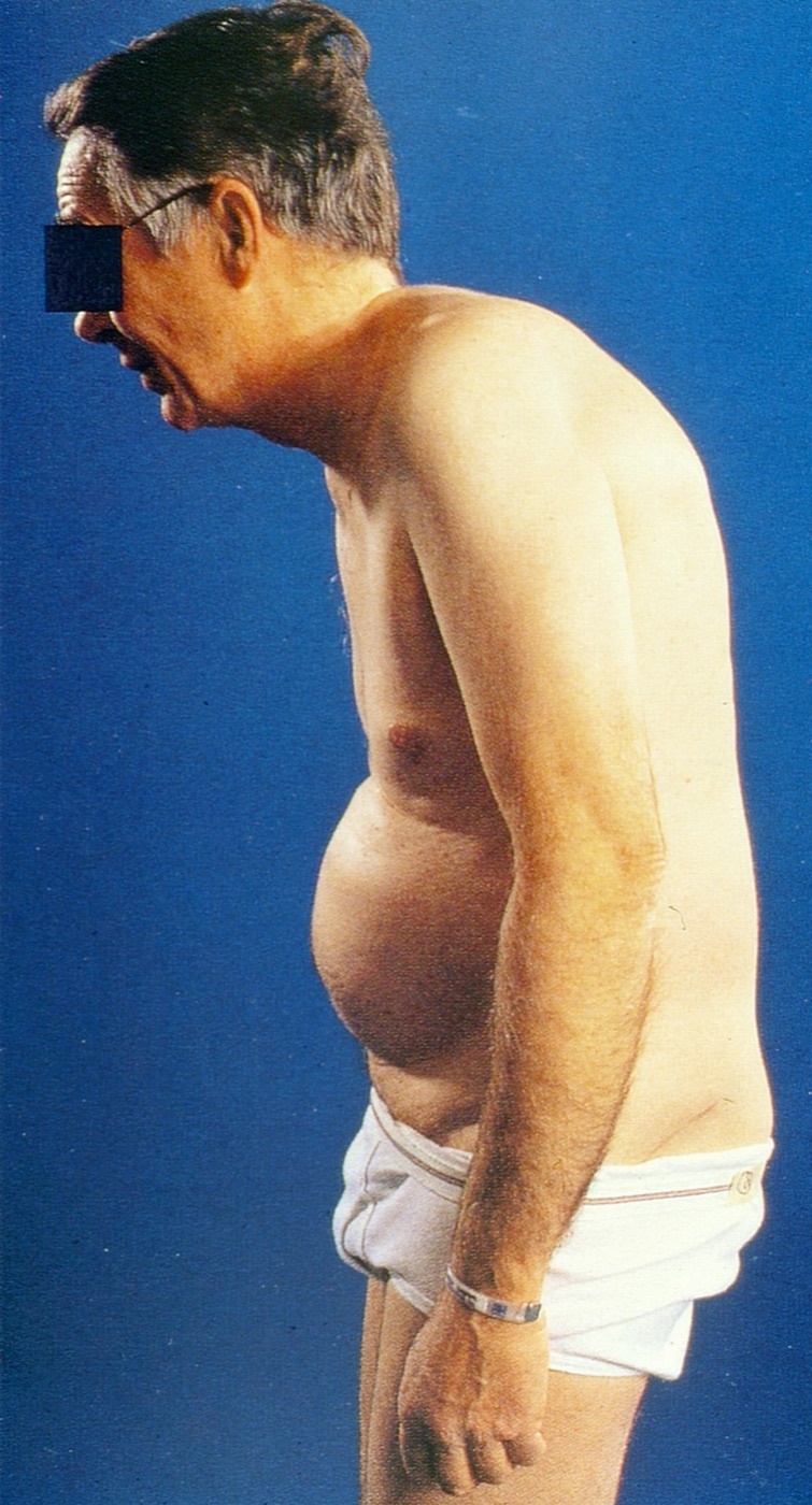
Fixed kyphotic posture of advanced ankylosing spondylitis - the stooped “question-mark” stance with a protuberant abdomen. Image: Mehlauge, CC BY-SA 3.0, via Wikimedia Commons.
Fixed kyphotic posture of advanced ankylosing spondylitis - the stooped “question-mark” stance with a protuberant abdomen. Image: Mehlauge, CC BY-SA 3.0, via Wikimedia Commons.
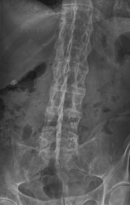
“Bamboo spine” of ankylosing spondylitis. AP lumbar radiograph with flowing marginal syndesmophytes bridging the vertebral bodies. Image: Stevenfruitsmaak, CC BY-SA 3.0, via Wikimedia Commons.
“Bamboo spine” of ankylosing spondylitis. AP lumbar radiograph with flowing marginal syndesmophytes bridging the vertebral bodies. Image: Stevenfruitsmaak, CC BY-SA 3.0, via Wikimedia Commons.
Psoriatic arthritis
Psoriatic arthritis follows the five patterns of Moll & Wright (asymmetric oligoarthritis, symmetric [RA-like] polyarthritis, a DIP-predominant pattern, an axial/spondylitic pattern, and the destructive arthritis mutilans) and is characteristically RF-negative, with dactylitis (“sausage digit”), enthesitis, nail pitting and onycholysis, and the radiographic “pencil-in-cup” deformity.[44] It is classified by the CASPAR criteria (inflammatory arthritis plus three points from psoriasis, nail disease, negative RF, dactylitis and juxta-articular new bone).[45] Treatment runs from NSAIDs and methotrexate to TNF, IL-17 and IL-12/23 inhibitors.[46]
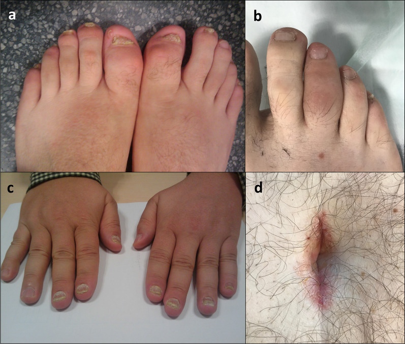
Psoriatic arthritis: nail disease and dactylitis. Nail pitting and onychodystrophy with a “sausage digit.” Crespo-Rodríguez et al., Insights Imaging 2021, CC BY 4.0.
Psoriatic arthritis: nail disease and dactylitis. Nail pitting and onychodystrophy with a “sausage digit.” Crespo-Rodríguez et al., Insights Imaging 2021, CC BY 4.0.
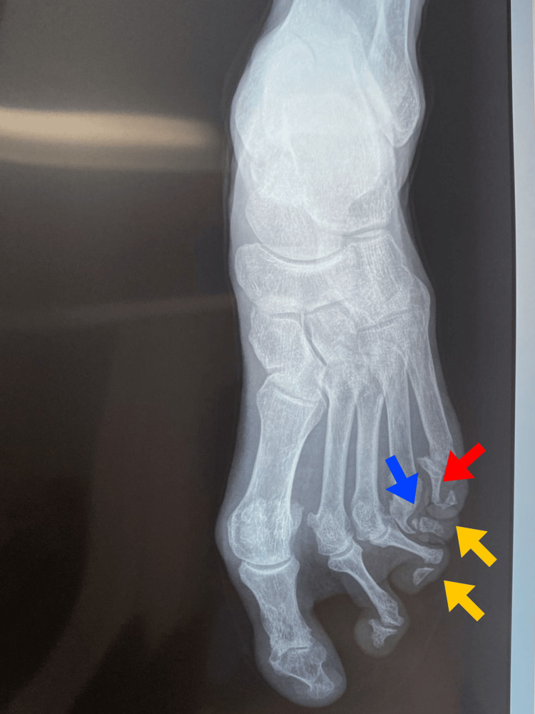
Pencil-in-cup deformity in psoriatic arthritis mutilans (foot radiograph): osteolysis whittles the proximal phalanx into the cupped base of its neighbour. Altamirano De La Cruz et al., Cureus 2025, CC BY 4.0.
Pencil-in-cup deformity in psoriatic arthritis mutilans (foot radiograph): osteolysis whittles the proximal phalanx into the cupped base of its neighbour. Altamirano De La Cruz et al., Cureus 2025, CC BY 4.0.
Reactive and enteropathic arthritis
Reactive arthritis follows a gastrointestinal (Salmonella, Shigella, Campylobacter, Yersinia) or genitourinary (Chlamydia) infection, with a sterile, asymmetric lower-limb oligoarthritis and the classic triad of arthritis, urethritis/cervicitis and conjunctivitis (with keratoderma blennorrhagicum and circinate balanitis), strongly linked to HLA-B27.[47] Enteropathic arthritis accompanies Crohn’s disease and ulcerative colitis: a type I oligoarthritis parallels gut activity and is self-limiting, whereas a type II polyarthritis runs independently of the bowel; NSAIDs are avoided as they flare the gut.[48]
Part VIII - Crystal arthropathies
Gout
Gout is the commonest inflammatory arthritis, caused by monosodium urate (MSU) crystals deposited when the serum urate exceeds its solubility (> 6.8 mg/dL); about 90% of cases are due to renal underexcretion.[49] It evolves through asymptomatic hyperuricaemia, then acute gouty attacks separated by symptom-free intercritical intervals, and finally chronic tophaceous gout (Firestein groups the acute and intercritical phases together as three stages).[50] The first attack is monoarticular in 85-90% and classically strikes the first MTP joint (podagra), with an explosive, exquisitely tender, red joint, often overnight; chronic disease forms tophi (over the ear helix, olecranon and Achilles) and radiographic erosions with overhanging edges.[51] Diagnosis is definitive on polarised microscopy: needle-shaped, strongly negatively birefringent intracellular crystals; dual-energy CT and the ultrasound “double-contour sign” support it.[52] Acute attacks are treated by low-dose colchicine, NSAIDs (indomethacin) or corticosteroids (the sooner the better). Urate-lowering therapy, with allopurinol first-line (then febuxostat, a uricosuric, or pegloticase for refractory disease), is started later to a target serum urate below 6 mg/dL, under colchicine prophylaxis for at least six months, and asymptomatic hyperuricaemia is not treated.[53]
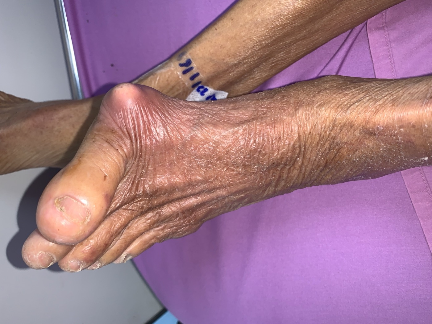
Tophaceous gout at the first MTP joint (podagra). A large erythematous tophus over the great-toe joint. Image: Chainwit., CC BY 4.0, via Wikimedia Commons.
Tophaceous gout at the first MTP joint (podagra). A large erythematous tophus over the great-toe joint. Image: Chainwit., CC BY 4.0, via Wikimedia Commons.
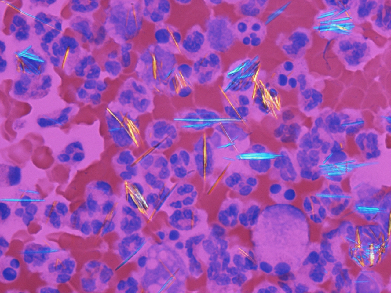
Monosodium urate crystals. Needle-shaped, strongly negatively birefringent crystals under compensated polarised light - the definitive diagnosis of gout. Image: Gabriel Caponetti, CC BY-SA 3.0, via Wikimedia Commons.
Monosodium urate crystals. Needle-shaped, strongly negatively birefringent crystals under compensated polarised light - the definitive diagnosis of gout. Image: Gabriel Caponetti, CC BY-SA 3.0, via Wikimedia Commons.
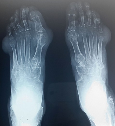
Chronic tophaceous gout (foot radiograph). Periarticular “punched-out” erosions, prominent at the first MTP joints. Image: N. Aradoini et al., CC BY, via Wikimedia Commons.
Chronic tophaceous gout (foot radiograph). Periarticular “punched-out” erosions, prominent at the first MTP joints. Image: N. Aradoini et al., CC BY, via Wikimedia Commons.
Calcium pyrophosphate deposition disease and basic calcium phosphate
Calcium pyrophosphate deposition (CPPD) disease produces rhomboid, weakly positively birefringent crystals and the radiographic finding of chondrocalcinosis (cartilage calcification), most often in the knee and the wrist (triangular fibrocartilage); it is associated with ageing, hyperparathyroidism, haemochromatosis and hypomagnesaemia, and the familial form maps to the ANKH gene.[54] It presents as acute “pseudogout” (a large joint, usually the knee, rarely the first MTP), or as a chronic pseudo-osteoarthritis affecting joints normally spared by OA (MCPs, wrists, elbows), and is treated like acute gout (NSAIDs, steroids, colchicine prophylaxis).[55] Basic calcium phosphate (hydroxyapatite) crystals are not birefringent (they stain with alizarin red) and cause calcific tendinitis of the rotator cuff and the destructive Milwaukee shoulder (rotator-cuff-tear arthropathy).[56]
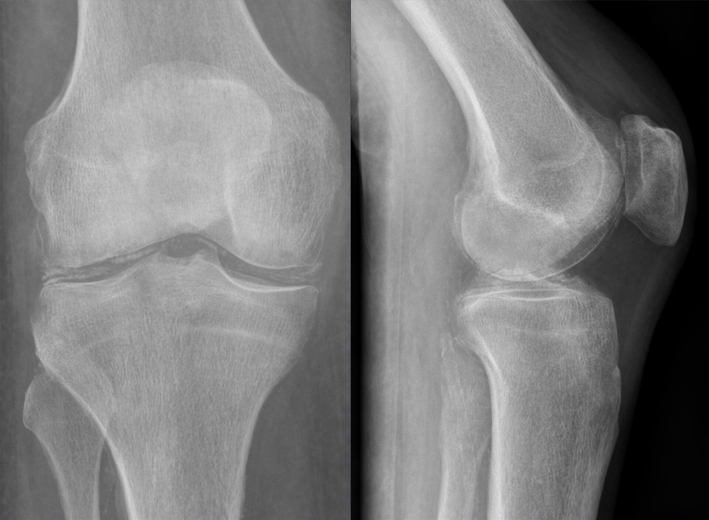
Chondrocalcinosis of the knee (CPPD). AP and lateral radiographs showing linear calcification of the menisci and articular cartilage. Image: Hellerhoff, CC BY-SA 4.0, via Wikimedia Commons.
Chondrocalcinosis of the knee (CPPD). AP and lateral radiographs showing linear calcification of the menisci and articular cartilage. Image: Hellerhoff, CC BY-SA 4.0, via Wikimedia Commons.
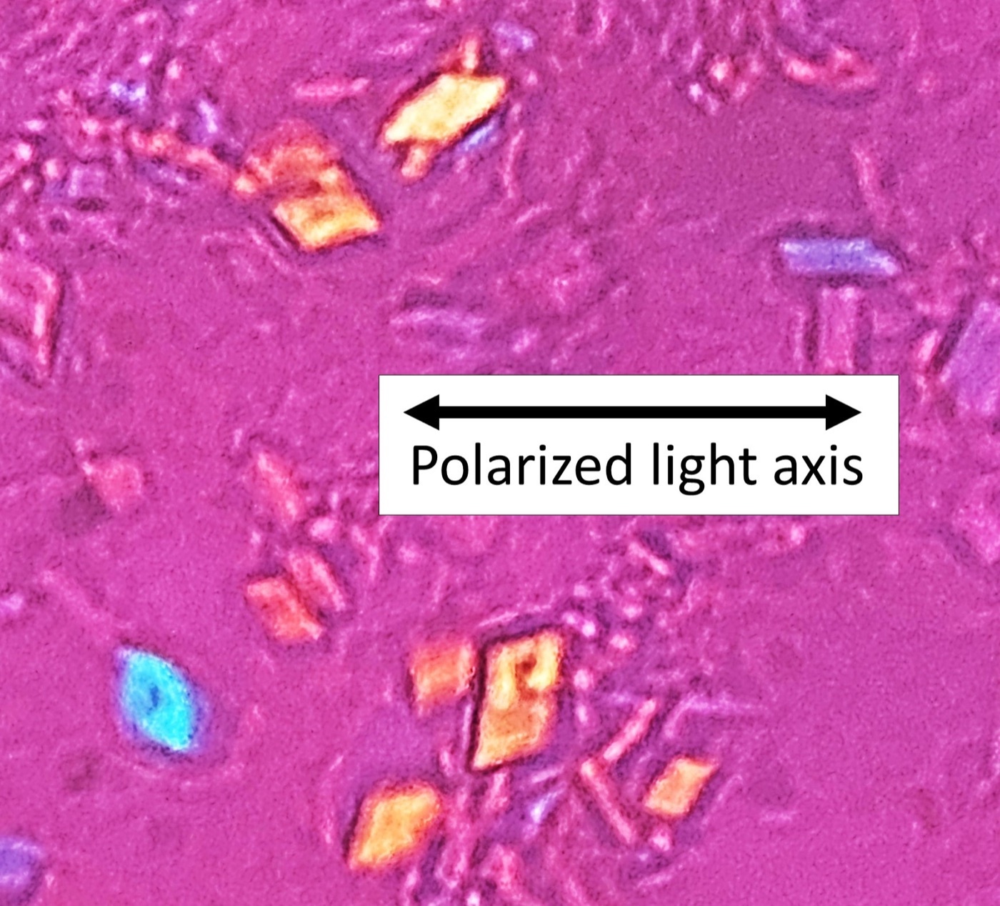
Calcium pyrophosphate crystals. Rhomboid, weakly positively birefringent crystals under compensated polarised light. Image: Mikael Häggström, CC0, via Wikimedia Commons.
Calcium pyrophosphate crystals. Rhomboid, weakly positively birefringent crystals under compensated polarised light. Image: Mikael Häggström, CC0, via Wikimedia Commons.
Part IX - Juvenile idiopathic arthritis
Juvenile idiopathic arthritis (JIA) is arthritis beginning before the sixteenth birthday, persisting for at least six weeks, of unknown cause, and is classified by the ILAR system into seven categories.[57] The key categories are: systemic JIA (Still’s disease) (about 10%; a quotidian high fever, an evanescent salmon-pink rash, hepatosplenomegaly and serositis, an autoinflammatory disease driven by IL-1 and IL-6, complicated by macrophage activation syndrome); oligoarticular JIA (the commonest; young, ANA-positive girls, the knee most often, and the highest risk of chronic anterior uveitis); polyarticular RF-negative and polyarticular RF-positive (the latter behaving like adult RA); psoriatic; enthesitis-related arthritis (HLA-B27-positive boys, a juvenile spondyloarthropathy); and undifferentiated.[58]
The chronic anterior uveitis is typically asymptomatic, so slit-lamp screening is essential, most frequently (every three months) in the young, ANA-positive, oligoarticular girl; untreated it causes band keratopathy, synechiae and cataract.[59] The dangerous mimics are leukaemia, septic arthritis and malignancy, and macrophage activation syndrome (a secondary haemophagocytic lymphohistiocytosis with a very high ferritin and a 20-30% mortality) is the feared complication of systemic disease.[60] Treatment uses NSAIDs, intra-articular and systemic steroids, methotrexate, and biologics (IL-1 and IL-6 inhibitors for systemic JIA, TNF inhibitors for polyarticular and enthesitis-related disease); the orthopaedic sequelae to anticipate are leg-length discrepancy (from knee overgrowth), micrognathia (from temporomandibular involvement), growth disturbance, joint contractures and atlantoaxial instability.[61]
To tie the topic together: rheumatoid arthritis is a symmetric, seropositive, small-joint synovitis whose pannus produces the ulnar-drift, swan-neck and boutonnière deformities, the rheumatoid forefoot and, dangerously, cervical instability; treat it early to target with methotrexate and biologics, and reconstruct the hand and foot in the right sequence. The spondyloarthropathies are HLA-B27-linked, seronegative, enthesitis-driven diseases with sacroiliitis and uveitis. Gout (negatively birefringent needles, podagra) and CPPD (positively birefringent rhomboids, chondrocalcinosis) are distinguished under the polarised microscope. And juvenile idiopathic arthritis demands slit-lamp screening for its silent uveitis.
Bulgarian terminology (Боев / Boychev tradition) - glossary
The following Bulgarian equivalents bridge the international literature with the terminology of the Bulgarian school of orthopaedics (in the tradition of Бойчо Бойчев / Boycho Boychev) used in the state examination.
| English term | Bulgarian term (Cyrillic) | Transliteration |
|---|---|---|
| Rheumatoid arthritis | Ревматоиден артрит | Revmatoiden artrit |
| Synovitis / pannus | Синовит / панус | Sinovit / panus |
| Rheumatoid factor | Ревматоиден фактор | Revmatoiden faktor |
| Anti-citrullinated peptide antibody | Анти-цитрулинирани антитела (anti-CCP) | Anti-tsitrulinirani antitela |
| Ulnar drift | Улнарна девиация на пръстите | Ulnarna deviatsiya na prastite |
| Swan-neck deformity | Деформитет тип “лебедова шия” | Deformitet tip “lebedova shiya” |
| Boutonnière deformity | Деформитет тип “бутониера” | Deformitet tip “butoniera” |
| Rheumatoid nodule | Ревматоиден възел | Revmatoiden vazel |
| Atlantoaxial subluxation | Атланто-аксиална сублуксация | Atlanto-aksialna subluksatsiya |
| Synovectomy | Синовектомия | Sinovektomiya |
| Methotrexate | Метотрексат | Metotreksat |
| Biologic agent | Биологичен агент | Biologichen agent |
| Spondyloarthropathy | Спондилоартропатия | Spondiloartropatiya |
| Ankylosing spondylitis | Анкилозиращ спондилит (болест на Бехтерев) | Ankilozirasht spondilit (bolest na Behterev) |
| Sacroiliitis | Сакроилиит | Sakroiliit |
| Syndesmophyte / bamboo spine | Синдесмофит / “бамбуков гръбнак” | Sindesmofit / “bambukov grabnak” |
| Enthesitis | Ентезит | Entezit |
| Psoriatic arthritis | Псориатичен артрит | Psoriatichen artrit |
| Reactive arthritis | Реактивен артрит | Reaktiven artrit |
| Gout | Подагра | Podagra |
| Monosodium urate crystal | Кристал на мононатриев урат | Kristal na mononatriev urat |
| Tophus | Тофус | Tofus |
| Calcium pyrophosphate (pseudogout) | Калциев пирофосфат (псевдоподагра) | Kaltsiev pirofosfat (psevdopodagra) |
| Chondrocalcinosis | Хондрокалциноза | Hondrokaltsinoza |
| Juvenile idiopathic arthritis | Ювенилен идиопатичен артрит | Yuvenilen idiopatichen artrit |
| Uveitis | Увеит | Uveit |
Image attributions
(Figure attributions and licences are listed in the figure MANIFEST and inserted with each image. All images are openly licensed [CC0 / Public Domain / CC BY / CC BY-SA] or used under their stated terms; any non-commercial [NC] item is flagged as such and must not be used in a commercial product.)
References
-
Kelley & Firestein’s Textbook of Rheumatology (10th ed.), p. 1264, p. 1269, p. 1323.
-
Firestein, p. 1267-1269.
-
Firestein, p. 1264-1266.
-
Firestein, p. 1278.
-
Firestein, p. 1264-1265, p. 1336.
-
Firestein, p. 1275-1276, p. 1325, p. 931.
-
Firestein, p. 1323, p. 1328-1329.
-
Firestein, p. 1325, p. 1328.
-
Firestein, p. 1329-1330.
-
Firestein, p. 1329.
-
Firestein, p. 1330.
-
Firestein, p. 1330.
-
Firestein, p. 1331.
-
Firestein, p. 1331.
-
Firestein, p. 1331-1332.
-
Firestein, p. 1334.
-
Firestein, p. 1334-1339.
-
Firestein, p. 1332.
-
Firestein, p. 1332-1333; Campbell’s Operative Orthopaedics, p. 4217-4218.
-
Firestein, p. 1325.
-
Firestein, p. 1326.
-
Firestein, p. 1327.
-
Firestein, p. 1326.
-
Firestein, p. 1343-1345.
-
Firestein, p. 1089, p. 1352.
-
Firestein, p. 1344, p. 1347, p. 1157, p. 1168.
-
Firestein, p. 1343, p. 1347.
-
Campbell’s, p. 4217.
-
Campbell’s, p. 4223; Mann’s Surgery of the Foot and Ankle (9th ed.), p. 881.
-
Campbell’s, p. 4268.
-
Campbell’s, p. 4268, p. 4223.
-
Campbell’s, p. 4239-4240.
-
Campbell’s, p. 4235, p. 4270-4272.
-
Campbell’s, p. 4224-4228, p. 4244-4245.
-
Mann’s, p. 886, p. 888, p. 893.
-
Mann’s, p. 883.
-
Firestein, p. 1426, p. 1414.
-
Firestein, p. 1425-1427.
-
Firestein, p. 1431-1432, p. 1435.
-
Firestein, p. 1434.
-
Firestein, p. 1433.
-
Firestein, p. 1426.
-
Firestein, p. 1438-1443.
-
Firestein, p. 1462-1465.
-
Firestein, p. 1468.
-
Firestein, p. 1476-1479.
-
Firestein, p. 1410-1411, p. 1425.
-
Firestein, p. 1496-1498.
-
Firestein, p. 1805, p. 1811, p. 1834.
-
Firestein, p. 1836-1840.
-
Firestein, p. 1836-1841.
-
Firestein, p. 1838, p. 1805.
-
Firestein, p. 1848-1854.
-
Firestein, p. 1863-1870.
-
Firestein, p. 1870-1878.
-
Firestein, p. 1869, p. 1876-1877.
-
Firestein, p. 2067, p. 2078.
-
Firestein, p. 2078-2082.
-
Firestein, p. 2088.
-
Firestein, p. 2077, p. 2082.
-
Firestein, p. 2084-2091.