Contents
- Orientation
- Part I - Applied Surgical Anatomy of the Thigh
- Part II - Surgical Approaches to the Femur
- Part III - Principles of Knee Arthroscopy
- Part IV - The Medial Side of the Knee
- Part V - The Lateral Side of the Knee
- Part VI - The Posterior Knee and the Popliteal Fossa
- References
Orientation
The thigh and the knee form a single mechanical column. To expose them safely you have to carry a map of three things at once: the muscular compartments of the thigh, the neurovascular highways that thread between them, and the layered soft-tissue envelope of the knee. This topic gathers all of it. It opens with the applied anatomy of the thigh, because every femoral approach is in the end a decision about which compartment to enter and which septum to respect. It then works through the four classical open approaches to the femur, the principles and portals of knee arthroscopy, and the open approaches to the knee from each of its four aspects (medial, lateral, posterior), finishing with the popliteal fossa, where a single careless retractor can cost a limb.[1]
A handful of structures recur as the villains of this region, and it is worth naming them before they appear. In the thigh, the perforating branches of the profunda femoris pierce the lateral intermuscular septum and, if torn flush, retract behind it and bleed where they cannot be reached. At the knee, the common peroneal nerve hugs the posterior border of the biceps femoris tendon on the lateral side, the infrapatellar branch of the saphenous nerve crosses every medial incision, the popliteal artery lies hard against the back of the joint capsule, and the lateral inferior genicular artery sits at the joint line waiting to be avulsed with the lateral meniscus. Hold these five in mind and most of what follows becomes a set of strategies for keeping clear of them.[2]
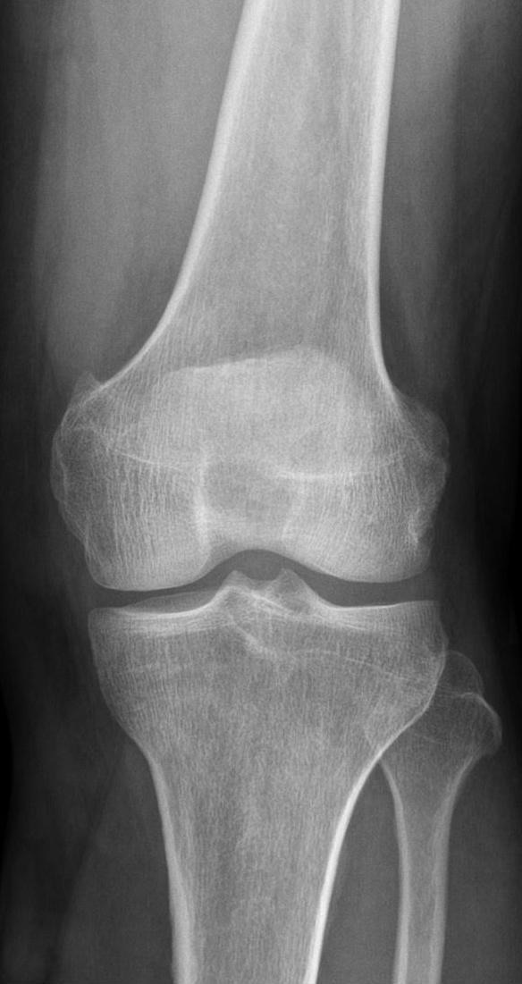
Figure 1. Normal anteroposterior radiograph of the knee, showing the femoral condyles, the tibial plateau and intercondylar eminence, and the proximal fibula. Hellerhoff, CC BY-SA 4.0, via Wikimedia Commons.
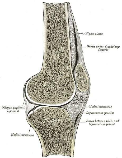
Figure 2. Sagittal section through the knee joint: the patella, suprapatellar bursa, infrapatellar fat pad, ligamentum patellae and the relations of femur, tibia and meniscus. Gray’s Anatomy (1918), Plate 350, Henry Vandyke Carter, public domain, via Wikimedia Commons.
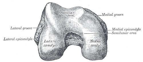
Figure 3. Distal end of the femur, inferior view: the patellar surface, the medial and lateral condyles and epicondyles, and the intercondylar fossa. Gray’s Anatomy (1918), Plate 246, public domain, via Wikimedia Commons.
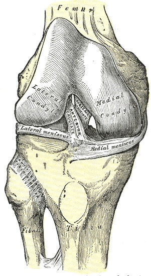
Figure 4. Right knee joint from the front, capsule removed: the anterior cruciate ligament, the medial and lateral menisci and the transverse ligament. Gray’s Anatomy (1918), Plate 347, public domain, via Wikimedia Commons.
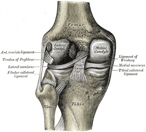
Figure 5. Right knee joint from behind, capsule removed: the anterior and posterior cruciate ligaments, the menisci, the tibial and fibular collateral ligaments and the popliteus tendon. Gray’s Anatomy (1918), Plate 348, public domain, via Wikimedia Commons.
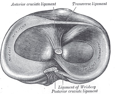
Figure 6. Superior surface of the tibial plateau: the medial and lateral menisci with the tibial attachments of the anterior and posterior cruciate ligaments. Gray’s Anatomy (1918), Plate 349, public domain, via Wikimedia Commons.
Part I - Applied Surgical Anatomy of the Thigh
The three compartments
The thigh is divided by intermuscular septa into three osteofascial compartments, and the cardinal fact for the surgeon is that each compartment carries its own nerve. The anterior compartment holds the quadriceps femoris and is supplied by the femoral nerve; the medial compartment holds the adductor group and is supplied by the obturator nerve; the posterior compartment holds the hamstrings and is supplied by the sciatic (tibial division) nerve. This one-compartment-one-nerve arrangement is what makes a true internervous plane possible only where two differently innervated compartments abut.[3]
A practical asymmetry follows from the septa themselves. The lateral intermuscular septum, running from the linea aspera to the iliotibial tract, is a tough, well-defined sheet: strong enough to be used as a landmark, and strong enough that vessels torn at its edge retract behind it. The medial intermuscular septum is thin and ill-defined by comparison, and the adductors and flexors share no true septum between them, which is precisely why there is no clean internervous plane on the medial and posterior aspects of the femur.[4]
One muscle deserves special mention because it sits across two compartments. The adductor magnus is the great composite muscle of the medial thigh: its adductor (ischiocondylar) portion is supplied by the obturator nerve, while its hamstring portion is supplied by the tibial division of the sciatic nerve. Its tendinous distal insertion onto the adductor tubercle leaves a gap, the adductor hiatus, through which the femoral vessels pass into the popliteal fossa.[5]
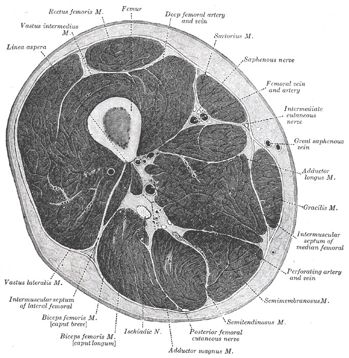
Figure 7. Transverse section through the middle of the thigh: the anterior (quadriceps), medial (adductor) and posterior (hamstring) compartments separated by the intermuscular septa, with the femoral and deep femoral vessels, the saphenous nerve and the sciatic nerve. Gray’s Anatomy (1918), Plate 432, public domain, via Wikimedia Commons.
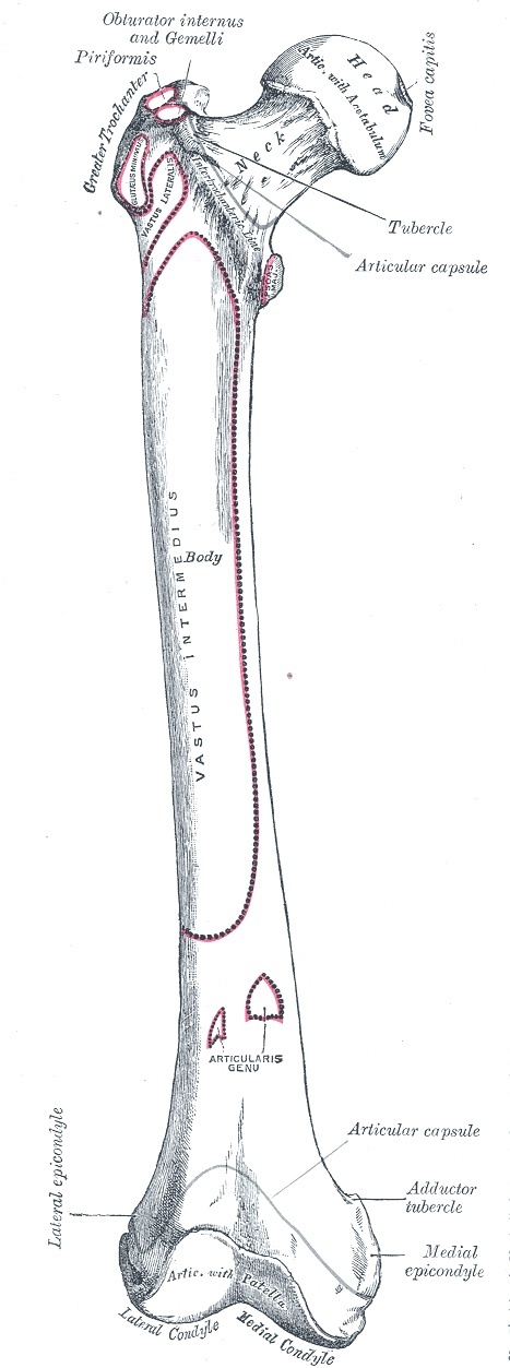
Figure 8. Right femur, anterior view, with muscle attachments outlined: head and fovea, neck, greater trochanter, shaft, condyles and adductor tubercle. Gray’s Anatomy (1918), Plate 244, public domain, via Wikimedia Commons.
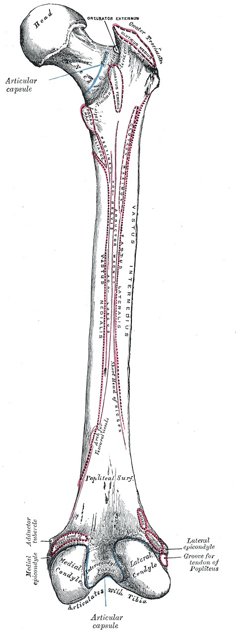
Figure 9. Right femur, posterior view: the linea aspera with the attachments of vastus lateralis, vastus medialis and adductor magnus, the gluteal tuberosity, popliteal surface and intercondylar fossa. Gray’s Anatomy (1918), Plate 245, public domain, via Wikimedia Commons.
The quadriceps and the anterior compartment
The quadriceps femoris is the largest muscle in the body and the workhorse of knee extension. Its four heads (rectus femoris, vastus lateralis, vastus medialis, and vastus intermedius) converge on the quadriceps tendon and thence, through the patella, onto the tibial tubercle by way of the patellar ligament. All four are supplied by the femoral nerve, and the branches enter proximally. This is why the vastus lateralis can be split longitudinally in its substance, or the interval between rectus femoris and vastus medialis opened, without denervating the muscle, provided the dissection stays distal.[6]
The adductor (subsartorial, Hunter’s) canal
The adductor canal, also called the subsartorial canal or Hunter’s canal, is the intermuscular tunnel that conveys the femoral vessels through the lower thigh. Its roof is the sartorius muscle and the overlying fascia; its anterolateral wall is the vastus medialis; its posteromedial wall (floor) is the adductor longus above and the adductor magnus below. The canal is the reason the femoral artery can intrude on the proximal extension of an anteromedial femoral approach, and the reason a saphenous nerve block is placed where it is.[7]
The contents are four and worth memorising as a set: the femoral artery, the femoral vein, the saphenous nerve, and the nerve to vastus medialis (the last present only in the upper half of the canal). At the lower end the femoral artery leaves the canal through the adductor hiatus, classically described as lying “one handbreadth above the knee,” and becomes the popliteal artery. The saphenous nerve does not follow it; instead it pierces the roof and emerges between the sartorius and gracilis to run subcutaneously down the medial leg with the long saphenous vein.[8]
The profunda femoris and its perforating branches
The profunda femoris (deep femoral) artery is the principal blood supply of the thigh. It gives off the medial and lateral femoral circumflex arteries proximally and then, as it descends, sends four perforating branches posteriorly through the medial (adductor) compartment; these wind around the femur and pierce the lateral intermuscular septum to re-enter the anterior compartment and supply the vastus lateralis. These perforators are the great surgical hazard of the lateral and posterolateral femoral approaches: they must be identified and ligated as the septum is divided, because a perforator torn flush with the septum retracts behind it, out of reach, and bleeds uncontrollably.[9]
The sciatic nerve and the posterior compartment
The sciatic nerve descends through the posterior compartment between the hamstrings, lying on the adductor magnus and crossed superficially by the long head of biceps femoris. Its surgical importance in the thigh is twofold: it is the structure sought in a posterior femoral approach (which exists chiefly to explore the nerve), and its relationship to the biceps femoris, in which the biceps “bridges” the nerve obliquely, dictates that a posterior exposure is taken lateral to biceps proximally and medial to biceps distally to keep the nerve in view throughout. The hamstrings (semimembranosus, semitendinosus, long head of biceps) and the ischiocondylar part of adductor magnus are its compartmental muscles.[10]
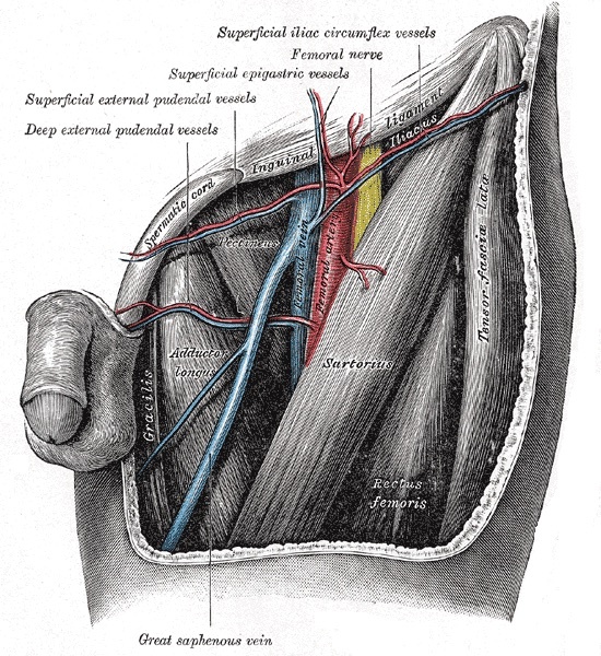
Figure 10. The femoral triangle: the femoral nerve, femoral artery and femoral vein bounded by the inguinal ligament, sartorius and adductor longus, with the great saphenous vein. Gray’s Anatomy (1918), Plate 549, public domain, via Wikimedia Commons.
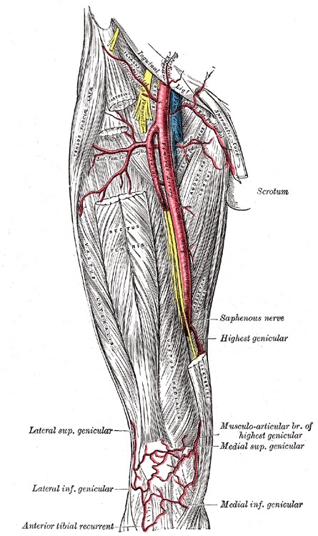
Figure 11. The femoral artery and profunda femoris descending the thigh, with the saphenous nerve accompanying the vessels in the adductor (subsartorial, Hunter’s) canal and the descending genicular branches at the knee. Gray’s Anatomy (1918), Plate 550, public domain, via Wikimedia Commons.
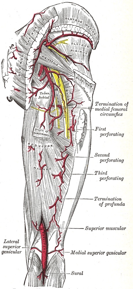
Figure 12. Arteries of the gluteal region and back of the thigh: the profunda femoris with its first, second and third perforating branches, the medial femoral circumflex artery, and the sciatic nerve (shown in yellow) descending the posterior compartment. Gray’s Anatomy (1918), Plate 544, public domain, via Wikimedia Commons.
Part II - Surgical Approaches to the Femur
Hoppenfeld describes four open approaches to the femoral shaft. The single most important examination point is that only one of them, the posterolateral approach, uses a true internervous plane. The others are intermuscular or muscle-splitting, and each is chosen for what it exposes and what it puts at risk.
The lateral approach (the workhorse)
The lateral approach is the most commonly used exposure of the femur, the standard for plating and for open reduction and internal fixation of intertrochanteric and shaft fractures. It has no internervous plane: it splits the vastus lateralis in the line of its fibres, or reflects it from the lateral intermuscular septum. This is safe because the femoral nerve supplies the vastus lateralis proximally, so a distal split does not denervate it. The hazard is vascular, not neural: as the vastus lateralis is reflected, the perforating branches of the profunda must be secured where they cross the lateral intermuscular septum.[11]
The posterolateral approach (the one true plane)
The posterolateral approach is the only femoral exposure with a genuine internervous plane: between the vastus lateralis (femoral nerve) anteriorly and the hamstrings (sciatic nerve) posteriorly, the plane being entered through the lateral intermuscular septum. The vastus lateralis is reflected forward off the septum. As in the lateral approach, the perforating branches crossing the septum are the danger, and they are ligated under direct vision rather than allowed to tear. The posterolateral approach gives good access to the shaft and is favoured where the anterior soft tissues are compromised.[12]
The anteromedial approach (distal two-thirds)
The anteromedial approach exposes the distal two-thirds of the femur and is used for medial-buttress plating of distal femoral fractures. It has no internervous plane, because the structures on either side of the working interval, the vastus medialis and the rectus femoris, are both supplied by the femoral nerve. Its particular hazard is the femoral vessels, which intrude on the proximal extension of the exposure as they run in the adductor (Hunter’s) canal; the more proximal the dissection, the closer the artery.[13]
The posterior approach (for the sciatic nerve)
The posterior approach to the femur is performed prone and exists chiefly for exploration of the sciatic nerve and for posterior skin problems, not for routine fracture work. Its plane lies between the vastus lateralis (femoral nerve) and the biceps femoris (sciatic nerve). Because the biceps crosses the nerve obliquely, the exposure is taken lateral to biceps proximally and medial to biceps distally, following the nerve. It addresses the middle three-fifths of the femur and is not extensile.[14]
Minimally invasive nailing
Closed antegrade or retrograde intramedullary nailing has displaced open plating for most diaphyseal femoral fractures. The shaft is bridged through small proximal or distal incisions over a guide wire and reamer under image intensification, preserving the fracture haematoma and the periosteal blood supply. The open approaches above remain essential for fractures requiring direct reduction, for nonunions, and for periarticular fractures where a plate is needed.[15]
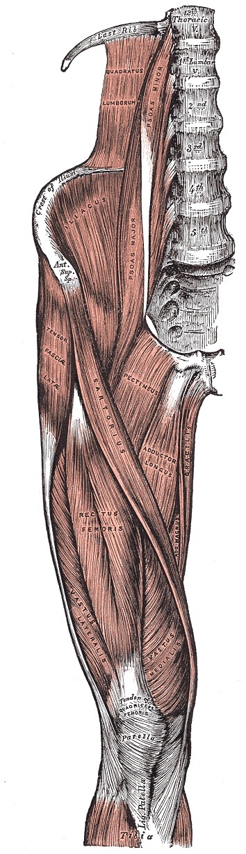
Figure 13. Muscles of the front of the thigh: sartorius, tensor fasciae latae, rectus femoris, vastus lateralis and vastus medialis converging on the quadriceps tendon, patella and ligamentum patellae. Gray’s Anatomy (1918), Plate 430, public domain, via Wikimedia Commons.
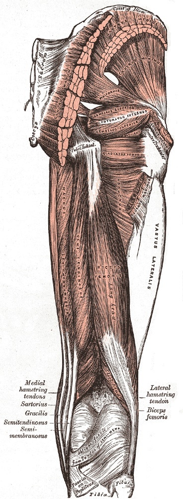
Figure 14. Muscles of the back of the thigh: biceps femoris (long and short heads), semitendinosus and semimembranosus, the hamstring tendons converging on the popliteal fossa. Gray’s Anatomy (1918), Plate 434, public domain, via Wikimedia Commons.
Part III - Principles of Knee Arthroscopy
Why the knee suits the arthroscope
The knee is “a large unconstrained hinge joint that is often described as subcutaneous,” and its anteromedial and anterolateral coverings consist largely of fibrous tissue: the patellar retinaculum and joint capsule. Incisions through these coverings can be made safely because no major nerves are present in these areas. There is, correspondingly, no internervous plane in arthroscopic approaches, which are simply stabs through retinaculum and capsule. Arthroscopy has largely replaced open surgery for meniscal resection and repair, removal of loose bodies, cruciate reconstruction, synovial biopsy and synovectomy, debridement and microfracture of early osteoarthritis, treatment of osteochondritis dissecans, and arthroscopically assisted fixation of tibial plateau fractures.[16]
The two standard portals
Hoppenfeld describes in detail the two portals used in the great majority of procedures. The anterolateral portal is the principal viewing portal: with the knee flexed to 90 degrees, the surgeon palpates the soft spot in the recess between the lateral border of the patellar tendon and the lateral joint line and makes an 8-mm transverse stab roughly 1 to 1½ cm above the lateral joint line. The anteromedial portal is the principal working/instrument portal: an 8-mm stab about 1½ cm above the medial joint line, through which the probing hook and instruments are passed. Because the lateral tibial plateau sits slightly lower than the medial, the lateral portal lies slightly lower than the medial one too.[17]
The accessory portals are not described in Hoppenfeld’s chapter but are standard teaching and carry the region’s nerve hazards. The superolateral and superomedial portals, placed into the suprapatellar pouch, serve inflow/outflow and assessment of patellar tracking. The posteromedial portal endangers the saphenous nerve and the long saphenous vein and is made in the soft spot between the posterior border of the MCL and the medial head of gastrocnemius, under transillumination. The posterolateral portal endangers the common peroneal nerve and is made in the interval bounded by the iliotibial band anteriorly and the biceps femoris tendon posteriorly; staying anterior to biceps protects the nerve.[18]
Dangers and the thermal-safety rule
Hoppenfeld’s explicitly enumerated dangers are articular cartilage and meniscus. Cartilage is damaged either by the capsular incision or by forceful insertion of the scope, and the rule is unambiguous: if you meet resistance while manipulating the arthroscope, you are damaging cartilage. The classic aphorism is worth retaining verbatim: “Ten seconds of careless use of an arthroscope within the knee may create the equivalent of 10 years of wear in that joint.” The meniscus is injured if the portal incision is made too close to the joint line, which is the reason for the 1-1½ cm clearance. One technical pearl governs setup: switch on the irrigation fluid before the light source to avoid thermal damage to the synovium.[19]
The diagnostic sweep
Every arthroscopy must examine all parts of the knee, not merely the suspected lesion, and “manipulation of the knee is the key to success in visualizing all portions of the joint.” The standard ten-view sweep runs: (1) suprapatellar pouch, (2) patellofemoral joint, (3) lateral recess/gutter, (4) popliteus insertion/popliteal hiatus (a common site for loose bodies), (5) anterior horn of the lateral meniscus, (6) medial femoral recess, (7) medial compartment, (8) posterior horn of the medial meniscus, (9) intercondylar notch and the cruciates, and (10) the entire lateral compartment in the figure-of-eight position. The compartment-opening maneuvers are the examiner’s favourite pair: valgus plus external rotation opens the medial side; varus plus internal rotation (the figure-of-eight) opens the lateral side.[20]
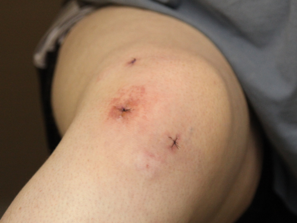
Figure 15. The front of a flexed knee showing the standard anterolateral and anteromedial arthroscopy portal sites flanking the patellar tendon, with a superior (suprapatellar) portal. Tim1965, CC BY-SA 3.0, via Wikimedia Commons.
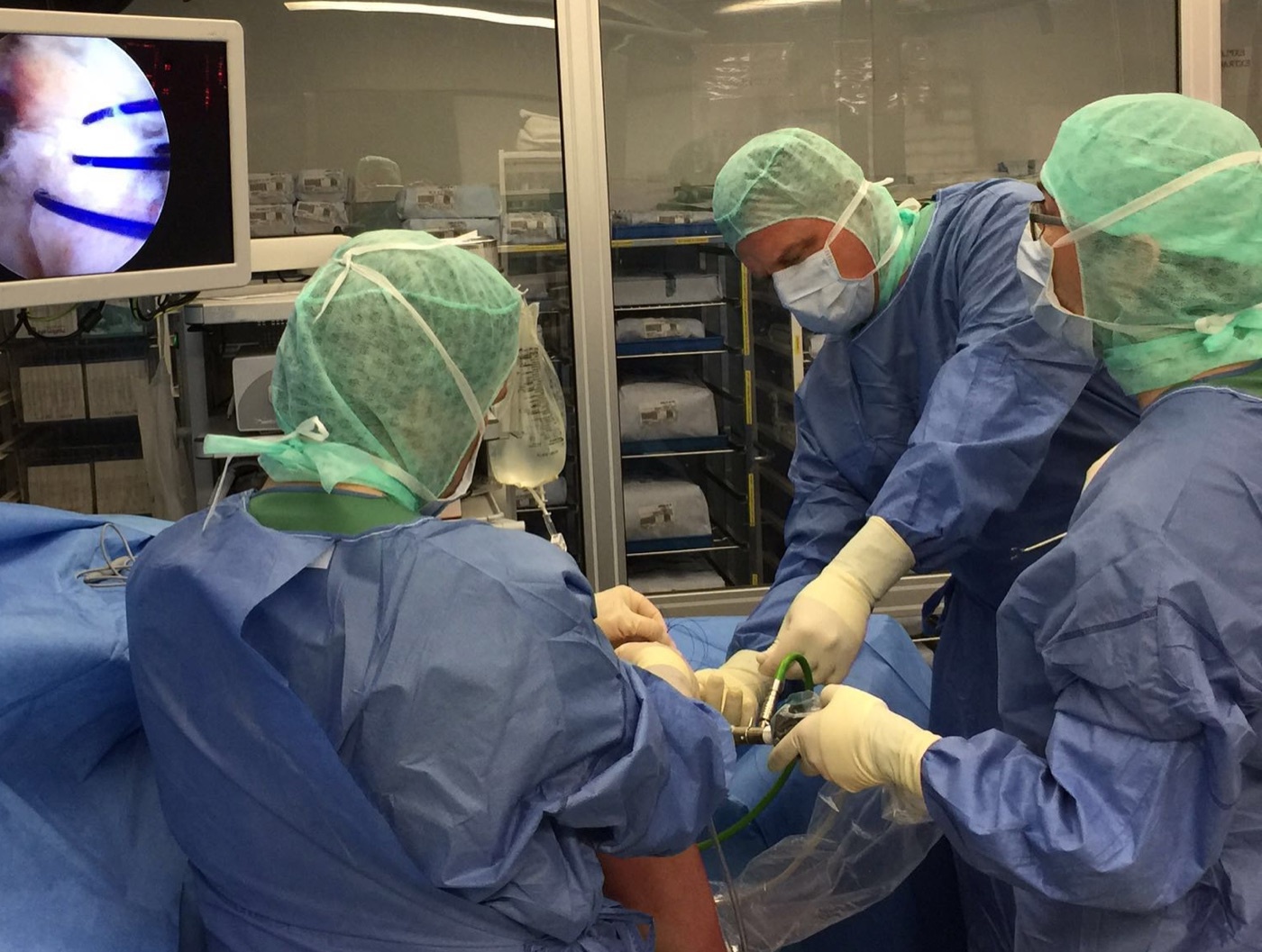
Figure 16. Knee arthroscopy in progress: the arthroscope and instruments inserted through the anterior portals, with the intra-articular image displayed on the tower monitor. Ligamentaxis, CC BY-SA 4.0, via Wikimedia Commons.
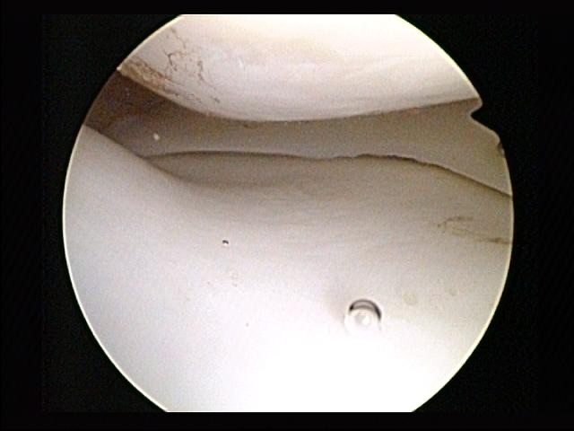
Figure 17. Arthroscopic intra-articular view of a normal medial meniscus seated between the femoral condyle and the tibial plateau. Arthroscopist, CC BY-SA 4.0, via Wikimedia Commons.
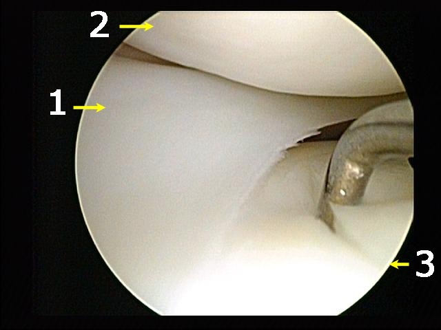
Figure 18. Annotated arthroscopic view inside the knee: the meniscus (1), the femoral condyle articular cartilage (2) and the arthroscopic probe (3) against damaged tibial cartilage. Arthroscopist / S. Fruitsmaak, CC BY-SA 3.0, via Wikimedia Commons.
Part IV - The Medial Side of the Knee
The medial parapatellar approach - the workhorse
The medial parapatellar approach is “the workhorse approach to the knee” and the standard exposure for total knee arthroplasty. A straight midline skin incision runs from about 5 cm above the superior pole of the patella to below the tibial tubercle; the capsule is entered medial to the patella, leaving a cuff of capsular tissue medial to the patella for closure, the quadriceps tendon is split in the midline into the suprapatellar pouch, and the incision is carried down the medial side of the patellar tendon. With the patella dislocated laterally, rotated 180 degrees, and the knee flexed, it gives “the widest possible exposure of the entire knee joint”: femoral condyles, tibial plateau, menisci, and cruciates.[21]
There is no internervous plane, but the superior extension into the interval between vastus medialis and rectus femoris is safe because both are supplied by the femoral nerve well proximal to the dissection. The cardinal technical warning is to avoid avulsing the patellar ligament from the tibial tubercle during eversion, because reattachment is difficult and leads to delayed mobilisation and residual stiffness; where eversion is impossible, the tubercle can be detached with a block of bone. The recurring nerve casualty is the infrapatellar branch of the saphenous nerve, which is “often cut.” It should be resected and its end buried in fat to prevent a painful neuroma, not repaired, because the resulting numbness is trivial.[22]
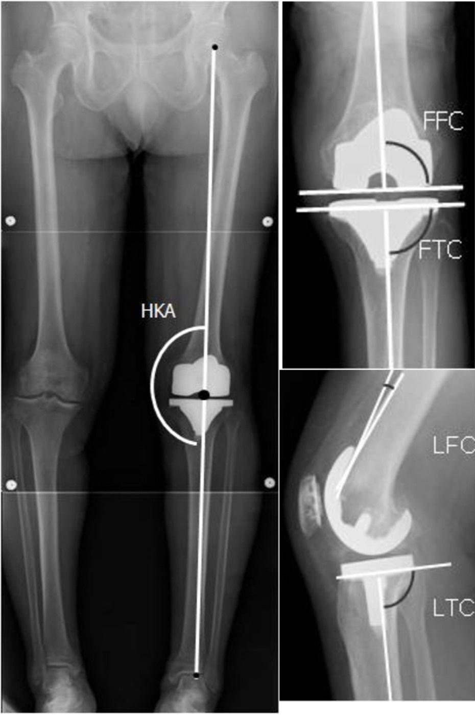
Figure 19. Radiograph of a total knee replacement with limb-alignment measurement: standing full-length view with zoomed anteroposterior and lateral views of the femoral and tibial components. Inui et al. (2013), CC BY 2.0, via Wikimedia Commons.
The approach for medial meniscectomy (the anteromedial approach)
The open anteromedial approach for medial meniscectomy has been largely abolished by arthroscopy but remains a clean illustration of the safe-arthrotomy principle. The skin incision begins at the inferomedial corner of the patella and ends about 1 cm below the joint line, no lower, or it cuts the infrapatellar branch of the saphenous nerve. The single most important rule is to enter the joint well above the joint line: opening the synovium above the line spares the medial meniscus, the coronary ligament, and the intrasynovial fat pad.[23]
Two positioning and protection points recur in vivas. The supporting sandbag must sit under the thigh, never under the popliteal fossa, because a sandbag in the fossa presses the popliteal artery against the posterior capsule and into harm’s way during work on the posterior horn. Flexing the knee further protects the artery, because the posterior capsule (and the artery with it) falls away from the bone. The other danger is the superficial medial collateral ligament, which an over-posterior incision may divide as it crosses the joint.[24]
The medial approach and the three-layer anatomy
The broad medial approach to the knee gives the widest exposure of the medial ligamentous structures and the posteromedial corner, used mainly for the superficial medial ligament, the medial capsule, and medial meniscal/ligamentous repair. The patient is supine, the knee flexed about 60 degrees with the hip abducted and externally rotated. There is no true internervous plane; the motor nerves lie safely posterior in the popliteal fossa, and the only cutaneous nerve at risk is the saphenous nerve and its branches. The infrapatellar branch is sacrificed and buried in fat, but the saphenous nerve trunk (emerging between gracilis and sartorius) and the long saphenous vein must be preserved.[25]
The medial side is best understood, after Warren and Marshall, as three layers, which the surgeon incises in sequence from outside to inside. The outer layer (Layer I) is the deep fascia of the thigh; it encloses the sartorius, blends anteriorly with vastus medialis fibres to form the medial patellar retinaculum, and is continuous posteriorly with the fascia over gastrocnemius. The middle layer (Layer II) is the superficial medial (tibial) collateral ligament: quadrangular, attached just below the adductor tubercle, fanning out to insert on the subcutaneous tibia 6 to 7 cm below the joint line, and giving off the medial patellofemoral ligament above. The deep layer (Layer III) is the true joint capsule, thickened on the medial side only by the deep medial ligament, which runs from the medial epicondyle to the medial meniscus and anchors the meniscus to the tibia by the coronary ligament.[26]
The pes anserinus and the posteromedial corner
The pes anserinus (“goose foot”) is the conjoined insertion of three muscles onto the subcutaneous medial tibia, and the examiner’s pleasure is that all three come from different nerves and different pelvic origins. From front to back they are sartorius (femoral nerve, from the anterior superior iliac spine), gracilis (obturator nerve, from the inferior pubic ramus), and semitendinosus (sciatic nerve, from the ischial tuberosity). Acting together they flex the knee and internally rotate the tibia; the semitendinosus and gracilis run between the superficial and middle layers before joining the outer layer beneath sartorius.[27]
The posteromedial corner is reinforced by three expansions of the semimembranosus tendon: the tough oblique popliteal ligament crossing the fossa to the lateral femoral condyle, an anterior (tibial) expansion passing forward beneath the superficial medial ligament, and a thin popliteus expansion. The fibrous tissue here (the semimembranosus-derived middle layer fused with the posteromedial capsule) corresponds to what modern texts call the posterior oblique ligament, and these dynamic stabilisers should be reattached anatomically when the corner is injured. The medial inferior genicular artery is the vessel at risk here, easily torn when the medial head of gastrocnemius is lifted off the posterior capsule, the bleeding sometimes unmasked only when the tourniquet is released.[28]
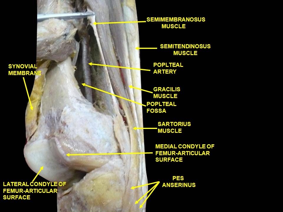
Figure 20. Cadaveric dissection of the medial aspect of the knee: the pes anserinus formed by sartorius, gracilis and semitendinosus, with the popliteal vessels behind. Anatomist90, CC BY-SA 3.0, via Wikimedia Commons.
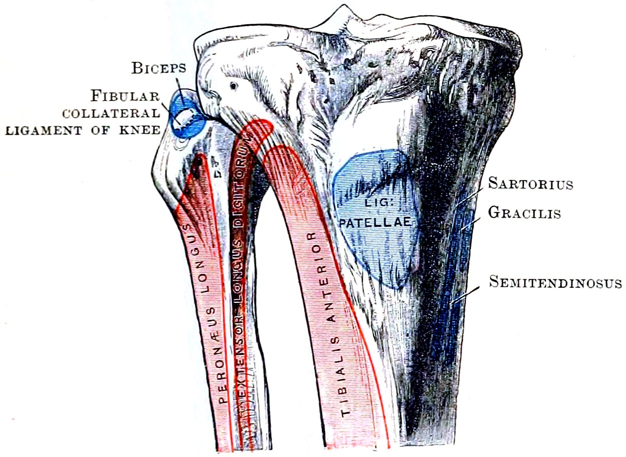
Figure 21. Attachments on the upper tibia and fibula: the pes anserinus footprint on the medial tibia, the ligamentum patellae, and the fibular collateral ligament and biceps on the fibular head. Cunningham’s Text-book of Anatomy (1914), Fig. 248, public domain, via Wikimedia Commons.
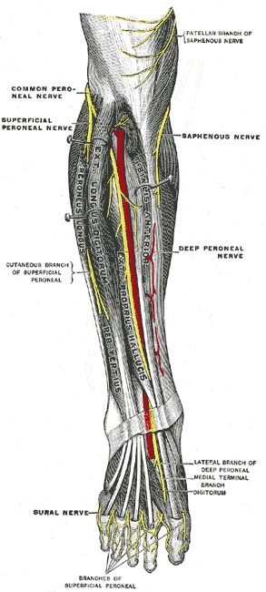
Figure 22. Cutaneous nerves of the front of the knee and leg: the saphenous nerve and its infrapatellar (patellar) branch on the medial side, with the common, superficial and deep peroneal nerves laterally. Gray’s Anatomy (1918), Plate 835, public domain, via Wikimedia Commons.
Part V - The Lateral Side of the Knee
The lateral meniscectomy approach
The open lateral meniscectomy approach survives mainly where arthroscopy is unavailable, but it teaches one rule that governs all lateral work: all incisions enter the lateral compartment anterior to the superficial lateral (fibular collateral) ligament. The oblique incision starting at the inferolateral corner of the patella offers the most leeway. There is no internervous plane; the incision divides lateral retinaculum and capsule. The danger that defines the operation is the lateral inferior genicular artery, which runs at the joint line beside the peripheral attachment of the lateral meniscus and, if the meniscus is detached with a strip of capsule, produces a massive postoperative haemarthrosis; it is not endangered by the exposure itself, only by the meniscectomy.[29]
The lateral approach and its internervous plane
The full lateral approach exposes all the supporting structures of the lateral side and is used chiefly to assess ligamentous damage. Its internervous plane lies between the iliotibial band and the biceps femoris: the iliotibial band is the aponeurosis of tensor fasciae latae (superior gluteal nerve) and gluteus maximus (inferior gluteal nerve), and the biceps femoris is supplied by the sciatic nerve, so the band-to-biceps interval qualifies as internervous by virtue of the band’s muscular origin. The single defining step of the whole approach is to incise that interval while avoiding the common peroneal nerve on the posterior border of the biceps tendon.[30]
The common peroneal nerve is “the structure most at risk during this approach.” It lies on the posterior border of the biceps tendon, must be found early, traced from a normal area into any zone of injury, and protected throughout. The other lateral hazards are the lateral inferior genicular artery (ligated for full posterolateral exposure; the tourniquet should be released before closure to confirm haemostasis) and the popliteus tendon, which lies intracapsularly and can be cut during a posterior arthrotomy if the capsule is not opened well above the joint line.[31]
The three layers of the lateral side and the posterolateral corner
The lateral side, like the medial, falls into three layers. The outer layer is the iliotibial band (inserting on Gerdy’s tubercle), the biceps femoris, and the lateral patellar retinaculum. The middle layer is the superficial lateral (fibular collateral) ligament, running from the lateral epicondyle to the fibular head and tight in extension, together with the recently described anterolateral ligament (distinct from the LCL and capsule, associated with the Segond fracture). The deep layer is the true capsule, the popliteus tendon within it, and the poorly developed short (deep) lateral ligament. A pivotal contrast: unlike the medial side, the lateral ligament does not attach to the meniscus, which is why the lateral meniscus is far more mobile, and, by standard teaching, less often torn, than the medial.[32]
The posterolateral corner comes into view once the iliotibial band is retracted anteriorly and the biceps femoris (with the peroneal nerve) posteriorly. Hoppenfeld names the LCL, the popliteus and its tendon (which can hide the corner), the short lateral ligament, and the posterolateral capsule. The fuller posterolateral-corner complex of standard teaching adds the popliteofibular ligament and the arcuate complex (arcuate and fabellofibular ligaments); the popliteus tendon enters the joint through the popliteus hiatus beneath the arcuate ligament. Injury to the corner produces varus and external-rotation laxity, and is classically combined with cruciate injury.[33]
The popliteus
The popliteus is one of the few muscles whose origin (the popliteal surface of the tibia above the soleal line) lies distal to its insertion (the lateral femoral condyle and posterior aspect of the lateral meniscus); it is supplied by the tibial nerve. Functionally it unlocks the fully extended (“screw-home”) knee by rotating the femur laterally on the fixed tibia, draws the lateral femoral condyle backward, and pulls the lateral meniscus posteriorly so it is not trapped between tibia and femur.[34]
The over-the-top approach for the ACL
The lateral approach to the distal femur, the “over-the-top” approach, is used together with a medial parapatellar approach for cruciate reconstruction and is never an isolated incision. It exposes the posterior intercondylar notch by passing over the top of the posterior aspect of the lateral femoral condyle, and provides access to the lateral condyle for femoral drill holes. Its internervous plane is between the vastus lateralis (femoral nerve) and the biceps femoris (sciatic nerve); note the contrast with the band-to-biceps plane of the lateral knee approach. The lateral superior genicular artery beneath vastus lateralis must be ligated; the common peroneal nerve is endangered if dissection strays behind biceps; and the popliteal artery is protected by staying subperiosteal and flexing the knee to 90 degrees as the notch is reached, letting the artery fall posteriorly with the capsule.[35]
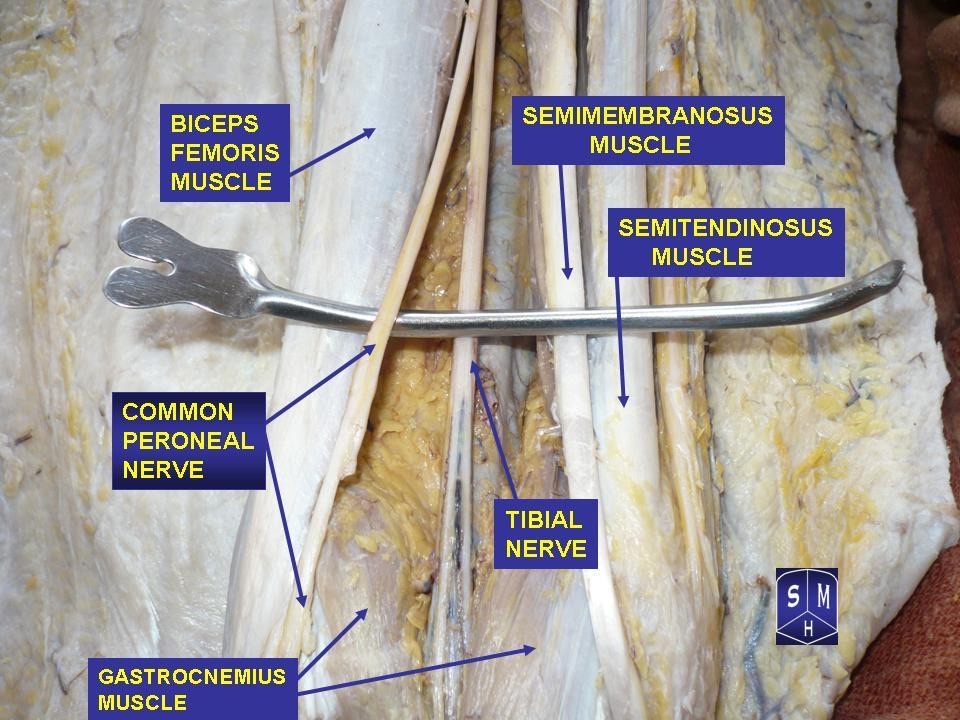
Figure 23. Cadaveric dissection at the knee: the sciatic nerve dividing into the tibial and common peroneal (fibular) nerves, with biceps femoris marking the lateral boundary and the common peroneal nerve passing toward the fibular neck. Anatomist90, CC BY-SA 3.0, via Wikimedia Commons.
Part VI - The Posterior Knee and the Popliteal Fossa
The posterior approach
The posterior approach to the knee is “primarily a neurovascular approach” and is rarely needed for orthopaedic work, because the medial and lateral approaches each reach half the posterior capsule. Its indications are repair of the popliteal neurovascular structures in trauma, reattachment of an avulsed tibial insertion of the posterior cruciate ligament, gastrocnemius recession for contracture, hamstring lengthening, and excision of a Baker (popliteal) cyst. The patient is prone; a tourniquet is used for all but vascular repairs; and because the operation is unfamiliar and done prone, the fibular head is marked with an “L” for orientation.[36]
The curved (“lazy-S”) incision starts laterally over the biceps femoris, crosses the fossa obliquely, and turns down over the medial head of gastrocnemius; its near-parallel relation to the flexion crease at this level avoids a flexion contracture on healing. There is no true internervous plane; the dissection works among the boundary muscles and the popliteal contents. The key superficial guide is the small (short) saphenous vein with the medial sural cutaneous nerve on its lateral side: the vein leads to the nerve, and the nerve (a branch of the tibial nerve) leads up to the tibial nerve and so into the fossa.[37]
The popliteal fossa
“The anatomy of the posterior approach to the knee is the anatomy of the popliteal fossa.” The diamond has four muscular borders: superomedially the semimembranosus and semitendinosus, superolaterally the biceps femoris, inferomedially the medial head of gastrocnemius, inferolaterally the lateral head of gastrocnemius. The roof is the popliteal fascia (the outer layer of the knee’s coverings), and the floor is the posterior femur, the posterior joint capsule, and the popliteus overlying the proximal tibia.[38]
The contents lie in a strict superficial-to-deep order that must be recited correctly: tibial nerve, then popliteal vein, then popliteal artery, the artery deepest, hard against the posterior capsule on the floor. The tibial nerve is the continuation of the sciatic and runs vertically through the fossa, leaving between the two gastrocnemius heads; its only cutaneous branch, the sural nerve, is the classic graft donor. The common peroneal nerve separates from the tibial at the fossa apex and slopes laterally along the posterior border of biceps toward the fibular neck, where it is vulnerable to compression; hence the rule to pad the fibular head in positioning. The popliteal vein lies between the artery and the tibial nerve and must be handled gently, since intimal damage may cause thrombosis.[39]
The genicular arteries
The popliteal artery gives five genicular branches around the knee: two superior, two inferior, and one middle. The lateral superior is ligated in the posterolateral/over-the-top exposure; the medial superior is ligated when the medial gastrocnemius is detached for the posteromedial corner. The middle genicular pierces the posterior capsule to supply the cruciates, so a traumatic cruciate rupture bleeds briskly into the joint, and crucially it tethers the popliteal trunk to the posterior capsule, which is why the artery is so readily injured in knee dislocations and in posterior dissection; flexing the knee lets the capsule and artery fall away from bone and protects it. The two inferior genicular arteries pass deep to the collateral ligaments at the joint line, and the lateral inferior genicular is the one most commonly damaged in lateral meniscectomy.[40]
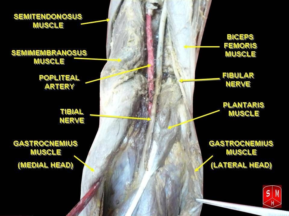
Figure 24. Dissection of the popliteal fossa: the muscular boundaries (semitendinosus and semimembranosus medially, biceps femoris laterally, the two heads of gastrocnemius inferiorly) and the popliteal artery, tibial nerve and common peroneal nerve. Anatomist90, CC BY-SA 3.0, via Wikimedia Commons.
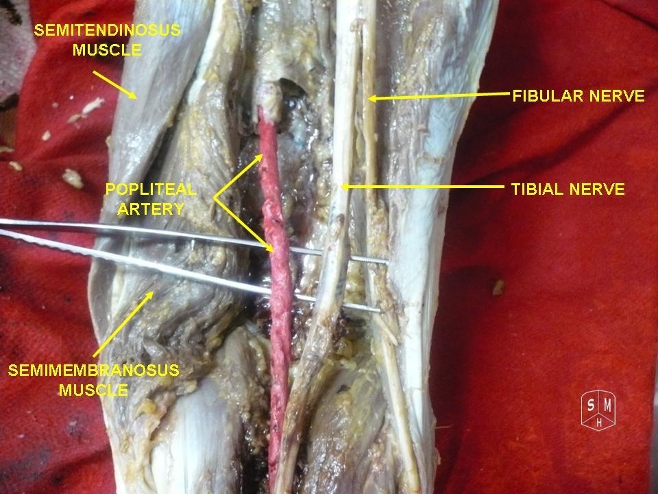
Figure 25. A second popliteal fossa dissection showing the tibial and fibular (common peroneal) nerves and the popliteal artery; the popliteal vein lies between the tibial nerve and the artery in the superficial-to-deep order. Anatomist90, CC BY-SA 3.0, via Wikimedia Commons.
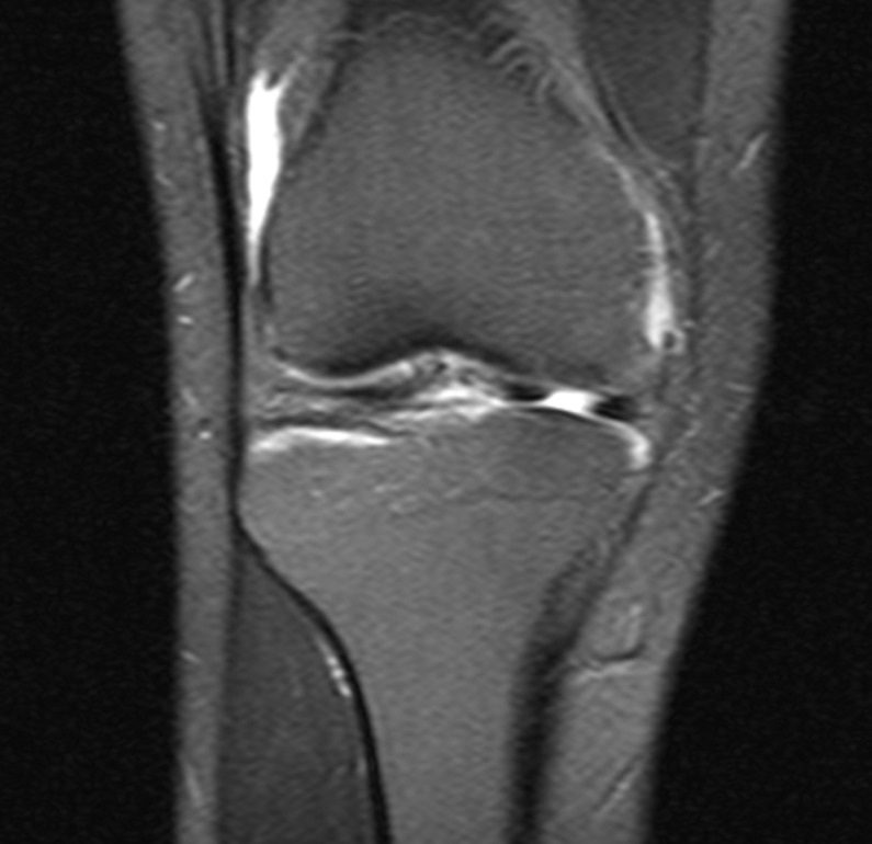
Figure 26. Magnetic resonance image of the knee demonstrating a meniscal tear. Ciernik M, CC BY-SA 4.0, via Wikimedia Commons.
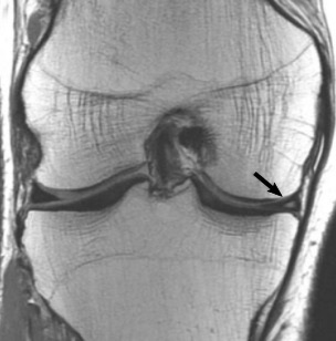
Figure 27. Coronal proton-density MRI of a grade-2 medial meniscal tear (arrow). Lefevre et al. (2016), CC BY 4.0, via Wikimedia Commons.
References
-
Scope and sequencing of this summary follow Hoppenfeld, Surgical Exposures in Orthopaedics: The Anatomic Approach, 5th ed. (2016), Chapter 9 (The Femur, pp.834-900) and Chapter 10 (The Knee, pp.901-1011); the popliteal fossa is treated as “the anatomy of the posterior approach to the knee” (Hoppenfeld p.994). General teaching that supplements the source is flagged in-line as standard teaching and carries no page citation.
-
The five recurring danger structures are drawn together from across the chapters: profunda perforators (Hoppenfeld p.858 region, thigh applied anatomy); common peroneal nerve “the structure most at risk during this approach” (Hoppenfeld p.973); infrapatellar branch of the saphenous nerve (Hoppenfeld p.938, p.922); popliteal artery “lies directly behind the posterior capsule of the knee joint” (Hoppenfeld p.995); lateral inferior genicular artery, most commonly damaged in lateral meniscectomy (Hoppenfeld p.996).
-
Hoppenfeld describes the three thigh compartments - anterior (quadriceps, femoral nerve), medial (adductors, obturator nerve), posterior (hamstrings, sciatic nerve) - in the applied anatomy of the thigh (Hoppenfeld ch.9 thigh anatomy, p.858 region). The functional principle that internervous planes exist between differently innervated muscle groups is stated throughout Hoppenfeld’s general introduction.
-
The tough lateral intermuscular septum versus the thin medial septum, and the absence of a septum shared by the adductors and flexors, are detailed in Hoppenfeld’s applied surgical anatomy of the thigh (Hoppenfeld ch.9). The lateral septum is the structure pierced by the profunda perforating branches.
-
The dual innervation of adductor magnus (obturator nerve to the adductor part, sciatic/tibial to the ischiocondylar “hamstring” part) and its role in forming the adductor hiatus are standard descriptive anatomy underlying Hoppenfeld’s account of the medial compartment and the adductor canal (Hoppenfeld ch.9 thigh anatomy).
-
Quadriceps femoris as the largest muscle, its four heads and femoral nerve supply, and the safety of splitting vastus lateralis or opening the rectus-vastus medialis interval distally (because the nerve enters proximally) are from Hoppenfeld’s thigh applied anatomy and the lateral/anteromedial femoral approaches (Hoppenfeld ch.9, pp.835-857, p.858 region). The same principle underlies the safe superior extension of the medial parapatellar knee approach (Hoppenfeld p.917).
-
The boundaries of the adductor (subsartorial/Hunter’s) canal - roof sartorius and fascia, anterior wall vastus medialis, posterior wall adductor longus (superiorly) and adductor magnus (inferiorly) - are from Hoppenfeld’s thigh applied anatomy (Hoppenfeld ch.9). The femoral vessels’ intrusion on the proximal anteromedial femoral approach (Hunter’s canal) is noted in the anteromedial femur approach (Hoppenfeld ch.9, p.858 region).
-
Adductor canal contents - femoral artery, femoral vein, saphenous nerve, and the nerve to vastus medialis (upper half) - and the femoral artery passing through the adductor hiatus “one handbreadth above the knee” to become the popliteal artery are from Hoppenfeld’s thigh applied anatomy (Hoppenfeld ch.9). The saphenous nerve emerging between sartorius and gracilis with the long saphenous vein is stated in the medial knee approach (Hoppenfeld p.938, p.944).
-
The profunda femoris as the main arterial supply of the thigh; its four perforating branches passing posteriorly through the adductor compartment, winding around the femur, and piercing the lateral intermuscular septum to re-enter the anterior compartment and supply the vastus lateralis (Hoppenfeld p.888); and the danger that a perforator torn flush with the septum retracts behind it and bleeds uncontrollably (Hoppenfeld p.846, posterolateral approach Dangers; lateral approach Dangers p.840).
-
The sciatic nerve’s course in the posterior compartment, its relationship to biceps femoris (the muscle bridging the nerve, dictating lateral-to-biceps proximally and medial-to-biceps distally in the posterior femoral approach), and the posterior approach existing chiefly for sciatic exploration are from Hoppenfeld’s femur chapter (Hoppenfeld ch.9 femur, pp.835-857, posterior approach; thigh applied anatomy p.858 region).
-
The lateral approach to the femur as the most common (ORIF of intertrochanteric and shaft fractures), with no internervous plane, splitting or reflecting vastus lateralis (safe because the femoral nerve enters proximally), and the perforating profunda branches as the principal danger at the lateral intermuscular septum are from Hoppenfeld’s lateral femoral approach (Hoppenfeld ch.9, pp.835-857).
-
The posterolateral approach as the only femoral approach with a true internervous plane - vastus lateralis (femoral nerve) and hamstrings (sciatic nerve), entered through the lateral intermuscular septum with vastus lateralis reflected off the septum - and the perforating profunda branches as the danger are from Hoppenfeld’s posterolateral femoral approach (Hoppenfeld ch.9, pp.835-857).
-
The anteromedial approach to the distal two-thirds of the femur (medial buttress plating), with no internervous plane because vastus medialis and rectus femoris are both femoral-nerve supplied, and the femoral vessels intruding on the proximal extension via Hunter’s canal are from Hoppenfeld’s anteromedial femoral approach (Hoppenfeld ch.9, p.858 region). This is the approach into which the medial parapatellar knee exposure extends superiorly (Hoppenfeld p.924).
-
The posterior approach to the femur - prone, for sciatic nerve exploration and skin problems, plane between vastus lateralis (femoral nerve) and biceps femoris (sciatic nerve), taken lateral to biceps proximally and medial to biceps distally, addressing the middle three-fifths, not extensile - is from Hoppenfeld’s posterior femoral approach (Hoppenfeld ch.9, pp.835-857).
-
Minimally invasive intramedullary nailing of the femoral shaft, preserving the fracture haematoma and periosteal supply, is described in Hoppenfeld’s femur chapter (Hoppenfeld ch.9, MIS nailing, p.858 region). The continued role of open approaches for direct reduction, nonunion, and periarticular plate fixation is standard teaching.
-
The knee as a “large unconstrained hinge joint… often described as subcutaneous,” its fibrous anteromedial/anterolateral coverings allowing safe incision with “no major nerves,” the absence of an internervous plane (Hoppenfeld p.903, p.908), and the list of procedures arthroscopy has replaced open surgery for (Hoppenfeld p.903) are from Hoppenfeld’s general principles of knee arthroscopy.
-
The anterolateral portal as the main viewing portal and anteromedial as the main working/instrument portal, the soft-spot localization, the 8-mm stab incisions ~1-1½ cm above the joint line, and the lateral portal lying slightly lower (lateral plateau is lower) are from Hoppenfeld’s arthroscopic approaches (Hoppenfeld pp.903-907). Usually the scope is in the anterolateral and instruments in the anteromedial, “however, either portal can be used for either purpose” (Hoppenfeld p.904).
-
Standard teaching (not in Hoppenfeld’s ch.10 arthroscopy extract, which describes only the two anterior portals): the superolateral/superomedial portals for inflow-outflow and patellar tracking; the posteromedial portal’s danger to the saphenous nerve and long saphenous vein (safe zone between posterior MCL and medial gastrocnemius, with transillumination); and the posterolateral portal’s danger to the common peroneal nerve (iliotibial band anteriorly, biceps femoris posteriorly as boundaries). Hoppenfeld’s text states only “No major nerves are present in these areas” for the two anterior portals (Hoppenfeld p.908).
-
The two explicitly listed dangers - articular cartilage (damaged by the capsular incision or forceful scope insertion; “if you meet with resistance… you are damaging the articular cartilage,” and “Ten seconds of careless use… 10 years of wear,” Hoppenfeld pp.909-910) and meniscus (if the incision is too close to the joint line, Hoppenfeld p.912) - plus the thermal-safety rule to switch on irrigation before the light source (Hoppenfeld p.908) are from Hoppenfeld’s arthroscopy section. Neurovascular dangers (saphenous nerve, common peroneal nerve, popliteal and geniculate vessels) are standard teaching, not enumerated in this extract.
-
The principle of examining all portions of the knee (Hoppenfeld p.908), “manipulation of the knee is the key to success” (Hoppenfeld p.912), the ten-view diagnostic sequence (Views 1-10, Hoppenfeld pp.908-909, Figs. 10-3 to 10-8), the popliteal hiatus as a common recess for loose bodies (Hoppenfeld pp.908-909), and the compartment-opening maneuvers - valgus + external rotation for the medial compartment, varus + internal rotation / figure-of-eight for the lateral (Hoppenfeld pp.904, 909, 912) - are all from Hoppenfeld’s arthroscopic exploration of the knee.
-
The medial parapatellar approach as “the workhorse approach to the knee” and the standard TKA exposure, the midline skin incision (5 cm above the superior pole of the patella to below the tibial tubercle), the medial capsular cuff left for closure, midline quadriceps-tendon split into the suprapatellar pouch, and the patella dislocated/everted with the knee flexed giving “the widest possible exposure of the entire knee joint” are from Hoppenfeld’s medial parapatellar approach (Hoppenfeld pp.915-922).
-
No internervous plane but a safe superior extension between vastus medialis and rectus femoris (both femoral-nerve supplied well proximally, Hoppenfeld p.917); the warning not to avulse the patellar ligament from the tibia (difficult to reattach, delayed mobilization, residual stiffness; bone-block detachment as the salvage, Hoppenfeld p.918, pp.921-924); and the infrapatellar branch of the saphenous nerve being “often cut,” to be resected and buried in fat rather than repaired (Hoppenfeld p.922) are from Hoppenfeld’s medial parapatellar approach.
-
The open anteromedial approach for medial meniscectomy, its skin incision from the inferomedial corner of the patella ending ~1 cm below the joint line (lower risks the infrapatellar branch of the saphenous nerve, Hoppenfeld pp.926-927), and the safe-arthrotomy rule of entering the joint well above the joint line to spare the medial meniscus, coronary ligament, and fat pad (Hoppenfeld p.927, p.930) are from Hoppenfeld’s approach for medial meniscectomy.
-
The sandbag under the thigh and never under the popliteal fossa (where it pushes the popliteal artery against the posterior capsule, Hoppenfeld p.925, pp.929-930), flexing the knee to let the posterior capsule and artery fall away from bone (Hoppenfeld p.929), and the superficial medial (tibial collateral) ligament at risk from an over-posterior incision (Hoppenfeld p.930) are from Hoppenfeld’s medial meniscectomy approach.
-
The medial approach giving the widest exposure of the medial ligamentous structures and posteromedial corner (Hoppenfeld p.936), the supine position with the knee flexed ~60° and hip abducted/externally rotated (Hoppenfeld p.936), no true internervous plane with the motor nerves safely posterior and only the saphenous nerve and branches at risk (Hoppenfeld pp.936-937), and the sacrifice of the infrapatellar branch (buried in fat) while preserving the saphenous trunk and long saphenous vein (Hoppenfeld pp.938-939, p.944) are from Hoppenfeld’s medial approach to the knee.
-
The three-layer concept of the medial knee (Warren and Marshall, “condensations within tissue planes,” incised outside-to-inside, Hoppenfeld p.950): outer layer (Layer I) = deep fascia enclosing sartorius, blending with vastus medialis to form the medial patellar retinaculum (Hoppenfeld p.950); middle layer (Layer II) = superficial medial (tibial collateral) ligament, quadrangular, from just below the adductor tubercle to the subcutaneous tibia 6-7 cm below the joint, giving off the MPFL (Hoppenfeld pp.950-951); deep layer (Layer III) = true capsule thickened only by the deep medial (meniscofemoral) ligament from medial epicondyle to medial meniscus, anchoring the meniscus by the coronary ligament (Hoppenfeld p.951). The explicit Layer I/II/III numerals are the standard Warren-Marshall equivalents; Hoppenfeld uses outer/middle/deep.
-
The pes anserinus (“goose foot”) - sartorius (femoral nerve, ASIS), gracilis (obturator nerve, inferior pubic ramus), and semitendinosus (sciatic nerve, ischial tuberosity), all crossing hip and knee, together flexing the knee and internally rotating the tibia, with semitendinosus and gracilis running between the superficial and middle layers before joining the outer layer under sartorius - is from Hoppenfeld’s applied anatomy of the medial side (Hoppenfeld p.951, p.960).
-
The three semimembranosus expansions (oblique popliteal ligament to the lateral femoral condyle, anterior/tibial expansion beneath the superficial medial ligament, and a thin popliteus expansion), their importance for dynamic stabilization and anatomic reattachment (Hoppenfeld pp.950-951), the posteromedial-corner fibrous tissue corresponding to the posterior oblique ligament (Hoppenfeld p.959; the term itself is standard teaching), and the medial inferior genicular artery torn when the medial gastrocnemius is lifted off the posterior capsule with bleeding unmasked on tourniquet release (Hoppenfeld p.944) are from Hoppenfeld’s medial-side applied anatomy. The popliteal artery lies against the posterior capsule in the midline, adjacent to the medial gastrocnemius (Hoppenfeld p.945).
-
The lateral meniscectomy approach, the rule that all incisions enter anterior to the superficial lateral (fibular collateral) ligament (Hoppenfeld p.961), the oblique incision from the inferolateral patellar corner offering the most leeway (Hoppenfeld p.964), no internervous plane (Hoppenfeld p.964), and the lateral inferior genicular artery at the joint line beside the lateral meniscus causing massive haemarthrosis if torn during meniscectomy (“not in danger during the approach,” Hoppenfeld p.967) are from Hoppenfeld’s lateral meniscectomy approach.
-
The lateral approach exposing all lateral supporting structures, chiefly for assessing ligamentous damage (Hoppenfeld p.969), and its internervous plane between the iliotibial band (tensor fasciae latae via superior gluteal nerve + gluteus maximus via inferior gluteal nerve) and biceps femoris (sciatic nerve), the band counting as internervous by its muscular origin (Hoppenfeld p.970), with the key step of incising the interval while avoiding the common peroneal nerve on the posterior border of biceps (Hoppenfeld p.970) are from Hoppenfeld’s lateral approach to the knee.
-
The common peroneal nerve as “the structure most at risk during this approach,” on the posterior border of biceps, to be found early and traced from normal into abnormal (Hoppenfeld pp.973-974); the lateral inferior genicular artery requiring ligation for full posterolateral exposure with tourniquet release before closure (Hoppenfeld p.974); and the popliteus tendon (intracapsular) at risk during posterior arthrotomy unless the capsule is opened well above the joint line (Hoppenfeld p.972, p.974) are from Hoppenfeld’s lateral approach.
-
The three layers of the lateral knee - outer (iliotibial band to Gerdy’s tubercle, biceps femoris, lateral patellar retinaculum), middle (superficial lateral/fibular collateral ligament, lateral epicondyle to fibular head, tight in extension; plus the anterolateral ligament, distinct from the LCL and capsule, associated with the Segond fracture), deep (true capsule, intracapsular popliteus tendon, short/deep lateral ligament) - and the key contrast that the lateral ligament does not attach to the meniscus, making the lateral meniscus far more mobile (Hoppenfeld pp.976-980) are from Hoppenfeld’s applied anatomy of the lateral side. The lateral meniscus being less commonly torn than the medial is standard teaching; Hoppenfeld supplies the mobility mechanism, not the explicit tear-frequency statement.
-
The posterolateral corner becoming visible after retracting the iliotibial band anteriorly and biceps femoris (with the peroneal nerve) posteriorly (Hoppenfeld p.970), the Hoppenfeld-named PLC structures (LCL, popliteus/tendon hiding the corner, short lateral ligament, posterolateral capsule, Hoppenfeld p.972, p.980), and the popliteus tendon entering through a gap beneath the arcuate ligament (Hoppenfeld p.999) are from the source; the popliteofibular and arcuate/fabellofibular components and the varus + external-rotation laxity pattern of PLC injury are standard teaching.
-
The popliteus - origin on the popliteal surface of the tibia above the soleal line, insertion on the lateral femoral condyle and posterior aspect of the lateral meniscus, tibial nerve supply, one of the few muscles whose origin is distal to its insertion (Hoppenfeld p.999, p.1001), and its functions of unlocking the screw-home knee, drawing the lateral femoral condyle backward, and pulling the lateral meniscus back out of harm’s way (Hoppenfeld p.1001) - is from Hoppenfeld’s lateral and posterior knee sections.
-
The lateral approach to the distal femur (“over-the-top”), used with the medial parapatellar approach for ACL reconstruction and never isolated, exposing the posterior intercondylar notch and the lateral condyle for drill holes (Hoppenfeld pp.1001-1002); its internervous plane between vastus lateralis (femoral nerve) and biceps femoris (sciatic nerve) (Hoppenfeld pp.1002-1003); and its dangers - the lateral superior genicular artery to be ligated, the common peroneal nerve if dissection strays behind biceps, and the popliteal artery protected by staying subperiosteal and flexing the knee to 90° to let it fall posteriorly (Hoppenfeld p.1005) - are from Hoppenfeld’s lateral approach to the distal femur. The ACL originates from the posteromedial wall of the lateral femoral condyle in the notch with anteromedial and posterolateral bundles (standard teaching).
-
The posterior approach as “primarily a neurovascular approach,” rarely needed because the medial/lateral approaches each reach half the posterior capsule (Hoppenfeld p.982), its indications (neurovascular repair in trauma, reattachment of the avulsed tibial PCL insertion, gastrocnemius recession, hamstring lengthening, Baker cyst excision, access to the posterior capsule, Hoppenfeld p.982, p.990), the prone position with a tourniquet for all but vascular repairs (Hoppenfeld p.982), and marking the fibular head “L” for orientation (Hoppenfeld p.983) are from Hoppenfeld’s posterior approach to the knee.
-
The curved (“lazy-S”) incision (laterally over biceps, obliquely across the fossa, down over the medial gastrocnemius) and its near-parallel relation to the flexion crease avoiding contracture (Hoppenfeld p.983, p.994), no true internervous plane (Hoppenfeld p.985), and the small saphenous vein with the medial sural cutaneous nerve (a branch of the tibial nerve) on its lateral side serving as the guide to the tibial nerve and the fossa (Hoppenfeld p.985, p.995) are from Hoppenfeld’s posterior approach.
-
“The anatomy of the posterior approach to the knee is the anatomy of the popliteal fossa” (Hoppenfeld p.994); the four muscular borders (superomedial semimembranosus/semitendinosus, superolateral biceps femoris, inferomedial and inferolateral heads of gastrocnemius), the roof (popliteal fascia) and floor (posterior femur, posterior capsule, popliteus over the proximal tibia) are from Hoppenfeld’s applied anatomy of the popliteal fossa (Hoppenfeld p.994, p.999).
-
The superficial-to-deep order of contents - tibial nerve, popliteal vein, popliteal artery (deepest, “directly behind the posterior capsule of the knee joint,” Hoppenfeld p.995, p.998) - the tibial nerve as the continuation of the sciatic leaving between the gastrocnemius heads, the sural nerve as its cutaneous branch used in grafting (Hoppenfeld p.995), the common peroneal nerve separating at the apex and sloping along the posterior border of biceps toward the fibular neck with the instruction to pad the fibular head (Hoppenfeld p.987, p.995), and the popliteal vein lying between the artery and tibial nerve with the caution that intimal damage may cause thrombosis (Hoppenfeld p.987, p.998) are from Hoppenfeld’s posterior approach and popliteal-fossa anatomy. (The source contains a minor internal inconsistency over whether the vein enters medial or lateral to the artery; it agrees the vein then lies directly posterior to the artery in the fossa.)
-
The five genicular branches (two superior, two inferior, one middle), the lateral superior ligated in the posterolateral approach and the medial superior when the medial gastrocnemius is detached (Hoppenfeld pp.995-996), the middle genicular supplying the cruciates (brisk intra-articular bleeding on rupture) and tethering the popliteal trunk firmly to the posterior capsule so it is easily injured in dislocations and posterior dissection - protected by flexing the knee (Hoppenfeld p.996) - and the inferior genicular arteries passing deep to the collateral ligaments at the joint line, the lateral inferior being most commonly damaged in lateral meniscectomy (Hoppenfeld p.996), are from Hoppenfeld’s applied anatomy of the popliteal fossa.
-
Three thigh compartments and their nerves, with adductor magnus dual innervation, from Hoppenfeld’s thigh applied anatomy (Hoppenfeld ch.9, p.858 region).
-
Posterolateral femoral approach as the only true internervous plane (vastus lateralis/hamstrings via the lateral intermuscular septum), from Hoppenfeld’s femoral approaches (Hoppenfeld ch.9, pp.835-857).
-
Profunda perforating branches at the lateral intermuscular septum and the retraction-behind-septum hazard, from Hoppenfeld’s thigh applied anatomy and lateral femoral approach (Hoppenfeld ch.9).
-
Adductor canal contents and walls, and the femoral artery passing through the adductor hiatus to become the popliteal artery, from Hoppenfeld’s thigh applied anatomy (Hoppenfeld ch.9).
-
Anterolateral (viewing) and anteromedial (working) portals, their placement, and interchangeability, from Hoppenfeld’s arthroscopic approaches (Hoppenfeld pp.903-907).
-
The two enumerated dangers (articular cartilage, meniscus), the “ten seconds / ten years” aphorism, and the irrigation-before-light rule, from Hoppenfeld’s arthroscopy section (Hoppenfeld pp.909-912, p.908).
-
The medial parapatellar approach and its role as the standard TKA exposure, with the safe superior extension, from Hoppenfeld’s medial parapatellar approach (Hoppenfeld pp.915-924).
-
The three layers of the medial knee (Warren-Marshall), from Hoppenfeld’s applied anatomy of the medial side (Hoppenfeld pp.950-951).
-
Pes anserinus components and their nerve supplies (sartorius-femoral, gracilis-obturator, semitendinosus-sciatic), from Hoppenfeld’s medial-side applied anatomy (Hoppenfeld p.951, p.960).
-
Common peroneal nerve as the structure most at risk on the lateral side (posterior border of biceps; found early, traced normal-to-abnormal; padding the fibular head), from Hoppenfeld’s lateral approach and popliteal-fossa anatomy (Hoppenfeld pp.970-974, p.995).
-
Popliteal fossa boundaries and the superficial-to-deep order of contents (tibial nerve, popliteal vein, popliteal artery), from Hoppenfeld’s applied anatomy of the popliteal fossa (Hoppenfeld p.994, pp.995-998).
-
The middle genicular artery tethering the popliteal trunk to the posterior capsule, the resulting vulnerability in dislocations and dissection, and protection by knee flexion (capsule and artery fall away) and a subperiosteal plane, from Hoppenfeld’s popliteal-fossa anatomy and medial meniscectomy approach (Hoppenfeld p.996, p.929, p.1005).