Contents
- Orientation
- Part I - Surgical Anatomy of the Shoulder Joint
- Part II - The Anterior (Deltopectoral) Approach
- Part III - The Anterolateral and Lateral (Deltoid-Split) Approaches
- Part IV - The Posterior Approach
- Part V - Arthroscopic Approaches (Portals)
- References
Orientation
The shoulder is the most mobile joint in the body, and one anatomical picture governs its surgery: a ball-and-shallow-socket joint wrapped in two muscular sleeves, an outer deltoid and an inner rotator cuff, with named nerves threaded between and around them that the surgeon must locate precisely.[1] Three problems bring the shoulder to operation, and they map onto the approaches: instability (recurrent anterior dislocation, approached from the front), rotator-cuff disease (approached laterally through a deltoid split or arthroscopically), and fractures of the proximal humerus (approached anteriorly or laterally). The examiner for this topic wants the same things as for the spine: the internervous plane of each approach, the danger structures at each depth, the position and landmarks, and the surgical anatomy behind them.
One nerve dominates the topic: the axillary nerve, which runs transversely on the deep surface of the deltoid about 5 to 7 cm below the tip of the acromion and passes through the quadrangular space with the posterior circumflex humeral artery. It limits the deltoid-split approaches, it lies in danger below the subscapularis in the anterior approach and below the teres minor in the posterior approach, and a low arthroscopy portal can catch it.[2] A Bulgarian glossary and a viva appendix close the document.
Part I - Surgical Anatomy of the Shoulder Joint
The two muscular sleeves and the glenohumeral joint
Two sleeves of muscle surround the shoulder: the outer sleeve is the deltoid, and the inner sleeve is the rotator cuff (subscapularis anteriorly, supraspinatus superiorly, infraspinatus and teres minor posteriorly), on which stability depends.[3] The deltoid, pectoralis major and latissimus dorsi supply most of the power for movement; the cuff, by contrast, holds the humeral head in the glenoid as the larger muscles move it. The supraspinatus initiates abduction, and the infraspinatus and teres minor are the only important external rotators. Approached from the front, a third, intermediate group lies between the sleeves: the short head of biceps, coracobrachialis and pectoralis minor, all attached to the coracoid and all retracted medially.
The glenohumeral joint is a large ball in a shallow socket. Its generous, redundant capsule (with an area roughly twice that of the humeral head) permits the enormous range of motion; the fibrocartilaginous glenoid labrum deepens the socket, and the four cuff muscles and three glenohumeral ligaments (capsular thickenings best seen arthroscopically) reinforce the capsule.[4] The capsule inserts around the anatomical neck except inferiorly, where it attaches about 1 cm below the articular margin, and it bridges the bicipital groove as the transverse humeral ligament; beneath that ligament the long head of biceps (intracapsular but extrasynovial) enters the joint. Anterior detachment of the labrum, with or without a bone fragment, is the Bankart lesion of recurrent anterior dislocation.
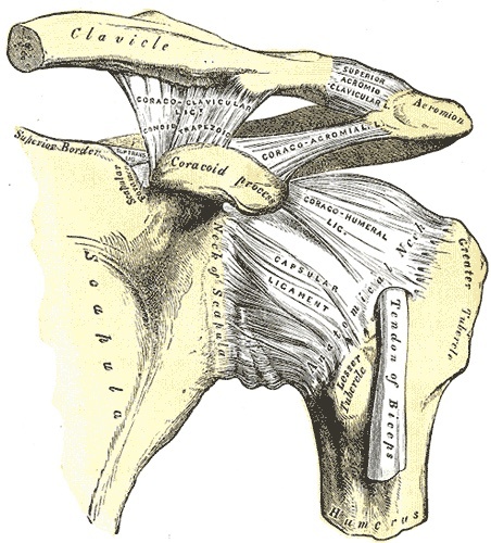
Figure 1. Anterior view of the shoulder showing the joint capsule and the coracohumeral and coracoacromial ligaments forming the coracoacromial arch. Gray’s Anatomy (1918), public domain, via Wikimedia Commons.
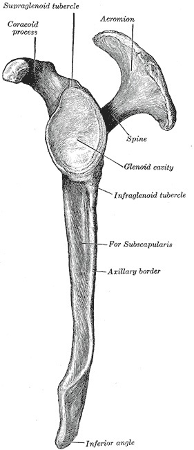
Figure 2. Lateral view of the scapula, showing the glenoid cavity, coracoid process, acromion and spine. Gray’s Anatomy (1918), public domain, via Wikimedia Commons.
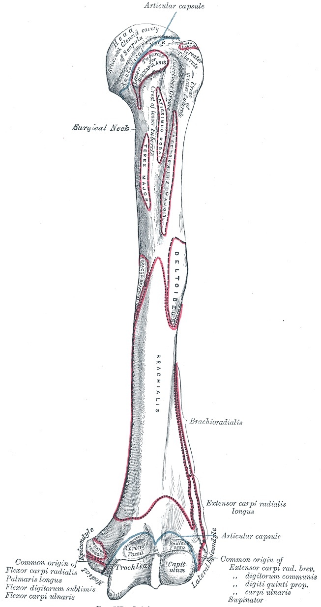
Figure 3. Anterior aspect of the proximal humerus, showing the head, the greater and lesser tuberosities, the bicipital groove and the surgical neck. Gray’s Anatomy (1918), public domain, via Wikimedia Commons.
The coracoid, the deltoid, and the subacromial bursa
The coracoid process is the key anterior landmark, palpable about 2.5 cm (one inch) below the anterior clavicle; it gives attachment to six clinically important structures, of which the surgically central ones are the conjoined tendon (short head of biceps and coracobrachialis), the pectoralis minor, the coracoacromial ligament, and the coracoclavicular (conoid and trapezoid) ligaments.[5] Behind the conjoined tendon and the pectoralis minor lie the cords of the brachial plexus and the second part of the axillary artery, and that is why the coracoid is treated as a danger line. The deltoid is multipennate, arising from the clavicle, acromion and scapular spine and inserting on the deltoid tuberosity; its tendinous bands let it be split longitudinally but make reattachment to the acromion difficult, so the surgeon avoids wide detachment.
The subacromial (subdeltoid) bursa separates the two sleeves, lets them glide and shields the supraspinatus from the overlying coracoacromial arch. It does not normally communicate with the joint, so a positive arthrogram (dye passing from joint to bursa) indicates a full-thickness cuff tear.[6] The painful arc of impingement, classically between 80 and 120 degrees of abduction, arises as the greater tuberosity and the undersurface of the acromion compress the inflamed bursa and supraspinatus between them.
The key nerves and the quadrangular space
Three nerves recur. The axillary nerve (posterior cord) runs on the posterior axillary wall over the subscapularis, passes back through the quadrangular space (bounded by teres minor above, teres major below, the long head of triceps medially and the surgical neck of the humerus laterally) alongside the posterior circumflex humeral artery, and winds onto the deep deltoid about 5 to 7 cm below the acromion.[7] The musculocutaneous nerve (lateral cord) supplies the arm flexors and enters the coracobrachialis on its medial side about 5 to 8 cm below the coracoid. The suprascapular nerve (upper trunk) supplies both supraspinatus and infraspinatus, curving around the base of the scapular spine from the supraspinous to the infraspinous fossa; over-medial retraction of the infraspinatus can stretch it there.
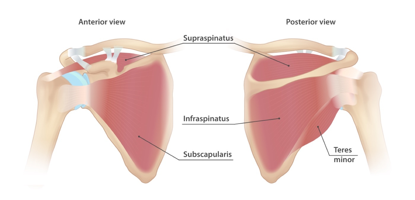
Figure 4. The four rotator cuff muscles: subscapularis anteriorly, and supraspinatus, infraspinatus and teres minor posteriorly, forming the inner muscular sleeve of the shoulder. InjuryMap, CC BY-SA 4.0, via Wikimedia Commons.
Part II - The Anterior (Deltopectoral) Approach
The anterior approach is the work-horse of shoulder surgery, giving wide access to the joint with its anterior, inferior and superior coverings and to the proximal humerus; its uses include reconstruction of recurrent dislocation (Bankart, Putti-Platt, Magnuson-Stack), arthroplasty, ORIF of proximal humeral fractures, biceps surgery, drainage of sepsis and tumour biopsy.[8] The patient lies supine with the head of the table raised 30 to 45 degrees and a sandbag under the medial scapula to push the side forward and let the arm fall back (the “beach-chair” position is standard teaching). The coracoid is the landmark, and the incision runs 10 to 15 cm along the deltopectoral groove from just above it (an axillary incision is the cosmetic alternative).
The internervous plane is between the deltoid (axillary nerve) and the pectoralis major (medial and lateral pectoral nerves), found by locating the cephalic vein in the deltopectoral groove and splitting the muscles apart; the cephalic vein may be retracted either way, but the surgeon preserves it where possible to avoid limb oedema.[9] Deep to this, the conjoined tendon (short head of biceps and coracobrachialis) is retracted medially, with the fascia incised on its lateral (safe) side because the musculocutaneous nerve enters the coracobrachialis medially. The arm stays adducted during coracoid work, since abduction draws the axillary artery and plexus against the coracoid. The subscapularis then comes into view as the last anterior cover; external rotation brings it into the wound and carries the axillary nerve away from its lower border. It is divided about 2 to 3 cm from the lesser tuberosity (2.5 cm spares its nerves) and tagged, after which the capsule is opened.[10]
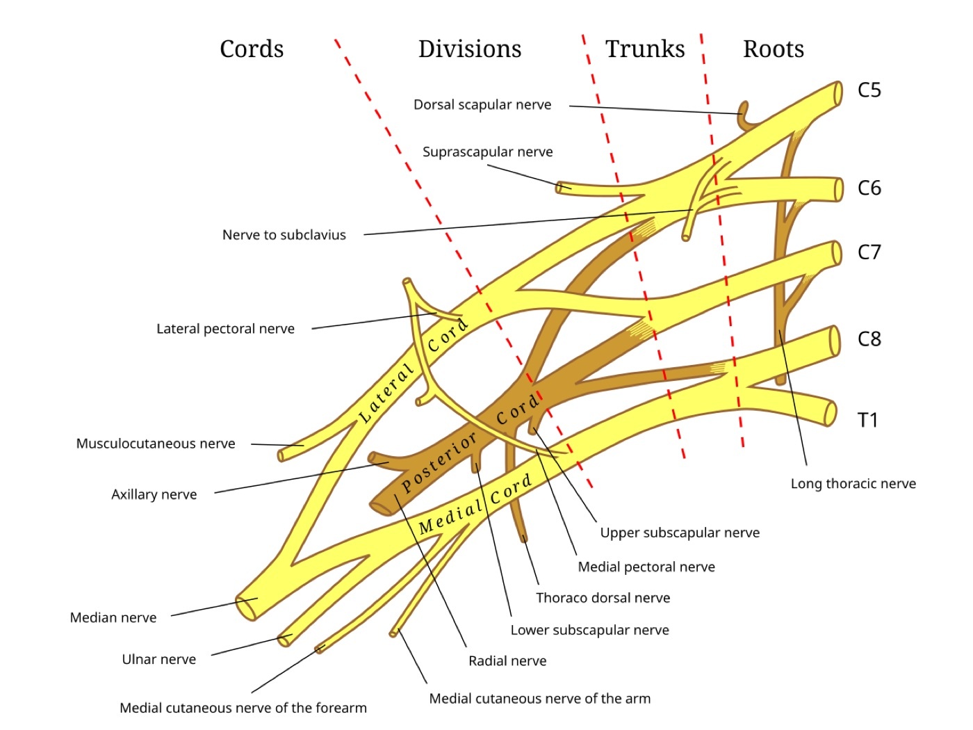
Figure 5. Schematic of the brachial plexus (roots, trunks, divisions, cords) and the origins of the suprascapular, musculocutaneous and axillary nerves; the cords and the axillary artery lie behind pectoralis minor, medial to the coracoid. Public domain, via Wikimedia Commons.
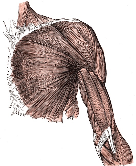
Figure 6. Superficial muscles of the anterior shoulder, the pectoralis major and deltoid, with the deltopectoral interval between them, the plane of the anterior approach. Gray’s Anatomy (1918), public domain, via Wikimedia Commons.
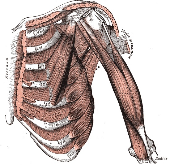
Figure 7. Deeper anterior dissection showing the coracoid process, pectoralis minor and subscapularis at the level of the clavipectoral fascia; the conjoined tendon arises from the coracoid and is retracted medially. Gray’s Anatomy (1918), public domain, via Wikimedia Commons.
Four dangers stand out. The musculocutaneous nerve is injured by vigorous medial or downward retraction of the conjoined tendon, since it enters coracobrachialis about 5 to 8 cm below the coracoid on its medial side. The axillary nerve lies just below the subscapularis and on the deep deltoid. The cephalic vein and the anterior humeral circumflex vessels are at risk superficially, and deep at the coracoid the axillary artery and brachial plexus sit behind pectoralis minor.[11] The approach extends distally into the anterolateral approach to the humerus, or proximally (with a middle-third clavicular osteotomy) to control the axillary artery. A brief but examined relative is the anterior approach to the clavicle, which has no internervous plane, threatens the supraclavicular nerves (with safe zones 2.5 cm from the sternoclavicular and 2 cm from the acromioclavicular joint) and, deep to the bone, the subclavian vessels lying immediately inferior in the middle and lateral thirds.[12]
Part III - The Anterolateral and Lateral (Deltoid-Split) Approaches
These approaches reach the rotator cuff, the subacromial space and the proximal humerus by splitting the deltoid, and they share one rule above all others: there is no internervous plane (the segmentally arranged deltoid is split in the line of its fibres), so the axillary nerve limits the split.[13] The anterolateral approach to the acromioclavicular joint and subacromial space serves rotator-cuff repair, subacromial decompression and AC-joint surgery: through a transverse incision from the anterolateral acromion to just lateral to the coracoid, the deltoid is split from the AC joint, the coracoacromial ligament is detached (often with a block of acromial bone), and the supraspinatus and subacromial bursa come into view. The lateral approach to the proximal humerus, through a longitudinal deltoid-splitting incision from the acromial tip, exposes the greater tuberosity and surgical neck for ORIF, cuff repair and nailing.
The cardinal danger is the axillary nerve, which runs transversely on the deep deltoid about 5 to 7 cm below the tip of the acromion. The deltoid split must not extend more than about 5 cm distal to the acromion, and a stay suture is placed at the inferior apex of the split to stop it propagating and dividing the nerve, which would denervate the anterior deltoid.[14] Where its position is in doubt, the nerve can be palpated on the undersurface of the muscle. The lateral approach extends proximally (by osteotomising the acromion and splitting the trapezius) to expose the whole supraspinatus, but the nerve means it cannot be extended distally through the deltoid; minimally invasive techniques work around this by using two windows on either side of the palpated axillary nerve, or a percutaneous deltoid split for antegrade humeral nailing (which inevitably injures part of the supraspinatus tendon and is repaired at closure).[15]
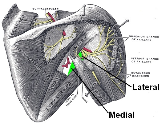
Figure 8. The posterior shoulder showing the suprascapular nerve and the axillary nerve passing through the quadrangular (quadrilateral) space with the posterior circumflex humeral artery, the key danger of the lateral and posterior approaches. After Gray’s Anatomy, public domain, via Wikimedia Commons.
Part IV - The Posterior Approach
The posterior approach reaches the posterior and inferior shoulder and is rarely needed, reserved for recurrent posterior instability, posterior glenoid and scapular-neck (floating shoulder) fractures, glenoid osteotomy, posterior fracture-dislocation, tumour and dependent drainage of sepsis.[16] The patient lies in the lateral decubitus position with the affected side up and the arm draped free; the spine of the scapula is the landmark, and the incision runs along it to the posterior corner of the acromion. The internervous plane is between the infraspinatus (suprascapular nerve) and the teres minor (axillary nerve), with a second superficial internervous plane between the deltoid (axillary nerve) and the infraspinatus. The deltoid is detached from the scapular spine, the infraspinatus-teres minor interval is opened by blunt finger dissection, and the posterior capsule is incised close to the glenoid.[17]
Two dangers define the safe limits. Straying below teres minor enters the quadrangular space and catches the axillary nerve and posterior circumflex humeral artery, paralysing the deltoid and causing bleeding that is hard to control, so the dissection must stay within the infraspinatus-teres minor interval.[18] Over-medial retraction of the infraspinatus stretches the suprascapular nerve around the unyielding lateral edge of the scapular spine, a common cause of infraspinatus wasting after this approach. Two further bleeding traps deserve mention: the circumflex scapular vessels in the triangular space (between teres minor, teres major and the long head of triceps), never entered electively, and the rich vascularity of the scapula, which lets a scapular fracture hide major blood loss. The approach cannot be usefully extended.[19]
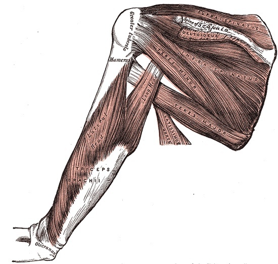
Figure 9. Muscles of the posterior shoulder, the deltoid, infraspinatus, teres minor and teres major, showing the infraspinatus-teres minor interval used in the posterior approach. Gray’s Anatomy (1918), public domain, via Wikimedia Commons.
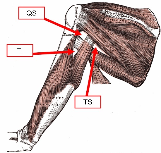
Figure 10. The quadrangular space (QS), triangular space (TS) and triangular interval (TI) of the posterior shoulder, defined by teres minor, teres major and the triceps; straying below teres minor enters the quadrangular space. Gray’s Anatomy (1918), annotated, public domain, via Wikimedia Commons.
Part V - Arthroscopic Approaches (Portals)
Shoulder arthroscopy is done in the beach-chair position (table raised 60 degrees, shoulder off the edge, arm free, which lowers venous pressure). Because the joint line cannot be palpated through the thick muscular cover, the portals are sited from bony landmarks: the acromion, the scapular spine, the clavicle and the coracoid.[20] The standard 30-degree arthroscope sees structures angled 30 degrees off its axis, so the surgeon changes the view by rotating it, angling it, or moving the joint rather than by advancing it.
Hoppenfeld describes two portals. The posterior (viewing) portal is an 8-mm stab 2 cm inferior and 1 cm medial to the posterolateral acromion (the “soft spot”), aimed at a finger on the coracoid and passing through the deltoid and the infraspinatus toward the joint just above its equator. The anterior (working) portal sits halfway between the coracoid tip and the anterior acromion, made under direct vision through the anterior triangle (biceps tendon, superior subscapularis and glenoid, the region of the rotator interval).[21] The governing safety rule is that the coracoid is the danger line: all anterior work stays superior and lateral to it, because the musculocutaneous nerve (2 to 8 cm distal to the coracoid), the brachial plexus and the axillary vessels lie medial to it. Posteriorly, a correctly sited portal lies about 3 cm above the axillary nerve and about 2 cm lateral to the suprascapular nerve, so only a too-inferior or too-medial portal endangers them.[22]
A diagnostic round through the posterior portal proceeds in order: the long head of biceps and its origin, the supraspinatus, the infraspinatus and teres minor (rotating the humerus), the anterior triangle, the anterior glenohumeral complex, the subscapularis recess (a frequent site for loose bodies), the posterior recess, and the humeral head and glenoid.[23]
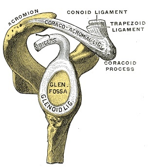
Figure 11. The glenoid fossa with the humerus removed, showing the glenoid labrum that deepens the socket and the origin of the long head of biceps, the central structures seen at arthroscopy. Gray’s Anatomy (1918), public domain, via Wikimedia Commons.
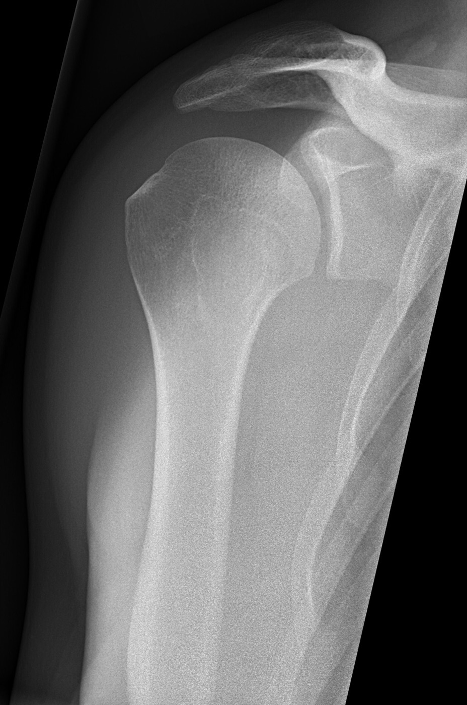
Figure 12. Normal shoulder radiograph in the AP glenoid (Grashey) projection, showing the humeral head congruent with the glenoid and an open joint space. Mikael Häggström, CC0, via Wikimedia Commons.
References
-
The single source is Hoppenfeld, de Boer and Buckley, Surgical Exposures in Orthopaedics: The Anatomic Approach (5th ed., 2016), chapter 1 (The Shoulder), pages 37 to 161. Every claim is page-cited. A few established facts Hoppenfeld does not state by name (the “beach-chair” position, the “three sisters” eponym for the anterior circumflex vessels, the Bigliani acromial morphology, the named glenohumeral ligaments, the lateral decubitus arthroscopy position, the Neviaser and other accessory portals) are flagged as standard teaching.
-
Hoppenfeld p.84, p.93, p.138. Because the axillary nerve is the sole supply to the deltoid, its injury is functionally devastating, and a stay suture at the apex of any deltoid split is the standard guard against it.
-
Hoppenfeld p.38, p.65. About one in four people over 65 ruptures the supraspinatus tendon; a complete tear abolishes the ability to initiate abduction (the drop-arm sign, with loss of control around 30 degrees) (Hoppenfeld p.117).
-
Hoppenfeld p.76-78. The individual superior, middle and inferior glenohumeral ligaments are standard teaching; Hoppenfeld notes the ligaments are hard to identify in open surgery and “appear to be of no clinical relevance,” obvious only arthroscopically.
-
Hoppenfeld p.66-67. The coracoacromial ligament is one of the few ligaments joining two parts of the same bone; with the coracoid and acromion it forms the coracoacromial arch implicated in impingement.
-
Hoppenfeld p.110-112. Acromial morphology varies, and certain shapes are associated with impingement (the Bigliani flat/curved/hooked classification is standard teaching).
-
Hoppenfeld p.135, p.138, p.139. The radial nerve leaves the axilla through the neighbouring triangular interval (teres major above, long head of triceps medially, humeral shaft laterally), and the circumflex scapular vessels occupy the triangular space (teres minor above, teres major below, long head of triceps laterally), a vessel leash not to be entered electively.
-
Hoppenfeld p.42-51. The approach is notorious for superficial bleeding, which must be controlled before the deeper layers are dissected.
-
Hoppenfeld p.48-50. The cephalic vein has no valves between it and the superior vena cava; a traumatised vein is ligated to avoid thromboembolism.
-
Hoppenfeld p.51-55. The lower border of subscapularis is marked by a transverse vascular leash, an artery flanked by two venae comitantes, the anterior humeral circumflex vessels (the “three sisters,” standard teaching); staying above this leash keeps the axillary nerve safe.
-
Hoppenfeld p.59-60, p.78.
-
Hoppenfeld p.39-41.
-
Hoppenfeld p.80, p.89-90. The deltoid is detached or split well proximal to its nerve supply, so the muscle itself is not denervated provided the split is not carried too far distally.
-
Hoppenfeld p.84, p.90, p.93.
-
Hoppenfeld p.94, p.96-97, p.101-105. In antegrade nailing the entry point is just lateral to the articular surface and medial to the greater tuberosity, and proximal locking bolts placed lateral-to-medial can also catch the axillary nerve.
-
Hoppenfeld p.121.
-
Hoppenfeld p.124-125. The teres minor’s parallel fibres distinguish it from the multipennate infraspinatus, helping to find the interval, which is genuinely hard to define.
-
Hoppenfeld p.126-127, p.138. The axillary nerve illustrates Hilton’s law: it supplies the deltoid and teres minor, sends a branch to the joint, and gives the upper lateral cutaneous nerve of the arm, whose sensory loss over the deltoid is good presumptive evidence of an axillary palsy after dislocation.
-
Hoppenfeld p.127, p.132, p.139. To place a posterior bone block the infraspinatus is detached 1 cm from its insertion and retracted, with care for the suprascapular nerve entering its undersurface just below the spine.
-
Hoppenfeld p.140, p.144-145. The lateral decubitus position with balanced traction is the standard alternative (standard teaching), distracting the joint at the cost of traction neurapraxia risk.
-
Hoppenfeld p.145-151. The subacromial space is entered by redirecting the posterior portal superiorly under the acromion with a separate deltoid penetration; the dedicated lateral subacromial portal is standard teaching.
-
Hoppenfeld p.156. The accessory portals (anterosuperior, Neviaser, portal of Wilmington, the low anterior 5-o’clock portal) are standard teaching not described by Hoppenfeld; the Neviaser portal in particular risks the suprascapular nerve.
-
Hoppenfeld p.150-155. The named superior, middle and inferior glenohumeral ligaments, the biceps anchor and SLAP region, and the Buford complex are standard arthroscopic landmarks not itemised by Hoppenfeld.
-
Hoppenfeld p.38, p.65.
-
Hoppenfeld p.48-50.
-
Hoppenfeld p.54, p.59, p.78.
-
Hoppenfeld p.54, p.60, p.76.
-
Hoppenfeld p.84, p.90, p.93.
-
Hoppenfeld p.135, p.138.
-
Hoppenfeld p.124-127, p.138.
-
Hoppenfeld p.139.
-
Hoppenfeld p.145, p.148, p.156.
-
Hoppenfeld p.142, p.149, p.156.
-
Hoppenfeld p.110-112.
-
Hoppenfeld p.78, p.125.