Contents
- Orientation
- Part I - Applied Surgical Anatomy of the Bony Pelvis
- Part II - Iliac Crest Bone Graft
- Part III - Anterior Approach to the Pubic Symphysis
- Part IV - Approaches to the Sacroiliac Joint
- Part V - Ilioinguinal Approach to the Acetabulum (Letournel)
- Part VI - Posterior (Kocher-Langenbeck) Approach to the Acetabulum
- References
Orientation
The pelvis is built from two innominate bones that meet the sacrum behind at the sacroiliac joints and each other in front at the pubic symphysis, closing a ring. Set into the lateral wall of each innominate bone is the acetabulum, carried on two thick pillars of bone, the anterior and posterior columns, that stand out like a fork when a dried hemipelvis is held to the light. Almost every decision in pelvic and acetabular surgery follows from two facts: which column or surface the fracture involves, and which great nerve or vessel guards the route to it. Hoppenfeld calls acetabular exposure “the most complex and demanding approaches a surgeon can be asked to perform,” since each approach reaches only part of the acetabulum and the right one must be matched to the fracture pattern.[1]
This summary opens with the applied anatomy of the bony pelvis and its dangerous neighbours, then works through the approaches in order of complexity: the iliac-crest bone-graft harvests (anterior and posterior), the anterior approach to the pubic symphysis, the anterior and posterior approaches to the sacroiliac joint, the ilioinguinal approach to the acetabulum, and the posterior (Kocher-Langenbeck) approach to the acetabulum. For each, it flags the internervous plane (where one exists) and the named dangers.[2]
Three dangers recur and are worth stating once. Behind, in the gluteal region, the danger is the sciatic nerve and the superior gluteal artery in the greater sciatic notch (a vessel that, if torn, retracts into the pelvis and bleeds catastrophically). In front, along the pelvic brim, it is the femoral nerve and the external iliac/femoral vessels, plus the corona mortis, the anastomosis crossing the superior pubic ramus that gives the “crown of death” its name. Medial to the sacroiliac joint, it is the L5 nerve root and lumbosacral trunk running on the sacral ala, which set the medial limit of the anterior SI exposure.[3]
Part I - Applied Surgical Anatomy of the Bony Pelvis
The innominate bone, the pelvic ring, and the acetabulum
Each innominate bone is formed by the fusion of three bones, the ilium, ischium, and pubis, which meet at the acetabulum. The two innominate bones plus the sacrum form the pelvic ring, joined posteriorly at the sacroiliac joints and anteriorly at the pubic symphysis (itself a secondary cartilaginous joint, not synovial). Surgical access exploits the subcutaneous parts: the iliac crest, from which the abdominal muscles and tensor fasciae latae arise, and the wing of the ilium, sandwiched between the glutei and tensor laterally and the iliacus medially. The safety principle behind all these approaches is to strip muscle subperiosteally off the bone, which keeps the dissection away from the vital structures and makes the exposures, in Hoppenfeld’s words, “extremely safe.”[4]
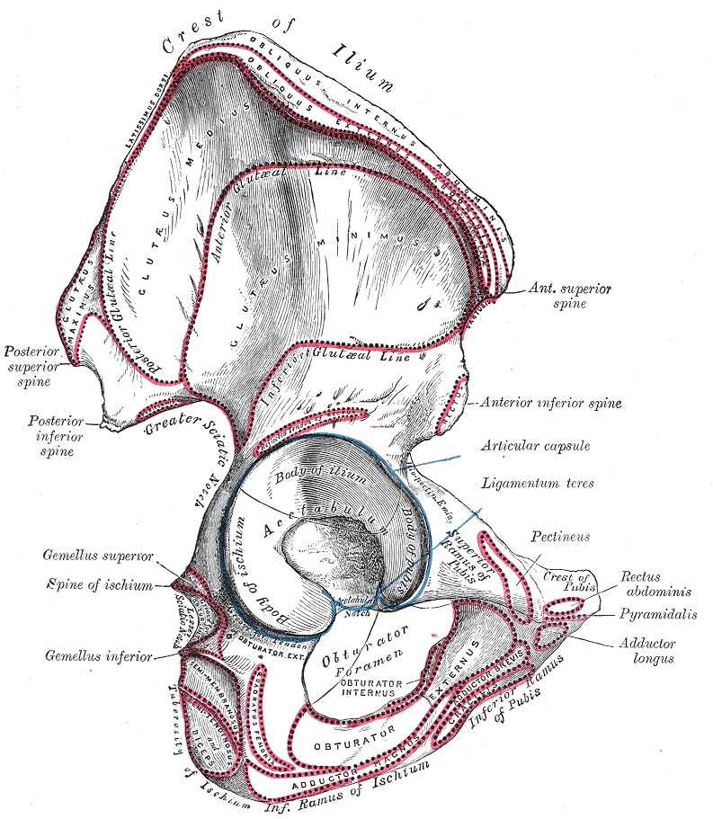
Figure 1. External (lateral) surface of the right hip bone, showing the ilium, ischium, pubis and the acetabulum. Gray’s Anatomy plate 235 (public domain), via Wikimedia Commons.
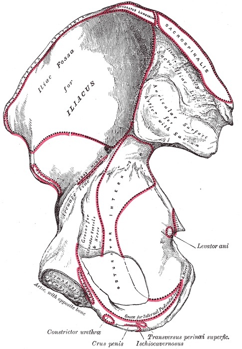
Figure 2. Internal (medial) surface of the right hip bone, showing the iliac fossa and the auricular (sacroiliac) surface. Gray’s Anatomy plate 236 (public domain), via Wikimedia Commons.
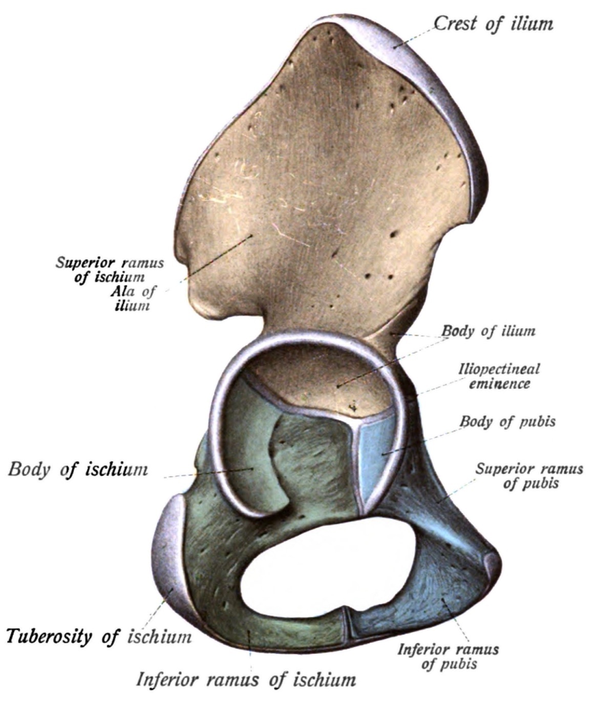
Figure 3. The hip bone subdivided into ilium, ischium and pubis. Sobotta’s Atlas of Human Anatomy, 1909 (public domain), via Wikimedia Commons.
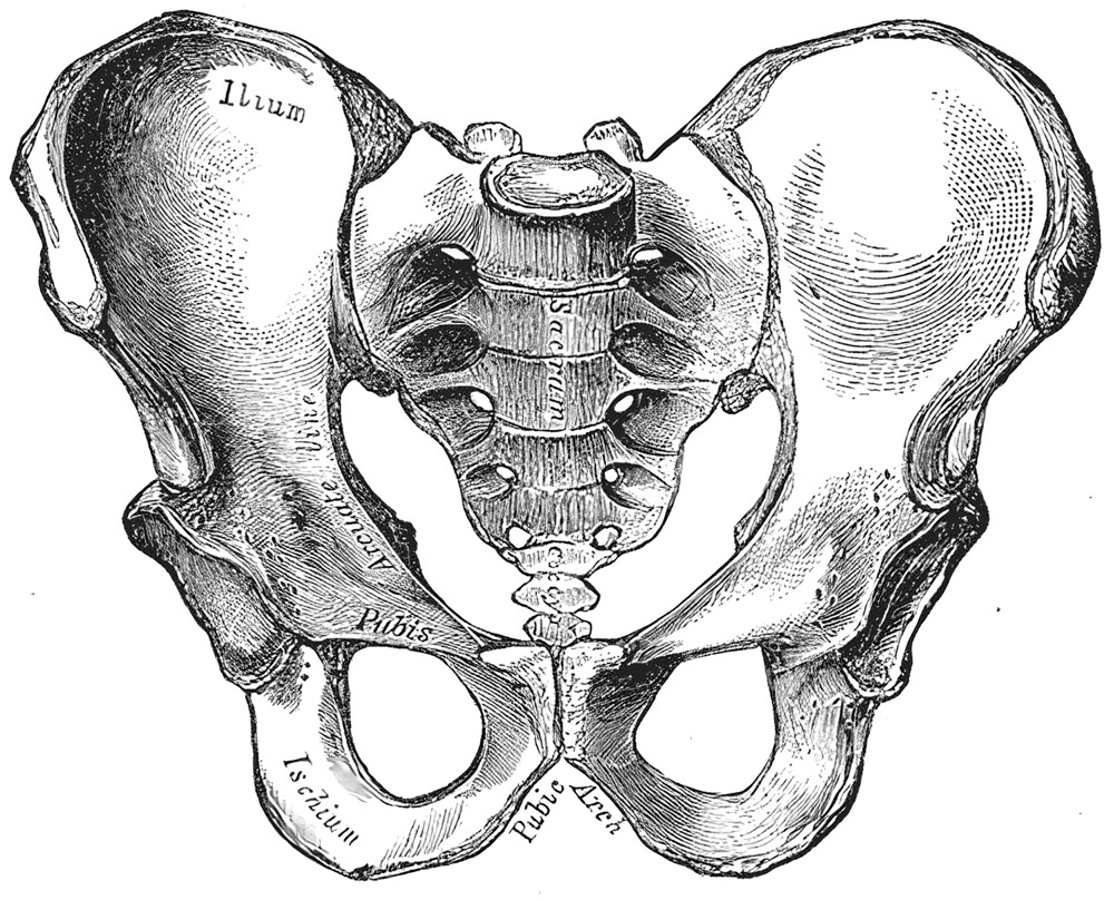
Figure 4. Anterior view of the bony pelvis, showing the sacrum, the sacroiliac joints and the pubic symphysis. Gray’s Anatomy plate 241 (public domain), via Wikimedia Commons.
The two acetabular columns
The acetabulum is suspended between two “massive columns” of bone set against the comparatively “thin central area of the wing of the ilium,” a contrast Hoppenfeld demonstrates by holding a hemipelvis up to a light source. The anterior (iliopubic) column runs from the iliac crest through the anterior acetabular wall to the superior pubic ramus and symphysis; the posterior (ilioischial) column runs from the greater sciatic notch through the posterior wall to the ischial tuberosity. Classically the two are conceived as an inverted “Y,” the apex at the weight-bearing acetabular dome (roof), with the flat medial wall between them, the quadrilateral surface (plate), forming the inner pelvic aspect of the acetabulum. This column concept is what maps a fracture to its approach: anterior/medial patterns go to the ilioinguinal approach, posterior patterns to the Kocher-Langenbeck, and complex both-column fractures to combined or extensile exposures.[5]
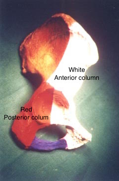
Figure 5. The two acetabular columns on a hip-bone model: anterior (iliopubic) column in white, posterior (ilioischial) column in red. Dr. C. J. Thakkar, CC BY-SA 4.0, via Wikimedia Commons.
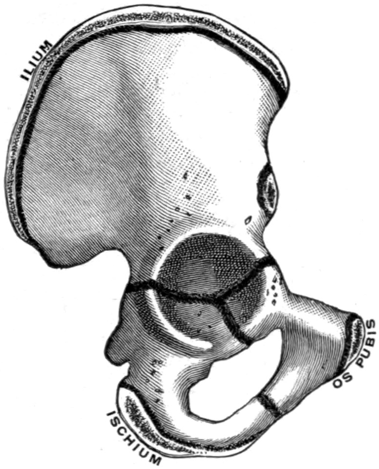
Figure 6. Union of the ilium, ischium and pubis at the triradiate cartilage of the acetabulum. Gerrish’s Text-book of Anatomy, 1902 (public domain), via Wikimedia Commons.
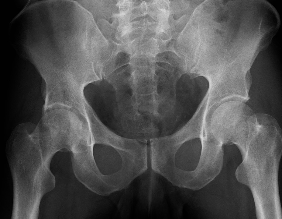
Figure 7. AP pelvic radiograph of a right acetabular fracture. James Heilman MD, CC BY-SA 4.0, via Wikimedia Commons.
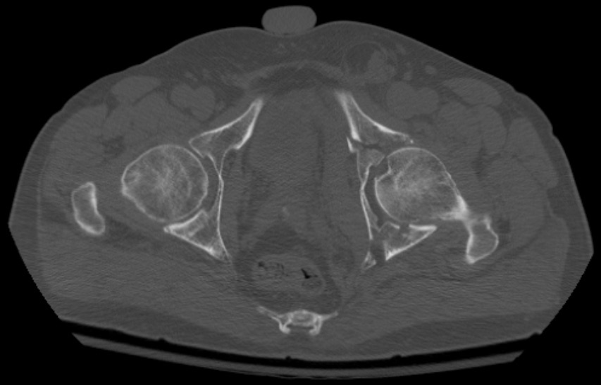
Figure 8. Axial CT (bone window) of a comminuted both-column acetabular fracture. Seannovak, CC BY-SA 3.0, via Wikimedia Commons.
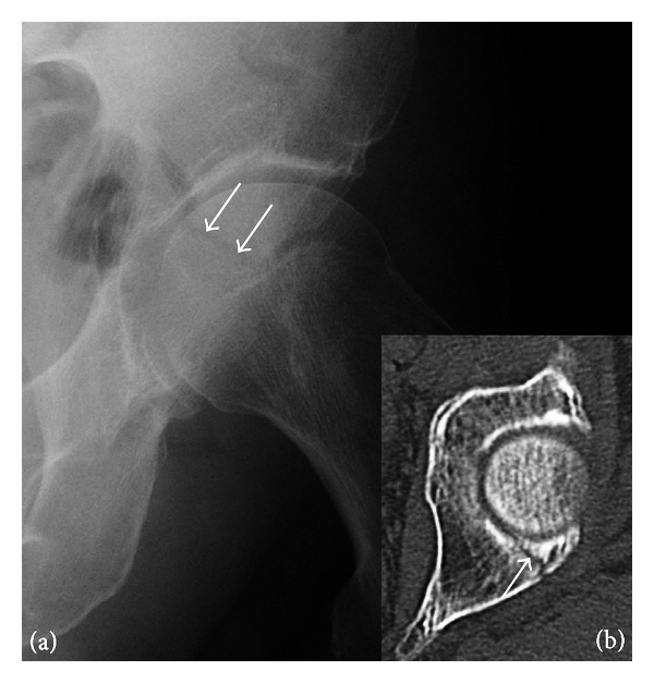
Figure 9. Subtle posterior acetabular wall fracture on radiograph (arrows) with corresponding CT. Jarraya et al., Radiol Res Pract 2013, CC BY 3.0, via Wikimedia Commons.
The sciatic notches, ligaments, and foramina
The posterior margin of the innominate bone carries the greater and lesser sciatic notches, separated by the ischial spine. The sacrotuberous ligament (to the ischial tuberosity) and the sacrospinous ligament (to the ischial spine) convert these notches into the greater and lesser sciatic foramina. The greater sciatic foramen is divided by the piriformis: above it pass the superior gluteal nerve and vessels; below it pass the inferior gluteal nerve and vessels, the sciatic nerve, the posterior femoral cutaneous nerve, the nerves to obturator internus and quadratus femoris, and the pudendal nerve with the internal pudendal vessels. The pudendal nerve and internal pudendal vessels then hook around the ischial spine and re-enter the pelvis through the lesser sciatic foramen, alongside the obturator internus tendon. During surgery, detaching part of the piriformis origin lets a finger pass through the greater sciatic notch to palpate the front of the sacroiliac joint.[6]
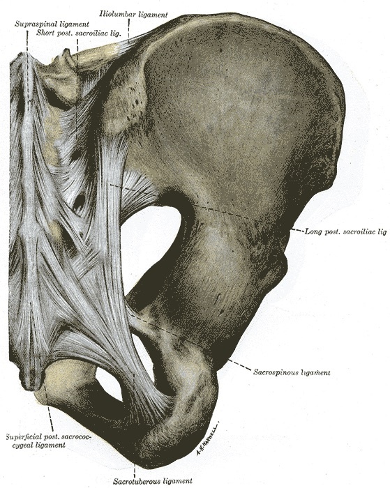
Figure 10. Posterior pelvic ligaments: the sacrotuberous and sacrospinous ligaments bounding the greater and lesser sciatic foramina, with the posterior sacroiliac ligaments. Gray’s Anatomy plate 320 (public domain), via Wikimedia Commons.
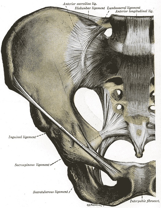
Figure 11. Anterior pelvic ligaments and the sacroiliac joint. Gray’s Anatomy plate 319 (public domain), via Wikimedia Commons.
The gluteal neurovascular bundles and the corona mortis
The superior gluteal artery, a branch of the internal iliac artery, leaves the pelvis through the greater sciatic notch above the piriformis and hugs the bone. It is the notorious bleeder of pelvic surgery, because a cut near the notch lets it retract into the pelvis where it is very hard to control. The inferior gluteal vessels and nerve leave below piriformis to supply gluteus maximus. Anteriorly, across the back of the superior pubic ramus, runs the corona mortis, an anastomosis between the obturator system and the external iliac/inferior epigastric system. It is present in a large proportion of pelves as an arterial form, a venous form, or both, and if it is torn during anterior exposure it produces brisk, hard-to-reach haemorrhage, hence the “crown of death.”[7]
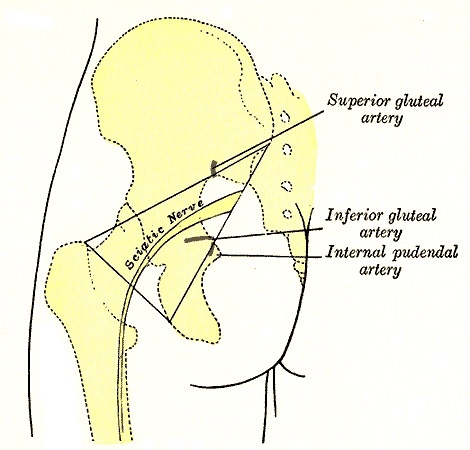
Figure 12. Surface markings of the gluteal region: the superior and inferior gluteal arteries, the internal pudendal artery and the sciatic nerve at the greater sciatic notch. Gray’s Anatomy plate 1244 (public domain), via Wikimedia Commons.
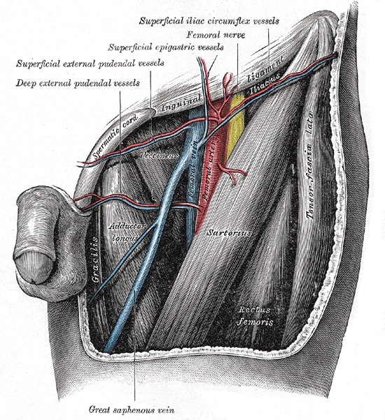
Figure 13. The femoral triangle and the superficial branches of the femoral vessels below the inguinal ligament (the inferior epigastric and external iliac systems that form the corona mortis anastomosis across the superior pubic ramus). Gray’s Anatomy plate 549 (public domain), via Wikimedia Commons.
Part II - Iliac Crest Bone Graft
Anterior approach to the iliac crest
The anterior iliac crest is the most commonly used bone-graft donor in orthopaedics. Being subcutaneous, it yields cortical, cancellous, or corticocancellous graft with ease, the largest cancellous reservoir lying at the iliac tubercle directly beneath the subcutaneous crest. The patient is supine with a sandbag under the buttock, and the incision parallels the crest, centred on the iliac tubercle. Because muscles take origin from or insert into the crest but do not cross it, the crest is a true internervous plane, so subperiosteal stripping denervates nothing. Two cardinal rules apply: do not carry the cut onto the anterior superior iliac spine (which would detach the inguinal ligament origin and risk a hernia), and leave the crest and spine intact for contour. The classic neurological danger, not named in this part of the text but standard teaching, is the lateral femoral cutaneous nerve near the anterior superior iliac spine, injury to which causes meralgia paraesthetica.[8]
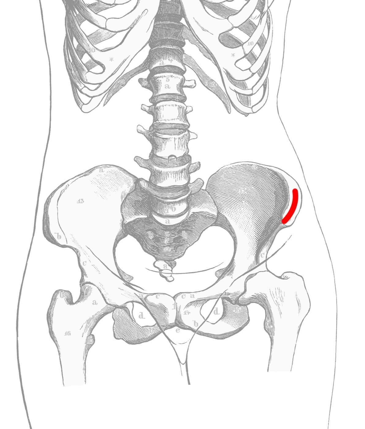
Figure 14. Donor site for anterior iliac-crest bone graft (highlighted). Public domain (after Kollmann), via Wikimedia Commons.
Posterior approach to the iliac crest
The posterior iliac crest is the workhorse donor for posterior spinal fusion and yields corticocancellous graft for any fusion. The patient is prone on longitudinal bolsters, and an oblique incision is centred on the posterior superior iliac spine in line with the crest. The outer border of the crest is again a true internervous plane. Three dangers govern the harvest. The superior cluneal nerves (posterior rami of L1-L3) cross the crest and are avoided by keeping the incision no more than 8 cm anterolateral to the posterior superior iliac spine. The sciatic nerve and, above all, the superior gluteal artery lie at the greater sciatic notch. A graft taken too close to the notch can cut the artery, which retracts into the pelvis, so all work is kept cephalad to a vertical line dropped from the posterior superior iliac spine, and the notch itself is never breached, which would also destabilise the pelvis.[9]
Part III - Anterior Approach to the Pubic Symphysis
This anterior approach is used almost exclusively for ORIF of a ruptured symphysis or a displaced superior pubic ramus fracture (and occasionally biopsy or osteomyelitis), and it exposes the symphysis, pubic crest, and superior pubic rami. Because widely displaced symphyseal injuries often go with urologic damage, a urethral catheter is placed before surgery (a full bladder obstructs the approach), and a urologic assessment, often including a retrograde urethrogram, is advisable. The patient is supine, and a curved 15 cm incision is made about 1 cm above the symphysis in the line of the skin crease. There is no internervous plane: the rectus abdominis is divided (it has a segmental supply, so division does not denervate it), the superficial epigastric vessels are ligated, and the rectus sheath is divided transversely about 1 cm above the symphysis.[10]
The deep dissection retracts the cut rectus superiorly to reveal the symphysis. If the back of the symphysis is needed, the bladder is swept gently off the bone to open the preperitoneal space of Retzius. The chief named danger is the bladder: if it was damaged in the original trauma, adhesions form behind the pubis, and mobilising the space of Retzius can cause inadvertent bladder rupture, so fixation in the presence of urologic injury is best done with an experienced urologist. The corona mortis (standard teaching here) crosses the superior pubic ramus in the retropubic space and must be sought and ligated, and the spermatic cord or round ligament is protected at the superficial inguinal ring. The approach extends laterally into the ilioinguinal approach to reach the whole anterior column.[11]
Part IV - Approaches to the Sacroiliac Joint
Anterior approach to the sacroiliac joint
Although the sacroiliac joint is one of the most posterior structures of the pelvis, the anterior approach gives better exposure and control than the posterior, because the joint is flat and directly available from in front whereas behind it is overhung by the posterior iliac crest. It is used for accurate placement of anterior plates across the joint and for fixation of associated iliac fractures. The patient is supine with a sandbag under the buttock and the table tilted 20° away so the pelvic contents fall clear, and a curved incision runs along the crest from about 7 cm behind the anterior superior iliac spine, then forward along the inguinal ligament line. There is no internervous plane: the iliacus is simply stripped subperiosteally off the inner wall of the iliac wing (with the anterior superior iliac spine sometimes mobilised on a bone block), and the lateral femoral cutaneous nerve, lying about 1 cm distal and medial to the anterior superior iliac spine, may have to be divided.[12]
The critical danger defines the medial limit of the whole operation: the L4 and L5 nerve roots and the lumbosacral trunk run on the anterior surface of the sacrum about 1.5 cm medial to the sacroiliac joint (Hoppenfeld’s figure; the classic teaching value is roughly 2-3 cm), and the dissection must not go more than that distance medial to the joint. When plating, only one screw hole may lie medial to the joint, because using two would endanger the nerve root. Usually only a single safe screw can be placed into the sacrum, and Hohmann retractor tips must not be driven into the sacral foramina, where the sacral roots emerge.[13]
Posterior approach to the sacroiliac joint
The posterior approach is simple and endangers no vital structure, but reduction (especially of vertical displacement) is hard, and it has largely been supplanted by percutaneous screw fixation. Vertical displacement is best corrected by preoperative longitudinal traction. The patient is prone, with care to exclude the anal region from the field, and a curved incision runs over the posterior crest from about 3 cm distal and lateral to the posterior superior iliac spine. There is no internervous plane: gluteus maximus and medius are partially detached, their neurovascular pedicles defining how far each can be mobilised. Inferior mobilisation of gluteus maximus is limited by the inferior gluteal nerve and artery entering its deep surface below piriformis, and forward elevation of gluteus medius by the superior gluteal nerve and vessels. Excessive retraction injures these nerves, and the superior gluteal artery at the greater sciatic notch is again the feared bleeder. Reduction is checked by passing a finger through the greater sciatic notch (after partial piriformis release) to feel the anterior joint, which is smooth when reduced; the thin posterior skin makes wound breakdown a real drawback.[14]
Part V - Ilioinguinal Approach to the Acetabulum (Letournel)
The ilioinguinal approach exposes the inner surface of the pelvis from the sacroiliac joint to the pubic symphysis, giving access to the anterior column and the medial (quadrilateral) surface of the acetabulum and allowing posterior-column screws. It is the standard anterior approach for anterior-column, anterior-wall, and both-column acetabular fractures. The patient is supine with the greater trochanter at the table edge (so gluteal soft tissue falls away) and a urinary catheter in place. A curved incision runs from about 5 cm above the anterior superior iliac spine along the crest and inguinal ligament to just past the midline. There is no internervous plane: the dissection lifts muscular, nervous, and vascular structures off the inner pelvis. The external oblique aponeurosis is split, the spermatic cord (or round ligament) slung, the rectus partly divided, and the posterior wall of the inguinal canal taken down (leaving a 1-2 mm cuff for repair), with the inferior epigastric vessels ligated at the deep ring.[15]
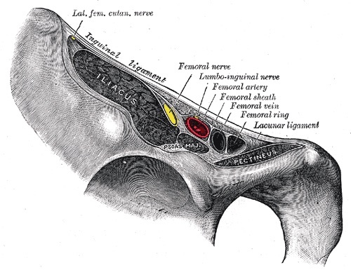
Figure 15. Structures passing behind the inguinal ligament: iliopsoas, the femoral nerve, the external iliac/femoral vessels and the lateral femoral cutaneous nerve, the contents of the windows of the ilioinguinal approach. Gray’s Anatomy plate 546 (public domain), via Wikimedia Commons.
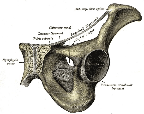
Figure 16. The superior pubic ramus with the inguinal, lacunar and pectineal (Cooper’s) ligaments, the obturator canal and the acetabulum. Gray’s Anatomy plate 435 (public domain), via Wikimedia Commons.
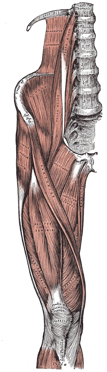
Figure 17. The iliopsoas (psoas major and iliacus) in the iliac fossa and anterior hip. Gray’s Anatomy plate 430 (public domain), via Wikimedia Commons.
The key to the deep exposure is the iliopectineal fascia, a thick layer over the iliacus that carries the femoral nerve on its surface, attaches deeply to the pubis, and separates the iliopsoas (and femoral nerve) from the external iliac vessels. Dividing it off the bone separates these two units and opens the approach. Sliding the slung iliopsoas-and-femoral-nerve bundle and the slung femoral vessels creates three windows: the lateral window (lateral to iliopsoas) reaches the internal iliac fossa and the front of the sacroiliac joint; the middle window (between iliopsoas and the external iliac vessels) reaches the pelvic brim, the quadrilateral surface, and the superior pubic ramus; and the medial window (medial to the vessels) reaches the superior pubic ramus, the space of Retzius, and the symphysis.[16]
The dangers are the femoral nerve, the femoral vessels, and the corona mortis. The femoral nerve is mobilised with the iliopsoas rather than dissected free, since stretching it paralyses the quadriceps; flexing the hip relaxes the muscle and nerve. The external iliac/femoral vessels are mobilised as a unit within their common femoral sheath (separate mobilisation of the vein risks thrombosis), and the femoral canal medial to the vein may contain a femoral hernia. The corona mortis is sought in the medial window and ligated before the vascular bundle is mobilised, because tearing it produces bleeding “difficult to control”; it is best seen with the surgeon standing on the opposite side of the table. The lateral femoral cutaneous nerve is often multiple and frequently must be divided, and the bladder is protected behind the symphysis. The approach extends proximally to the sacroiliac joint but no further distally.[17]
Part VI - Posterior (Kocher-Langenbeck) Approach to the Acetabulum
The posterior approach, eponymously the Kocher-Langenbeck, gives access to the posterior wall and posterior column of the acetabulum and the dorsocranial dome (through the fracture or a capsulotomy). It is the easiest of the acetabular approaches and is used for posterior-wall, posterior-column, transverse, and posterior fracture-dislocation patterns. The patient is positioned lateral (for posterior-wall/column work, and if a trochanteric osteotomy is planned) or prone (for transverse fractures, which the prone position helps to reduce). If traction is used, a distal femoral pin is placed with the knee flexed to spare the sciatic nerve. The incision is longitudinal, centred on the greater trochanter, from just below the iliac crest to about 10 cm beyond the trochanter (the classic description adds a limb toward the posterior superior iliac spine). There is no internervous plane: gluteus maximus is split in the line of its fibres, which does not significantly denervate it because its supply enters proximally.[18]
With the leg internally rotated, the deep dissection identifies the short external rotators, then detaches the obturator internus and gemelli (and, if a surgical dislocation is planned with a bifid sciatic nerve, the piriformis), dividing them about 1 cm from the femur (Hoppenfeld’s figure; the classic teaching is ≥1.5 cm, to protect the medial femoral circumflex artery, the femoral head’s blood supply). The quadratus femoris is identified but not detached, again to protect that artery. Retractors are placed gently into the greater and lesser sciatic notches, using the detached rotators as a cushion over the sciatic nerve. A trochanteric flip osteotomy gives greater exposure and access to the anterior column or a surgical dislocation; it is reattached with screws but carries a risk of heterotopic ossification.[19]
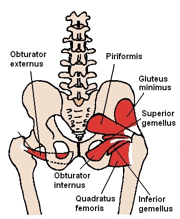
Figure 18. The short external rotators of the hip, piriformis, the gemelli, obturator internus and quadratus femoris, the deep layer encountered in the Kocher-Langenbeck approach. “Beth ohara”, CC BY-SA 3.0, via Wikimedia Commons.
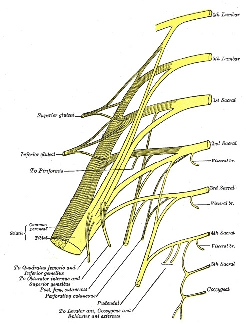
Figure 19. Plan of the sacral plexus: the superior and inferior gluteal nerves, the nerves to the short external rotators, and the sciatic nerve. Gray’s Anatomy plate 828 (public domain), via Wikimedia Commons.
The central danger is the sciatic nerve, often already contused by the injury; it lies on the short external rotators and usually exits anterior to piriformis into the greater sciatic notch. It is protected by keeping the knee flexed (and, by standard teaching, the hip extended) to relax it, by never retracting it forcibly, and by never pressing hard on the notch retractor, which would cause a palsy; the divided rotators shield it from direct trauma. The superior gluteal artery and nerve above piriformis tether gluteus medius (limiting proximal retraction, so the approach cannot be usefully extended proximally) and are at risk when the gluteus minimus-piriformis plane is developed. The inferior gluteal artery below piriformis, if torn, retracts into the pelvis and requires turning the patient supine and ligating the external iliac artery. The medial femoral circumflex artery (standard teaching) is protected by the 1 cm rotator division and by sparing quadratus femoris, since its injury causes femoral-head avascular necrosis.[20]
References
-
The pelvis is two innominate bones articulating with the sacrum at the SI joints and with each other at the pubic symphysis; the acetabulum is carried on two columns best seen by transilluminating a hemipelvis (Hoppenfeld, Surgical Exposures in Orthopaedics, 5th ed., ch.7, p.654-656). “Approaches to the acetabulum are the most complex and demanding… each approach only gives access to a limited part of the acetabulum” so the approach must match the fracture pattern (p.655).
-
The chapter covers anterior iliac-crest graft (p.657), posterior iliac-crest graft (p.661), the anterior approach to the pubic symphysis (p.666), the anterior (p.670) and posterior (p.677) SI joint approaches, the applied anatomy of the bony pelvis (p.684), the ilioinguinal approach (p.688) and its applied anatomy (p.703), and the posterior (Kocher-Langenbeck) approach (p.714).
-
Recurring dangers: the sciatic nerve and the superior gluteal artery at the greater sciatic notch posteriorly (the artery retracts into the pelvis if cut - Hoppenfeld p.665, p.724); the femoral nerve, external iliac/femoral vessels, and the corona mortis anteriorly (p.695, p.700-701); and the L4/L5 roots and lumbosacral trunk on the sacral ala medial to the SI joint (p.673, p.675).
-
Each innominate bone = ilium + ischium + pubis fused at the acetabulum (standard teaching); the two innominate bones + sacrum form the pelvic ring (SI joints posteriorly, pubic symphysis anteriorly, the symphysis a secondary cartilaginous joint) (Hoppenfeld p.654-655, p.688); subcutaneous access via the iliac crest and the iliac wing (sandwiched between glutei/tensor laterally and iliacus medially); subperiosteal stripping keeps the approaches safe (p.684-685, p.687).
-
The acetabulum sits between two “massive columns” against the “thin central area of the wing of the ilium,” seen by transilluminating a hemipelvis (Hoppenfeld p.656, Fig. 7-1); the ilioinguinal approach reaches the anterior column and medial acetabulum, the posterior approach the posterior column, posterior lip, and dome (p.655-656). The iliopubic/ilioischial column detail, the inverted-Y, the dome/tectum, the quadrilateral plate, and the Judet-Letournel classification are standard teaching not detailed in this extract.
-
The greater and lesser sciatic notches are separated by the ischial spine; the sacrotuberous (to the ischial tuberosity) and sacrospinous (to the ischial spine) ligaments convert them into foramina; the greater sciatic foramen is divided by piriformis (suprapiriform: superior gluteal nerve/vessels; infrapiriform: inferior gluteal nerve/vessels, sciatic nerve, posterior femoral cutaneous nerve, nerves to obturator internus and quadratus femoris, pudendal nerve + internal pudendal vessels), the latter re-entering via the lesser sciatic foramen with the obturator internus tendon (standard teaching); detaching part of piriformis lets a finger reach the anterior SI joint through the greater sciatic notch (Hoppenfeld p.684, Fig. 7-19).
-
The superior gluteal artery (from the internal iliac) leaves through the greater sciatic notch above piriformis and, if cut, retracts into the pelvis with catastrophic bleeding (Hoppenfeld p.665); the inferior gluteal vessels/nerve leave below piriformis to gluteus maximus (p.680, p.687). The corona mortis (obturator ↔ external iliac/inferior epigastric anastomosis across the superior pubic ramus, ~6 cm lateral to the symphysis, arterial and/or venous) is described in the ilioinguinal approach (p.695, p.700-701); the frequency figures are standard teaching.
-
The anterior iliac crest is the commonest graft donor (subcutaneous; cortical/cancellous/corticocancellous; largest cancellous bone at the iliac tubercle under the subcutaneous crest); supine with a sandbag under the buttock, incision parallel to the crest over the iliac tubercle; the crest is a true internervous plane (muscles origin/insert but do not cross it), so subperiosteal stripping denervates nothing; do not carry the cut onto the ASIS (inguinal-ligament detachment → hernia), leave crest and ASIS intact (Hoppenfeld p.657-660). The lateral femoral cutaneous nerve near the ASIS (meralgia paraesthetica) is standard teaching.
-
The posterior iliac crest is the workhorse donor for posterior spinal fusion; prone on longitudinal bolsters, oblique incision over the PSIS in line with the crest; the outer crest is a true internervous plane; the superior cluneal nerves (posterior rami L1-L3) are avoided by keeping the incision ≤8 cm anterolateral to the PSIS; the sciatic nerve and the superior gluteal artery lie at the greater sciatic notch, the artery retracting into the pelvis if cut, so work stays cephalad to a vertical line from the PSIS and the notch is not breached (which would destabilise the pelvis); control bone bleeding with bone wax (Hoppenfeld p.661-665).
-
The anterior symphysis approach is used almost exclusively for ORIF of a ruptured symphysis or displaced superior pubic ramus fracture, exposing the symphysis, pubic crest, and superior pubic rami; a urethral catheter is placed pre-op (full bladder obstructs the approach) and urologic assessment ± retrograde urethrogram is advisable; supine, curved 15 cm incision ~1 cm above the symphysis in the skin crease; no internervous plane (segmentally supplied rectus abdominis is divided without denervation), the superficial epigastric vessels ligated and the rectus sheath divided transversely ~1 cm above the symphysis (Hoppenfeld p.666-667).
-
Retract the cut rectus superiorly to reveal the symphysis; sweep the bladder gently off the bone to open the preperitoneal space of Retzius; the bladder is the chief named danger (trauma adhesions → inadvertent rupture; operate with a urologist if there is urologic injury); the approach extends laterally into the ilioinguinal approach to reach the whole anterior column (Hoppenfeld p.668-669). The corona mortis (retropubic, must be sought and ligated) and the spermatic cord/round ligament at the superficial inguinal ring are standard teaching for this approach.
-
The anterior SI approach gives better exposure than the posterior because the joint is flat and directly available anteriorly while overhung by the posterior iliac crest behind (Hoppenfeld p.670-671); uses = accurate anterior plating across the joint and fixation of associated iliac fractures; supine with a buttock sandbag, table tilted 20° away, curved incision from ~7 cm behind the ASIS forward along the inguinal-ligament line; no internervous plane (iliacus stripped subperiosteally off the inner iliac wing, the ASIS sometimes mobilised on a bone block); the lateral femoral cutaneous nerve, ~1 cm distal and medial to the ASIS, may need division, causing thigh numbness that can be permanent (p.671-675).
-
The L4/L5 roots and the lumbosacral trunk run on the anterior sacrum about 1.5 cm medial to the SI joint (Hoppenfeld’s stated figure, p.673, p.675; the classic ~2-3 cm value, with the L5 root over the sacral ala the most vulnerable, is standard teaching), and “do not continue the dissection more than 1.5 cm medial to the sacroiliac joint”; “this nerve root marks the medial limit of the approach”; place only one screw hole medial to the joint (two would endanger the root), usually only one safe sacral screw, and do not drive Hohmann tips into the sacral foramina (Hoppenfeld p.673, p.675).
-
The posterior SI approach is simple and endangers no vital structure but reduction is hard (correct vertical displacement by preoperative traction) and it is largely supplanted by percutaneous screws; prone, anal region excluded, curved incision over the posterior crest from ~3 cm distal and lateral to the PSIS; no internervous plane (gluteus maximus and medius partially detached); inferior mobilisation of maximus limited by the inferior gluteal nerve/artery (below piriformis), forward elevation of medius by the superior gluteal nerve/vessels, excessive retraction injuring them; reduction checked by a finger through the greater sciatic notch (smooth = reduced); sacral roots safe from the approach but at risk from inaccurate screws (two-plane C-arm mandatory) (Hoppenfeld p.677-682). The superior gluteal artery retracting into the pelvis and the thin-skin wound-breakdown rationale are standard teaching.
-
The ilioinguinal approach exposes the inner pelvis from the SI joint to the symphysis, the anterior column and medial/quadrilateral acetabulum, and allows posterior-column screws (Hoppenfeld p.688); supine with the greater trochanter at the table edge and a urinary catheter; curved incision from ~5 cm above the ASIS along the crest/inguinal ligament to just past the midline; no internervous plane (structures lifted off the inner pelvis); split the external oblique aponeurosis, sling the spermatic cord/round ligament, divide the rectus partly, take down the inguinal-canal posterior wall leaving a 1-2 mm cuff for repair, and ligate the inferior epigastric vessels at the deep ring (p.689-693). The classic Letournel indications are standard teaching.
-
The iliopectineal fascia (thick, over iliacus, carrying the femoral nerve, separating iliopsoas/femoral nerve from the external iliac vessels, attached to the pubis) must be divided off the bone to open the approach; sliding the slung iliopsoas+femoral nerve and the slung femoral vessels creates three windows: lateral (lateral to iliopsoas → internal iliac fossa, anterior SI joint), middle (between iliopsoas and the external iliac vessels → pelvic brim, quadrilateral surface, superior pubic ramus), and medial (medial to the vessels → superior pubic ramus, space of Retzius, symphysis) (Hoppenfeld p.693-695, p.702). The lacuna musculorum/vasorum terminology is standard teaching.
-
Dangers: the femoral nerve, mobilised with the iliopsoas not dissected free (stretching paralyses the quadriceps; flex the hip to relax it); the external iliac/femoral vessels mobilised as a unit in the common femoral sheath (separate vein mobilisation risks thrombosis; the femoral canal may hold a femoral hernia); the corona mortis, sought in the medial window and ligated before mobilising the vascular bundle (tearing it bleeds “difficult to control”), best seen from the opposite side of the table; the lateral femoral cutaneous nerve (often multiple, frequently divided) and the bladder; the approach extends proximally to the SI joint but not distally (Hoppenfeld p.695, p.699-701).
-
The posterior (Kocher-Langenbeck) approach reaches the posterior wall and column and the dorsocranial dome (the eponym is standard teaching); the easiest acetabular approach, for posterior-wall/column, transverse, and posterior fracture-dislocation patterns; positioned lateral (posterior-wall/column work and trochanteric osteotomy) or prone (transverse fractures, which prone helps reduce); a distal femoral traction pin is placed with the knee flexed to spare the sciatic nerve; longitudinal incision centred on the greater trochanter from just below the iliac crest to ~10 cm beyond it (the PSIS-based limb is the classic description, standard teaching); no internervous plane (gluteus maximus split, supply enters proximally) (Hoppenfeld p.714-716).
-
With the leg internally rotated, identify the short external rotators and detach obturator internus and the gemelli (and piriformis if a surgical dislocation is planned with a bifid sciatic nerve), dividing them ~1 cm from the femur (Hoppenfeld p.719-721; the classic ≥1.5 cm figure, to protect the medial femoral circumflex artery / femoral-head supply, is standard teaching); identify but do not detach quadratus femoris (to protect that artery - standard teaching); place retractors gently into the greater and lesser sciatic notches using the rotators as a cushion over the sciatic nerve; a trochanteric flip osteotomy (reattached with screws, associated with heterotopic ossification) gives greater exposure and access to the anterior column / surgical dislocation (p.720-722).
-
The sciatic nerve is the central danger (often contused by injury; on the short external rotators, usually anterior to piriformis into the greater sciatic notch), protected by knee flexion (and hip extension, standard teaching), no forcible retraction, and no heavy pressure on the notch retractor (which causes a palsy), the divided rotators shielding it; the superior gluteal artery/nerve above piriformis tether gluteus medius (limiting proximal extension) and are at risk in the gluteus minimus-piriformis plane; the inferior gluteal artery below piriformis, if torn, retracts into the pelvis (turn supine and ligate the external iliac artery); the medial femoral circumflex artery, protected by the 1 cm rotator division and sparing quadratus femoris, supplies the femoral head and its injury causes AVN (Hoppenfeld p.720-724; the MFCA course and AVN mechanism are standard teaching). The internal pudendal vessels at the lesser sciatic notch are standard teaching.
-
The acetabulum sits on two columns seen by transilluminating a hemipelvis (Hoppenfeld p.656); ilioinguinal for anterior/both-column, posterior approach for posterior column/wall (p.655-656); the iliopubic/ilioischial detail and quadrilateral surface are standard teaching.
-
Sacrotuberous (ischial tuberosity) and sacrospinous (ischial spine) ligaments convert the notches into foramina; greater foramen suprapiriform = superior gluteal nerve/vessels, infrapiriform = inferior gluteal nerve/vessels, sciatic nerve, posterior femoral cutaneous nerve, nerves to obturator internus and quadratus femoris, pudendal nerve + internal pudendal vessels (standard teaching; Hoppenfeld p.684 names piriformis and the greater sciatic notch).
-
The crest is a true internervous plane (muscles origin/insert but do not cross it); do not carry the cut onto the ASIS (inguinal-ligament detachment → hernia); leave crest and ASIS intact (Hoppenfeld p.658-660). The LFCN/meralgia paraesthetica is standard teaching.
-
Superior cluneal nerves avoided by keeping the incision ≤8 cm anterolateral to the PSIS; the superior gluteal artery and sciatic nerve at the greater sciatic notch, the artery retracting into the pelvis if cut, so work cephalad to a vertical line from the PSIS and do not breach the notch (Hoppenfeld p.664-665).
-
The anterior SI approach is preferred because the joint is flat and directly available anteriorly while overhung behind (Hoppenfeld p.670-671); the L4/L5 roots and lumbosacral trunk lie ~1.5 cm medial to the joint (classic ~2-3 cm, standard teaching), marking the medial limit, with only one screw hole medial to the joint (p.673, p.675).
-
The corona mortis is an obturator ↔ external iliac/inferior epigastric anastomosis across the superior pubic ramus, sought in the medial window and ligated before mobilising the vascular bundle, tearing it causing bleeding “difficult to control” (Hoppenfeld p.695, p.700-701); the arterial/venous frequency and ~6 cm location are standard teaching.
-
The iliopectineal fascia (over iliacus, carrying the femoral nerve, attached to the pubis) separates iliopsoas/femoral nerve from the external iliac vessels; dividing it off the bone separates the two units and opens the middle window onto the brim, quadrilateral surface, and superior pubic ramus (Hoppenfeld p.693, p.706).
-
Lateral window (lateral to iliopsoas) → internal iliac fossa and anterior SI joint; middle window (between iliopsoas and the external iliac vessels) → pelvic brim, quadrilateral surface, superior pubic ramus; medial window (medial to the vessels) → superior pubic ramus, space of Retzius, symphysis (Hoppenfeld p.694-695, p.702).
-
The femoral nerve is mobilised with the iliopsoas (stretching paralyses the quadriceps; flex the hip to relax it); the femoral vessels are mobilised together in their common sheath, separate vein mobilisation risking thrombosis (Hoppenfeld p.695, p.699, p.706).
-
The short external rotators are divided ~1 cm from the femur (Hoppenfeld p.721; classic ≥1.5 cm, standard teaching) and quadratus femoris is spared to protect the medial femoral circumflex artery (the femoral-head supply, course and AVN mechanism standard teaching), preventing AVN (p.719).
-
The sciatic nerve (often contused; on the short rotators, usually anterior to piriformis) is protected by knee flexion (and hip extension, standard teaching), no forcible retraction, no heavy notch-retractor pressure (causes palsy), and the divided rotators as a cushion; divide the piriformis tendon if the nerve is bifid/pierces it before a surgical dislocation (Hoppenfeld p.719-722).
-
The superior gluteal artery (from the internal iliac) leaves through the greater sciatic notch above piriformis and retracts into the pelvis if cut (Hoppenfeld p.665, p.724); the inferior gluteal artery behaves similarly, control requiring turning supine and ligating the external iliac artery (p.723-724).