Contents
- Orientation
- Part I - The Structure of the Growing Bone
- Part II - Why Children’s Bones Fail Differently
- Part III - The Unique Pediatric Fracture Patterns
- Part IV - Traumatic Epiphysiolyses: Physeal Injuries
- Part V - Growth Arrest and the Physeal Bar
- Part VI - Healing and Remodelling in the Child
- References
Orientation
A child’s skeleton is not a small adult’s. It is a living, growing structure that changes month by month, and that single fact governs everything in this topic: how a child’s bone is built, why it breaks in ways an adult’s never does, and why a fracture that would be trivial in an adult can, in a child, stop a limb from growing. The organising idea is the physis, the cartilaginous growth plate that sits between epiphysis and metaphysis. It is the engine of longitudinal growth and the mechanically weakest part of the growing bone, which makes it both the commonest special site of pediatric injury and the one whose damage carries the gravest long-term consequences.[1]
This summary builds the topic in the order the konspekt asks for it. It opens with the surgical anatomy of the growing bone, then explains why pediatric bone fails differently. From there it works through the unique pediatric fracture patterns (plastic deformation, the torus or buckle fracture, and the greenstick fracture), then the traumatic epiphysiolyses and their Salter-Harris classification. It closes with the two consequences that define pediatric fracture care: growth arrest from a physeal bar, and the remarkable capacity of the child to heal and remodel.[2]
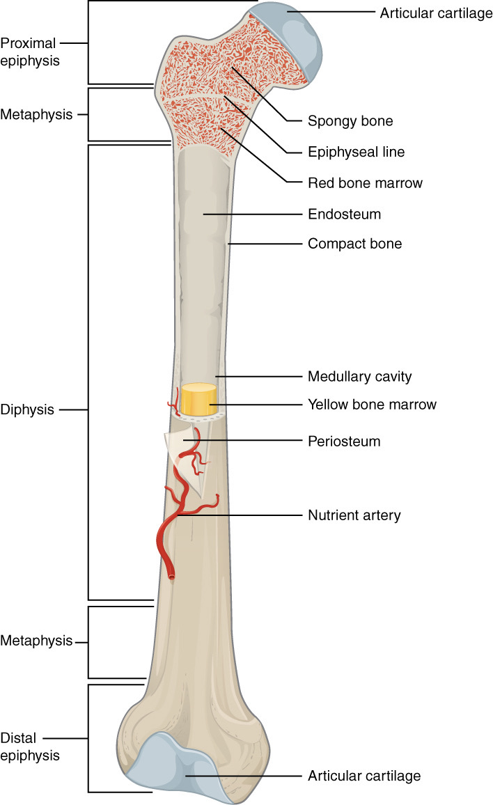
Figure 1. Regions of a long bone: the epiphysis, metaphysis and diaphysis, with the epiphyseal (growth) line, articular cartilage, periosteum and marrow cavity. OpenStax College, CC BY 3.0, via Wikimedia Commons.
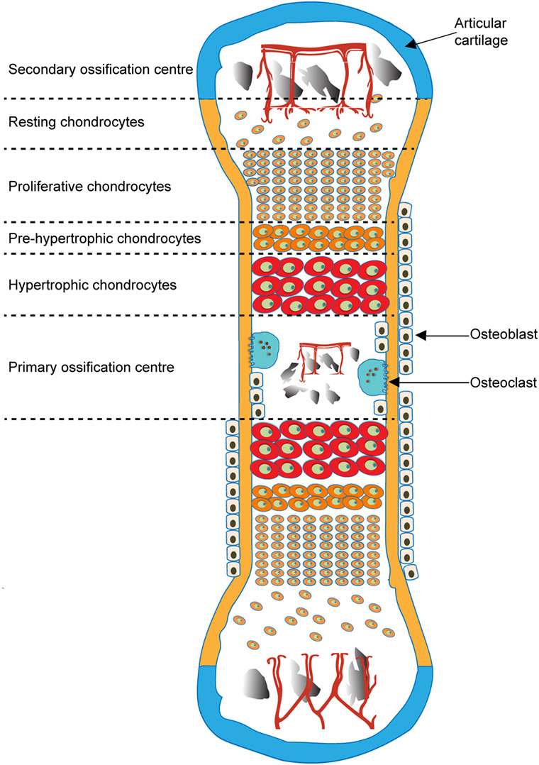
Figure 2. Endochondral ossification of a long bone, showing the primary (diaphyseal) and secondary (epiphyseal) ossification centres and the chondrocyte zones of the intervening growth plate. Javaheri et al., CC BY 4.0, via Wikimedia Commons.
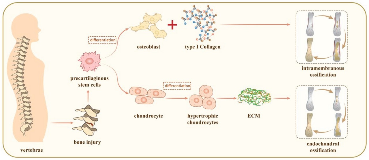
Figure 3. The two ossification pathways: intramembranous (direct) versus endochondral (through a cartilage model). Ge et al., CC BY 4.0, via Wikimedia Commons.
Part I - The Structure of the Growing Bone
The regions of a growing long bone
A growing long bone has four regions, each with its own behaviour and its own characteristic injuries. The diaphysis is the cortical shaft. The metaphysis is the flared end, with a thin cortex over abundant trabecular bone and many cortical fenestrations, which makes it the site of the torus (buckle) fracture and a region of rapid remodelling. The epiphysis is the end of the bone, cartilaginous at birth (the distal femur is the exception) and progressively ossified through a secondary ossification centre. Between epiphysis and metaphysis lies the physis, the cartilaginous growth plate responsible for endochondral longitudinal growth. A fifth region, the apophysis, is a traction (tension) variant of the physis at a tendon insertion; the tibial tuberosity is the prototype and Osgood-Schlatter disease its disorder.[3]
The physis and its zones
The growth plate is read from its epiphyseal side toward the metaphysis as a sequence of zones, and the surgeon’s reason for knowing them is mechanical. The reserve (resting/germinal) zone lies against the epiphysis, supplied by the epiphyseal vessels, and holds the stem cells; damage here is what threatens future growth. Next comes the proliferative zone, where chondrocytes divide into the longitudinal columns that lengthen the bone, fed by those same epiphyseal vessels. Then the hypertrophic zone, where the cells enlarge and the matrix calcifies in its lower part (the zone of provisional calcification), and finally the zone of transformation (primary spongiosa), where metaphyseal vessels invade the calcified cartilage and lay down bone. The cardinal point is that the hypertrophic and transformation zones are the mechanically weakest, so a physeal fracture characteristically runs through them, sparing the germinal and proliferative cells above and usually preserving growth.[4]
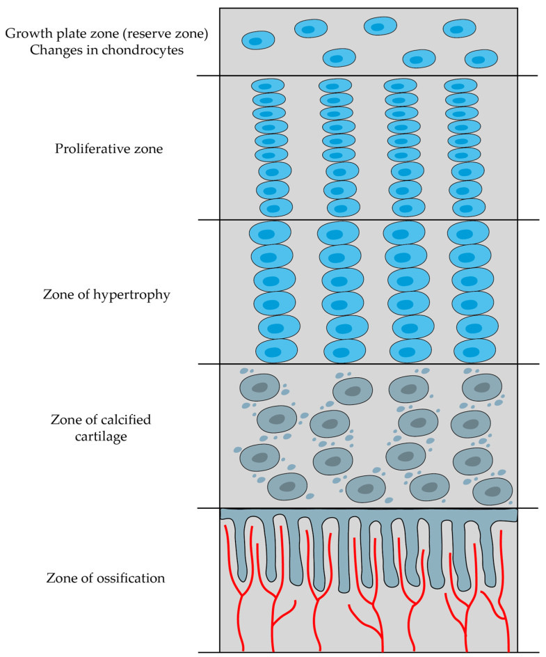
Figure 4. Zones of the growth plate (physis) from epiphysis to metaphysis: resting/reserve, proliferative, hypertrophic, calcified-cartilage and ossification zones, with vascular invasion at the metaphysis. Šromová, Sobola & Kaspar, CC BY 4.0, via Wikimedia Commons.
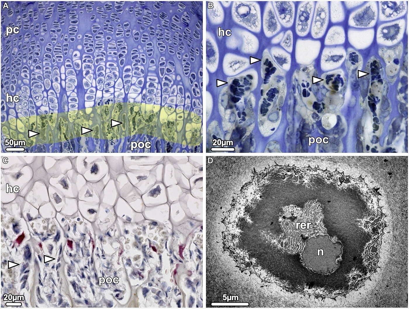
Figure 5. Histology of the epiphyseal growth plate at the chondro-osseous junction: proliferative (pc) and hypertrophic (hc) chondrocyte columns meeting the primary ossification front (poc). Khan, Clifton, Lorenzo, Hansen & Drissi, CC BY 4.0, via Wikimedia Commons.
The peripheral growth apparatus, the periosteum, and the blood supply
Around the rim of the physis sit two peripheral structures that an examiner likes to pair. The groove of Ranvier supplies the chondroblasts for latitudinal (width) growth of the plate, and the osseous ring of LaCroix, a downward extension of the metaphyseal cortex, mechanically braces the plate without impeding that width growth. The child’s periosteum is an advantage all its own: thick, strongly osteogenic, loosely attached over the shaft but densely fixed at the physeal periphery, and so strong that detaching an epiphysis took 550 pounds in one classic experiment but only 119 once the periosteum had been divided. Left intact, it forms a hinge and a sleeve that aids reduction and lays down rapid callus.[5]
The blood supply explains the worst complications of pediatric injury. The epiphyseal and metaphyseal circulations are separate, and vessels generally do not cross the physis. The epiphyseal vessels run as end-arterial cartilage canals without anastomoses, so their interruption produces segmental ischaemia, avascular necrosis, and angular growth deformity. That is why loss of the epiphyseal supply is serious while loss of the metaphyseal supply is minor and reversible. Bone grows in length at the physis and in width by periosteal apposition, and the physes close in a programmed order (physiologic epiphysiodesis) that explains transitional fractures such as the Tillaux.[6]
Part II - Why Children’s Bones Fail Differently
The immature skeleton differs from the adult’s in three linked ways: its anatomy, its physiology, and its biomechanics. Mechanically, pediatric bone is more porous and more elastic, with a lower modulus, so it absorbs more energy before failing and bends further before it breaks. The consequence is captured in one sentence: an adult’s bone usually fails first in tension, but a child’s bone may fail in tension, compression, or both. Its greater porosity tends to arrest a propagating crack, so comminution is uncommon, and incomplete failures such as bowing, buckling, and the greenstick are the rule rather than the exception.[7]
Two structures dominate pediatric fracture behaviour. The first is the physis, which is mechanically weaker than the surrounding ligaments and joint capsule. Where an adult would sprain a ligament, a child instead separates the growth plate, so the physeal fracture is in effect the child’s version of the ligament tear. The second is the periosteum, whose thick, intact hinge on the compression side both limits displacement and resists over-reduction, and whose osteogenic activity throws down subperiosteal new bone that can bridge defects without grafting.[8]
Part III - The Unique Pediatric Fracture Patterns
The incomplete and plastic failures of children’s bone produce a small family of fractures that have no real adult equivalent. Plastic (bowing) deformation is a macroscopic bend produced by innumerable microscopic failures without a discrete cortical break. It is seen in the ulna and fibula of younger children, may show no fracture line at all, and, because the bone is set in a bow, can block reduction of the paired bone. The torus (buckle) fracture is a compression failure at the metaphyseal-diaphyseal junction, where the porous metaphyseal cortex crumples into a stable little ring; the classic site is the distal radius and the fracture is inherently stable. The greenstick fracture fails the tension cortex while the compression cortex stays intact but bent, like a green twig. The practical problem is that the intact, springy cortex tends to re-angulate in plaster, so it is sometimes deliberately completed to allow a stable reduction. Children also sustain ordinary complete fractures (transverse, oblique, spiral), and a handful of special patterns such as the toddler’s fracture.[9]
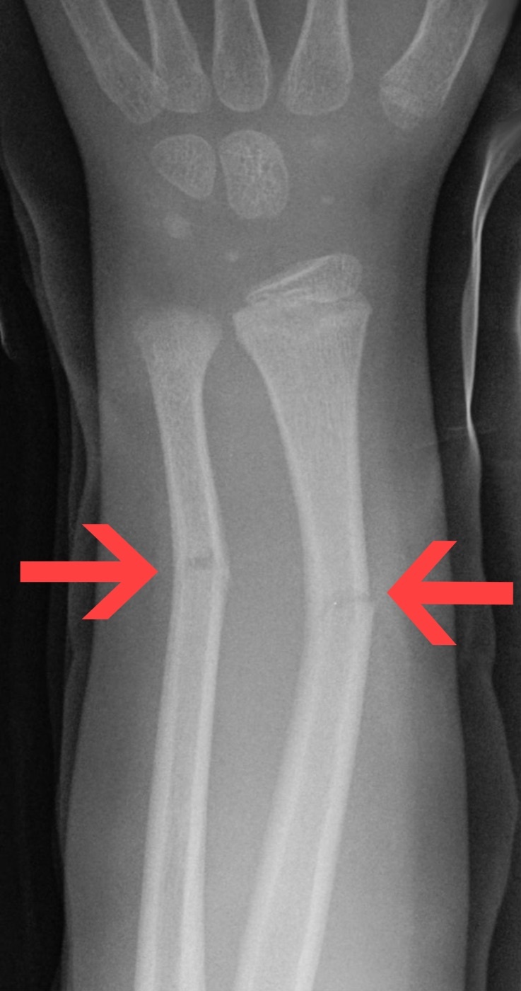
Figure 6. Greenstick fractures of the distal radius and ulna in a child (arrows): the cortex breaks on the convex (tension) side while the concave cortex remains intact. Hellerhoff, CC BY-SA 3.0, via Wikimedia Commons.
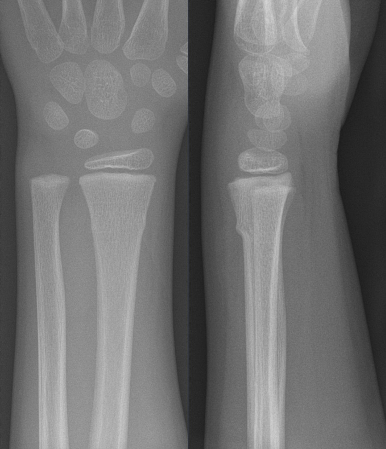
Figure 7. Torus (buckle) fracture of the distal radius in a 6-year-old: cortical buckling of the metaphysis in two planes, with the physis intact. Hellerhoff, CC BY-SA 4.0, via Wikimedia Commons.
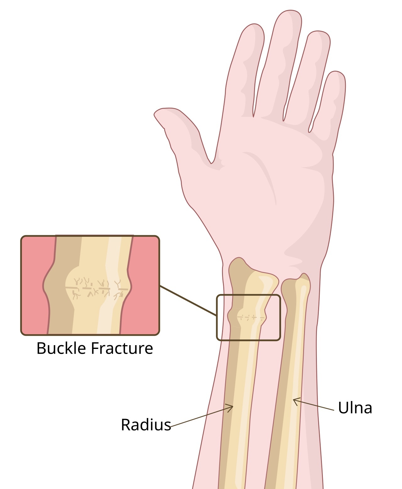
Figure 8. Diagram of a buckle (torus) fracture of the distal radius. RouDhi, CC BY-SA 4.0, via Wikimedia Commons.
Part IV - Traumatic Epiphysiolyses: Physeal Injuries
Epidemiology and the level of separation
A traumatic epiphysiolysis is a separation through the growth plate. Physeal injuries make up roughly 15 per cent of childhood fractures, are commonest in the hand (the phalanges), affect distal physes more than proximal ones and boys more than girls, and peak around ages 9 to 12 in girls and 12 to 15 in boys, the years when the physis is loosening before closure. The separation usually runs through the hypertrophic zone, at the junction of its calcified and uncalcified layers. That spares the germinal and proliferative cells, and it is the anatomical reason most physeal fractures do not arrest growth.[10]
The Salter-Harris classification
The working classification of physeal injuries is the Salter-Harris scheme of five types, which Ogden presents while noting that it was derived largely from radiographs. A Type I injury separates the epiphysis cleanly through the physis with no bony fragment, the pattern of the infant and of the slipped capital femoral epiphysis. A Type II, the commonest, runs through the physis and then exits through a corner of the metaphysis, carrying a triangular metaphyseal fragment, the Thurston-Holland fragment. A Type III passes through the physis and then through the epiphysis into the joint, so it is intra-articular. A Type IV crosses the metaphysis, the physis, and the epiphysis as a single vertical fracture line, again intra-articular. A Type V is a crush of the physis. By standard teaching the prognosis worsens down the list: Types I and II usually preserve growth with closed reduction, whereas Types III and IV demand anatomic (usually open) reduction because they are displaced and intra-articular, and Type V carries the worst prognosis for growth arrest. Ogden’s own larger classification refines this further, and he cautions pointedly that Salter-Harris Type II is not as uniformly benign as once thought, and that a true pure-crush Type V may be rare, the damage often being vascular.[11]
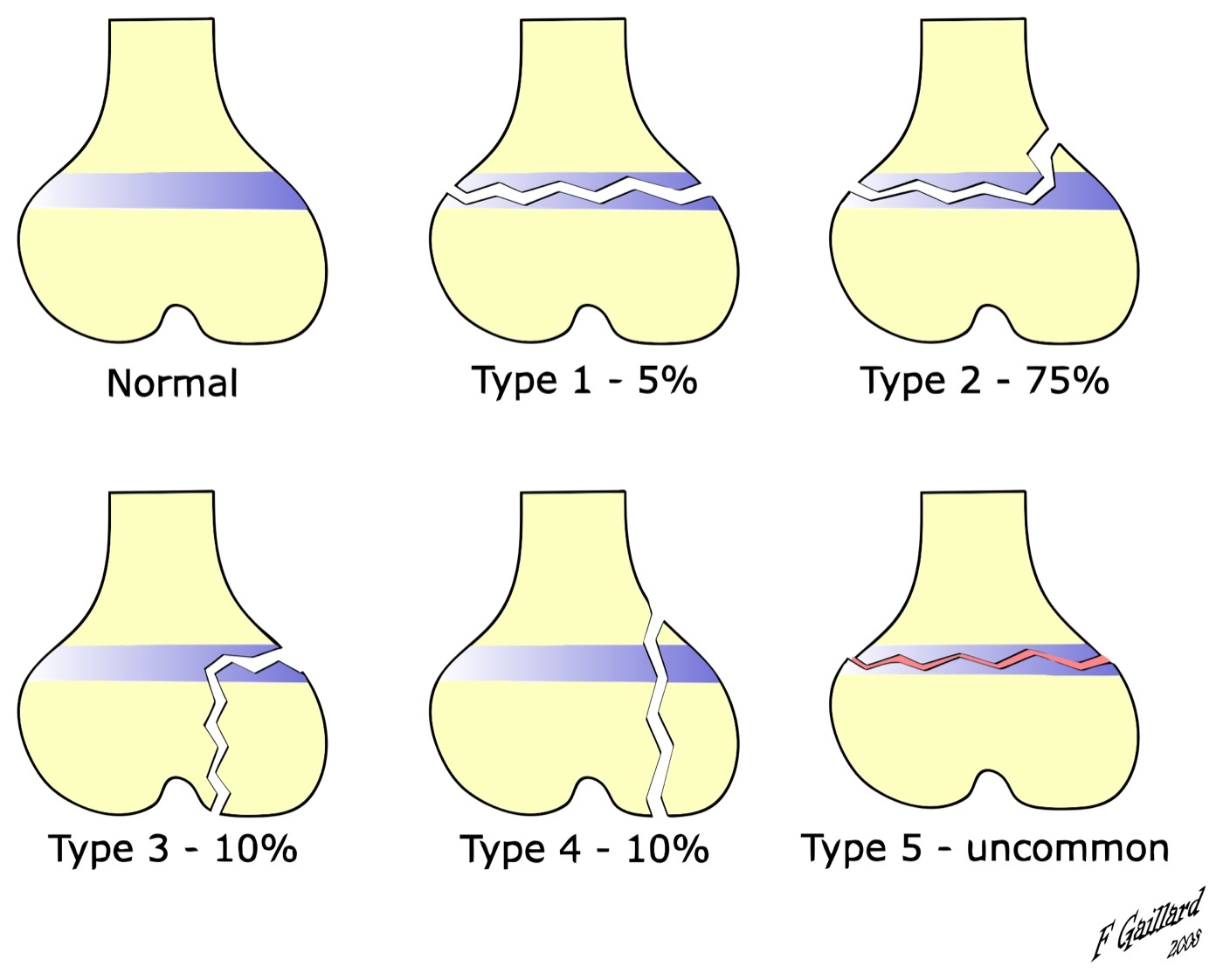
Figure 9. The Salter-Harris classification of physeal fractures (Types I-V) with their approximate frequencies. Dr Frank Gaillard, CC BY-SA 3.0, via Wikimedia Commons.
Prognosis and management
What threatens growth is damage to the germinal/reserve zone and to the epiphyseal blood supply, so the danger of an injury depends on its type, the displacement, the specific physis, and how much growth remains. The management principles follow from the anatomy. Reduction should be gentle and prompt, achieved with muscle relaxation and without repeated forceful manipulation, because grinding the fragments can itself injure the germinal cells and cause the very arrest one is trying to avoid. Intra-articular Type III and Type IV injuries need anatomic reduction, usually open, to restore both the joint surface and the physis. When fixation is required it should where possible avoid crossing the physis; if a smooth wire must cross it, the wire should be fine and removed early.[12]
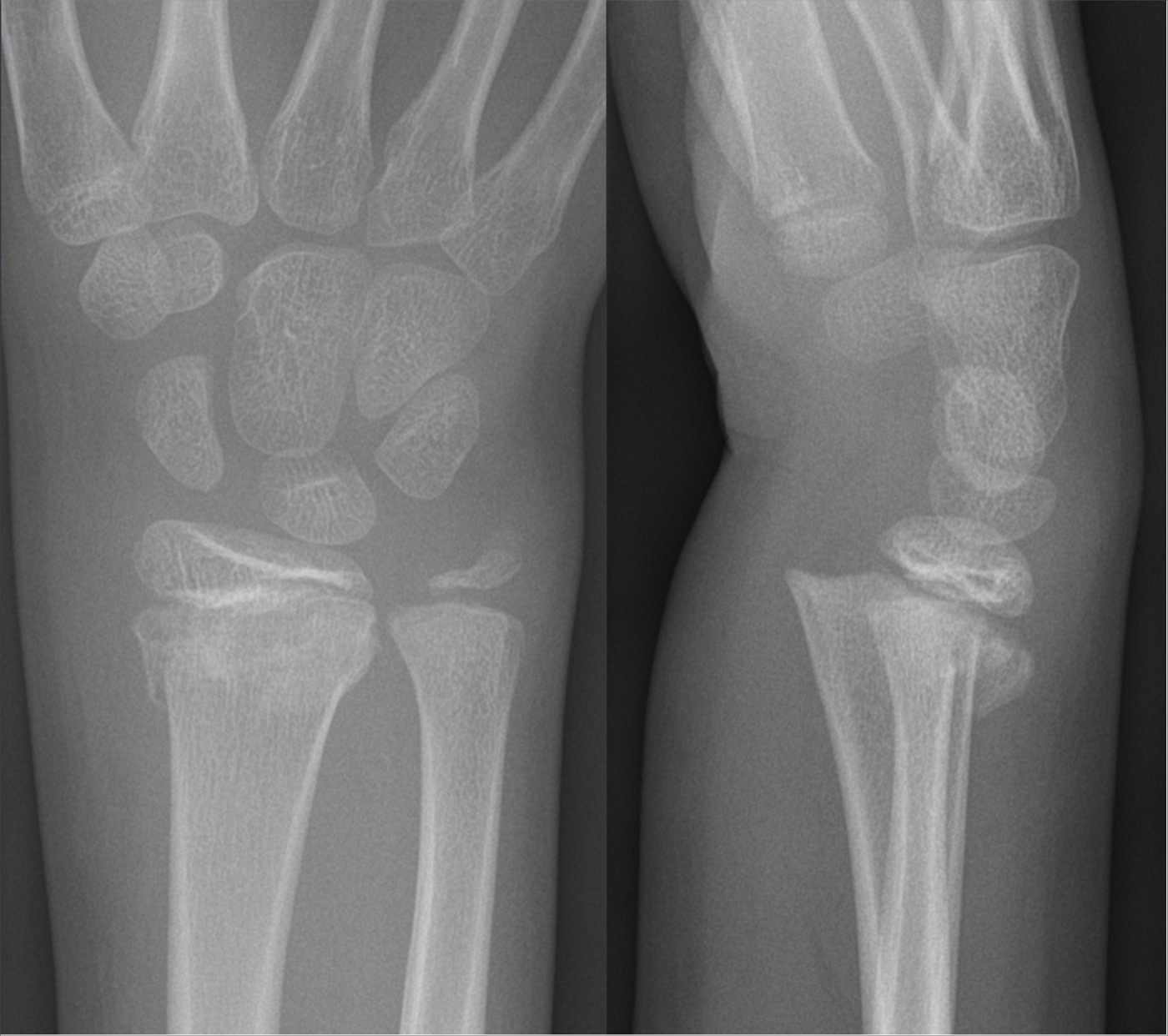
Figure 10. Salter-Harris type II fracture of the distal radius: physeal separation with the triangular metaphyseal (Thurston-Holland) fragment, two planes. Hellerhoff, CC BY-SA 4.0, via Wikimedia Commons.
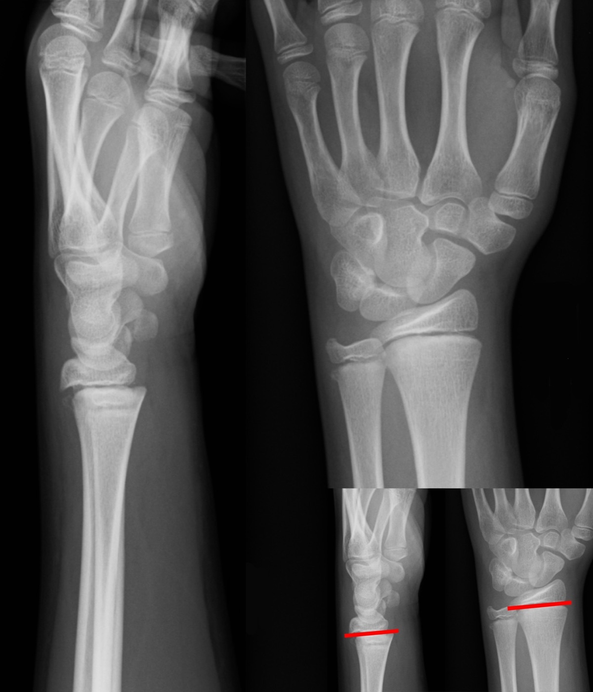
Figure 11. Salter-Harris type I physeal injury of the distal radius (lines), a separation straight through the physis with no bony fragment. Gilo1969, CC BY 3.0, via Wikimedia Commons.
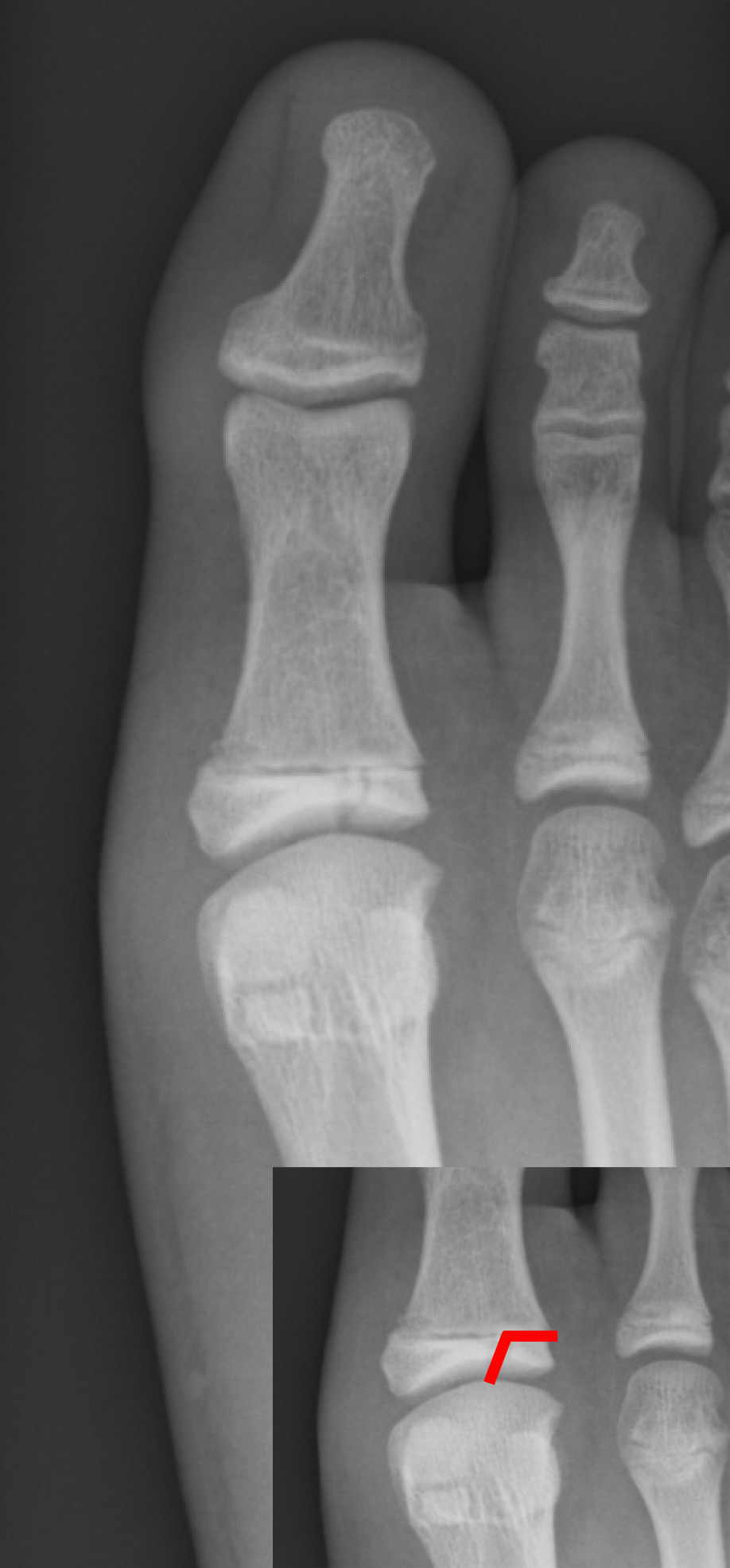
Figure 12. Salter-Harris type III fracture of the great-toe proximal phalanx, the fracture line passing through the physis into the epiphysis and joint. Gilo1969, CC BY 3.0, via Wikimedia Commons.
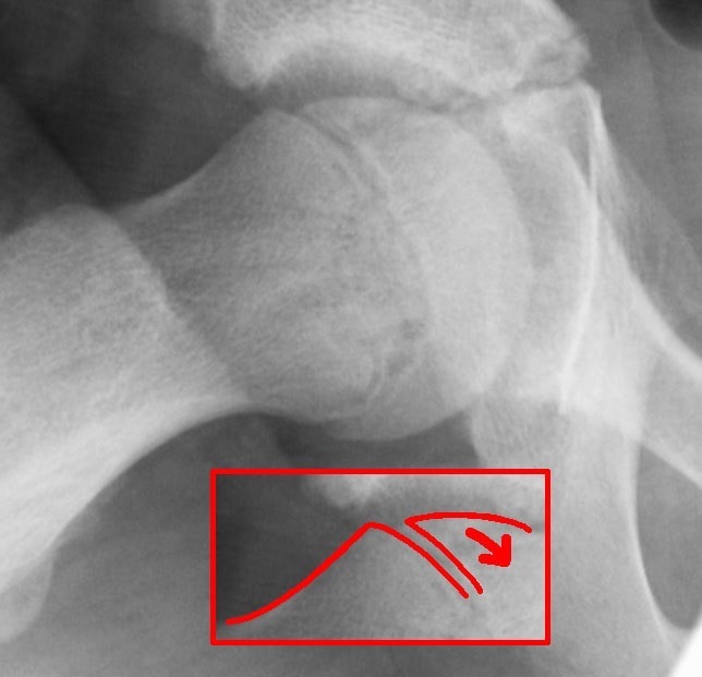
Figure 13. Slipped capital femoral epiphysis, a Salter-Harris type I-equivalent slip through the proximal femoral physis. Dr. Jochen Lengerke, CC0, via Wikimedia Commons.
Part V - Growth Arrest and the Physeal Bar
When a physeal injury heals badly, a bony bridge (physeal bar) can form across the plate, tethering it. The bridge matures from fibrous to fibrovascular to calcified-cartilage to a sclerotic osseous bar, and its effect depends on where and how large it is. A partial arrest, whether a peripheral or a central bar, tethers one part of the plate while the rest grows, producing a progressive angular deformity (and, with a central bar, a tented or coned physis). A complete arrest stops growth across the whole plate and produces limb-length discrepancy (shortening). A bar is mapped by imaging: plain films and the convergence of Harris growth-arrest lines give the first clue, and CT or MRI defines the area as a percentage of the physis.[13]
Treatment is chosen by the size of the bar and the growth remaining. A small bar, conventionally less than about half the physis in a child with substantial growth left, can be excised and the cavity filled with an interposition material (fat, silastic, or cement) to let the remaining plate resume growth, the Langenskiöld procedure. Established angular deformity is corrected by osteotomy. A large or complete bar with significant predicted discrepancy is managed by completing the arrest and performing a contralateral epiphysiodesis, or by limb lengthening or shortening to equalise length. The unifying prognostic factors are the bar’s size as a fraction of the physis and the patient’s age, that is, the growth that remains to be protected or lost.[14]
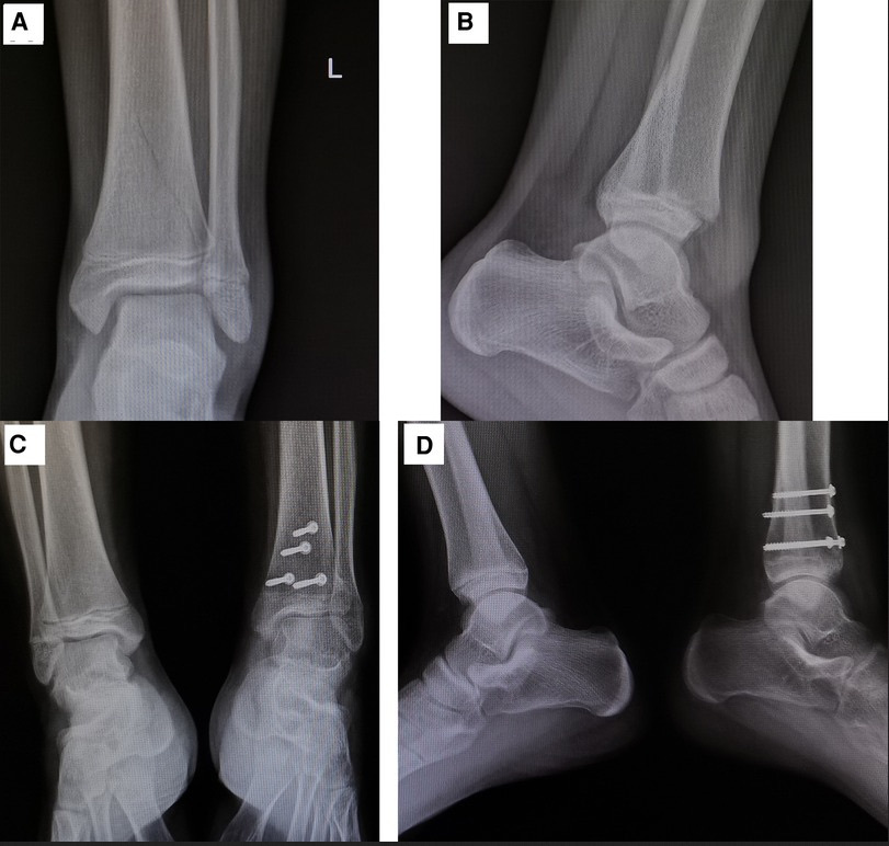
Figure 14. Premature physeal closure (growth arrest) of the distal tibia after a physeal fracture: the injured physis closes while the contralateral physis stays open. Wang et al., CC BY 4.0, via Wikimedia Commons.
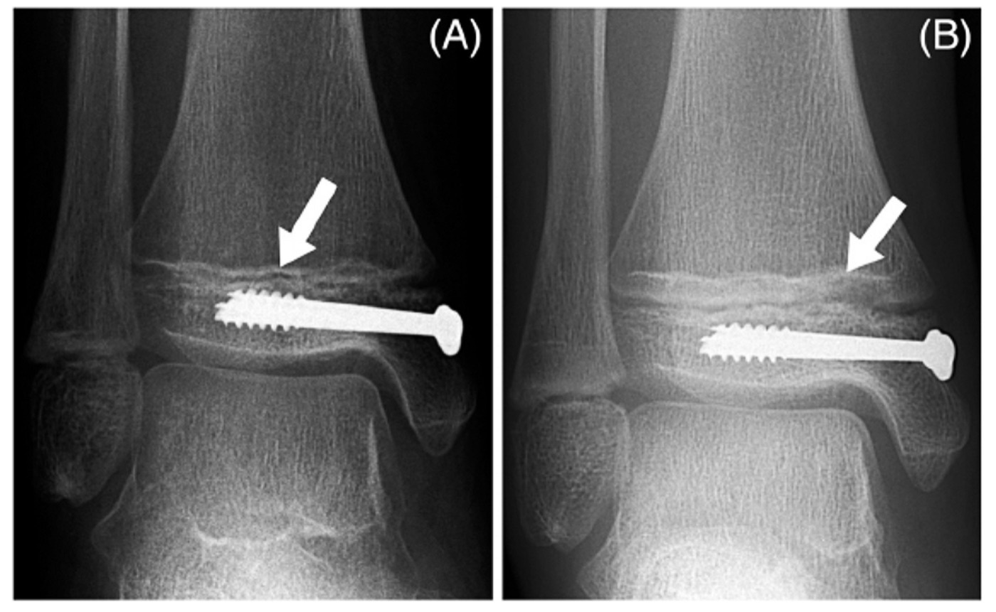
Figure 15. A Park-Harris growth-arrest line (arrows) that has grown away parallel to the distal tibial physis between two and seven months, indicating continued symmetric growth. Kennedy et al., CC BY 2.0, via Wikimedia Commons.
Part VI - Healing and Remodelling in the Child
Children heal faster and more reliably than adults, and the reason is again the periosteum: thick, osteogenic, and readily elevated, it throws down abundant external (periosteal) callus that bridges a fracture quickly, so that nonunion is rare in childhood. Healing passes through the familiar phases of inflammation (haematoma), reparative soft and then hard callus, and remodelling, but it runs faster and more vigorously in the child. The younger the child, the faster the union.[15]
The child’s other gift is remodelling, and its limits are as important as its power. A malunited fracture remodels best when the child is young, when the deformity is near a physis, and when the angulation lies in the plane of the joint’s motion. Remodelling occurs at the physis by apposition on the concave side and resorption on the convex side, not at the fracture itself. What does not remodel reliably is rotational malalignment, and to a degree displaced intra-articular incongruity. Finally, a diaphyseal fracture can stimulate the adjacent physis and cause overgrowth, classically about a centimetre in the femur, through the hyperaemia of healing and the release of the periosteal tether. That is why some shortening at the time of femoral fracture is accepted or even desired in the young child.[16]
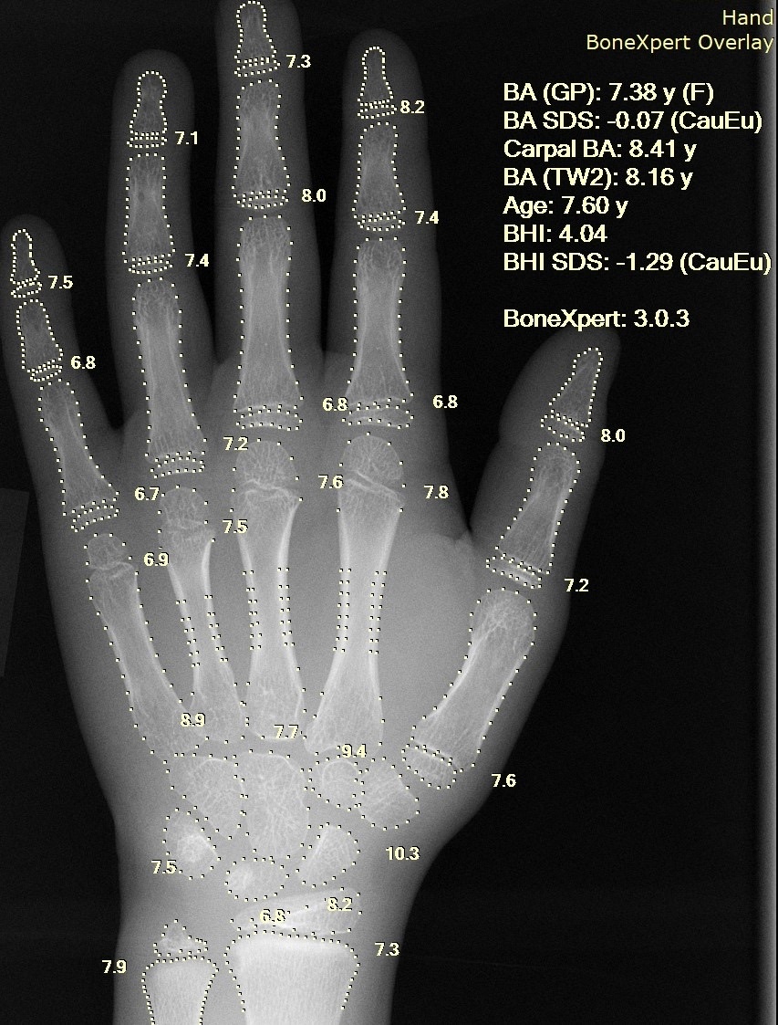
Figure 16. A pediatric hand radiograph showing open physes throughout, the reserve of remaining growth that gives the child the capacity to remodel a malunion. Setzner1337, CC0, via Wikimedia Commons.
References
-
The framing of the immature skeleton as biologically distinct and constantly changing, with the physis (growth plate) as the engine of longitudinal growth and a mechanical weak link, follows Ogden, Skeletal Injury in the Child, 3rd ed. (2000), Chapter 1 (Anatomy and Physiology of Skeletal Development) and Chapter 6 (Injury to the Growth Mechanisms). Page numbers in this summary refer to the printed pages of that edition. Established teaching that supplements the source (for example the standard Salter-Harris grading details) is flagged in-line as standard teaching and carries no page citation.
-
The sequence of topics (structure of the growing bone, biomechanics of failure, the unique pediatric fracture patterns, physeal injury and classification, growth arrest, and repair/remodelling) maps to Ogden Chapters 1, 2, 6, 7, and 8 respectively. The terms plastic deformation, torus/buckle, greenstick, and epiphysiolysis are all treated by Ogden in Chapters 2 and 6.
-
The four regions of the growing long bone (diaphysis, metaphysis, epiphysis, physis) plus the apophysis as a tension-loaded variant, the metaphysis as a thin-cortex trabecular flare with cortical fenestrations and the site of the torus fracture, the epiphysis being cartilaginous at birth except the distal femur, and the apophysis prototype (tibial tuberosity, Osgood-Schlatter) are from Ogden ch.1 (Ogden pp.3, 5-11). The physis as the mechanism of endochondral ossification is from Ogden ch.1 (Ogden p.10).
-
The physeal zones from the epiphyseal side (reserve/germinal supplied by the epiphyseal vessels and holding the stem cells, proliferative producing the longitudinal cell columns, hypertrophic with the zone of provisional calcification, and the zone of transformation/primary spongiosa invaded by metaphyseal vessels) and the cardinal fact that the hypertrophic and transformation zones are mechanically weakest, “the regions most often involved in physeal fractures,” with the germinal/proliferative cells usually spared, are from Ogden ch.1 (Ogden pp.12-13). That the physis is most resistant to traction and least resistant to torsion is from Ogden ch.1 (Ogden p.26).
-
The groove (zone) of Ranvier supplying chondroblasts for latitudinal/width growth and the osseous ring of LaCroix providing mechanical restraint without impeding width growth are from Ogden ch.1 (Ogden pp.11-13). The thick, vascular, strongly osteogenic periosteum, loosely attached over the diaphysis but densely fixed at the physeal periphery, and the experimental figures (550 lb to detach an epiphysis versus 119 lb after periosteal division) are from Ogden ch.1 (Ogden pp.5-7, 26).
-
That the epiphyseal and metaphyseal circulations are anatomically and functionally separate, that vessels generally do not cross the physis, that the epiphyseal supply runs as end-arterial cartilage canals without anastomoses (so its loss causes avascular necrosis and angular deformity), and that epiphyseal vascular compromise is serious whereas metaphyseal compromise is minor and reversible, are from Ogden ch.1 (Ogden pp.18-20). Longitudinal growth at the physis and width growth by periosteal apposition, and the programmed order of physeal closure (physiologic epiphysiodesis, the distal tibia closing medial to lateral and producing the Tillaux pattern), are from Ogden ch.1 (Ogden pp.16-18).
-
The three-part framework (anatomy, physiology, biomechanics) for why immature bone differs, the lower modulus and greater porosity and elasticity (more energy absorbed, more deformation before failure), the statement that “adult bone usually fails initially in tension, whereas a child’s bone may fail in either tension, compression, or both,” and that the greater porosity arrests crack propagation so comminution is uncommon, are from Ogden ch.2 (Ogden p.48).
-
That the physis is mechanically weaker than the adjacent ligaments and joint capsule, so a physeal separation replaces the adult ligament tear, is from Ogden ch.2 (Ogden p.57). The thick periosteal hinge limiting displacement and resisting over-reduction, and subperiosteal new bone bridging defects without grafting, are from Ogden ch.2 (Ogden p.56).
-
Plastic (bowing) deformation as a bend from microscopic failures without a discrete cortical break (ulna and fibula, younger children, may block reduction of the paired bone), the torus/buckle fracture as a stable compression failure at the metaphyseal-diaphyseal junction (classic distal radius), and the greenstick fracture as failure of the tension cortex with the compression cortex intact but bent (with the tendency to re-angulate in cast, sometimes requiring completion of the fracture for a stable reduction), together with complete fractures and the toddler’s fracture, are from Ogden ch.2 (Ogden pp.51-54).
-
That physeal injuries are about 15% of childhood fractures, are most common in the hand/phalanges, involve distal more than proximal physes and boys more than girls with peaks at 9-12 (girls) and 12-15 (boys), and that the separation usually passes through the hypertrophic zone at the calcified/uncalcified junction (sparing the germinal layer), are from Ogden ch.6 (Ogden pp.147, 152-153).
-
The five Salter-Harris types and their paths (I through the physis with no bony fragment; II through the physis exiting a metaphyseal corner with the Thurston-Holland fragment, the commonest; III through the physis into the epiphysis and joint; IV crossing metaphysis, physis and epiphysis as one intra-articular line; V a crush of the physis), Ogden’s note that the scheme was derived largely from radiographs, his caution that Type II is not uniformly benign (around 6.5% arrest in one population study) and that a pure-crush Type V may not exist (the mechanism often vascular), and Ogden’s own expanded classification, are from Ogden ch.6 (Ogden pp.150-175). The worsening prognosis down the Salter-Harris list, the slipped capital femoral epiphysis as a Type I equivalent, and the SALTR mnemonic are standard teaching consistent with Ogden’s account.
-
That growth disturbance follows damage to the germinal/reserve zone and the epiphyseal blood supply, with risk depending on injury type, displacement, the specific physis, and remaining growth, and the management principles (gentle prompt reduction under muscle relaxation, avoiding repeated manipulation that injures the germinal cells, anatomic usually-open reduction for intra-articular Types III and IV, and fixation that avoids crossing the physis or uses fine smooth wires removed early), are from Ogden ch.6 (Ogden pp.150-175).
-
The formation of a physeal bar maturing from fibrous through fibrovascular and calcified cartilage to a sclerotic osseous bridge (with the epiphyseal blood supply a key determinant), the distinction between partial arrest (peripheral or central bar) causing progressive angular deformity and complete arrest causing limb-length discrepancy/shortening, and the assessment of a bar by plain films with converging Harris growth-arrest lines and by CT/MRI mapping its area as a percentage of the physis, are from Ogden ch.7 (Ogden pp.209-234).
-
That bar treatment is chosen by bar size and remaining growth, that a small bar (conventionally under about half the physis) can be excised with interposition of fat, silastic or cement to restore growth (the Langenskiöld procedure), that established angular deformity is corrected by osteotomy and a large/complete arrest by completion plus contralateral epiphysiodesis or by limb lengthening/shortening, and that the prognostic factors are bar size as a percentage of the physis and the patient’s age/remaining growth, are from Ogden ch.7 (Ogden pp.209-234). The roughly 50% physeal-area threshold for bar excision is associated with the Carlson-Wenger mapping work cited by Ogden.
-
That children heal faster and more reliably than adults because of the thick, osteogenic, readily elevated periosteum and its abundant external callus (so nonunion is rare), and that healing passes through inflammatory/haematoma, reparative (soft then hard callus), and remodelling phases but faster in the younger child, are from Ogden ch.8 (Ogden pp.243-268).
-
That remodelling is greatest with youth, proximity to a physis, and deformity in the plane of joint motion, that it occurs at the physis by concave-side apposition and convex-side resorption rather than at the fracture, that rotation (and to a degree displaced intra-articular incongruity) does not remodel, and that diaphyseal fracture can stimulate overgrowth (classically about 1 cm in the femur, via the hyperaemia of healing and loss of the periosteal tether), are from Ogden ch.8 (Ogden pp.243-268) and Ogden ch.2 (Ogden pp.42, 57-58).
-
The four regions plus the apophysis, and the physis as the cartilaginous growth plate driving endochondral longitudinal growth, are from Ogden ch.1 (Ogden pp.3, 5-11).
-
The physeal zones and the fact that the hypertrophic and transformation zones are weakest (so fractures run there, sparing the germinal cells) are from Ogden ch.1 (Ogden pp.12-13).
-
The groove of Ranvier (width growth) and the ring of LaCroix (peripheral mechanical restraint without impeding width growth) are from Ogden ch.1 (Ogden pp.11-13).
-
The thick osteogenic periosteum, its attachment gradient, the 550-versus-119-pound figures, and its role as an intact hinge laying down rapid callus are from Ogden ch.1 (Ogden pp.5-7, 26) and ch.2 (Ogden p.56).
-
The separate epiphyseal and metaphyseal circulations, vessels not crossing the physis, the end-arterial epiphyseal canals causing avascular necrosis and angular deformity, and the metaphyseal loss being minor and reversible are from Ogden ch.1 (Ogden pp.18-20).
-
The lower modulus and greater porosity/elasticity, the “tension, compression, or both” failure statement, and the rarity of comminution are from Ogden ch.2 (Ogden p.48).
-
Plastic deformation, the torus/buckle fracture, and the greenstick fracture (with the re-angulation tendency and occasional deliberate completion) are from Ogden ch.2 (Ogden pp.51-54).
-
That an epiphysiolysis is a physeal separation, about 15% of childhood fractures and commonest in the hand, usually running through the hypertrophic zone at the calcified/uncalcified junction (sparing the germinal cells), is from Ogden ch.6 (Ogden pp.147, 152-153).
-
The five Salter-Harris types, the Thurston-Holland fragment in Type II, and the worsening prognosis are from Ogden ch.6 (Ogden pp.150-175); the SALTR mnemonic and “III/IV need anatomic reduction, V worst” framing are standard teaching consistent with Ogden.
-
The management principles (gentle prompt reduction, avoiding repeated manipulation, anatomic open reduction for Types III/IV, and fixation avoiding the physis or using fine smooth wires removed early) are from Ogden ch.6 (Ogden pp.150-175).
-
The partial-versus-complete arrest distinction (angular deformity versus shortening), the Langenskiöld bar excision with interposition for a small bar, and osteotomy/epiphysiodesis/length equalisation for established deformity or large arrest are from Ogden ch.7 (Ogden pp.209-234).
-
That the thick osteogenic periosteum gives fast union and rare nonunion, that remodelling is greatest with youth/proximity to a physis/deformity in the plane of motion (occurring by concave apposition and convex resorption at the physis), that rotation does not remodel, and that overgrowth (about 1 cm in the femur) can follow a diaphyseal fracture are from Ogden ch.8 (Ogden pp.243-268) and ch.2 (Ogden pp.42, 57-58).