Contents
- Orientation
- Part I - Applied Surgical Anatomy of the Leg
- Part II - Approaches to the Tibial Shaft
- Part III - Approaches to the Proximal Tibia and Tibial Plateau
- Part IV - The Approach to the Fibula
- Part V - Acute Compartment Syndrome and Fasciotomy
- Part VI - Intramedullary Tibial Nailing
- References
Orientation
The leg between knee and ankle is built around two very different bones and four unforgiving compartments. The tibia carries a broad subcutaneous surface down its whole medial length, which makes it easy to reach but cruelly exposed: the skin over it is thin, it blisters and sloughs after high-energy trauma, and wound breakdown is the complication that haunts every approach to it. The fibula is the opposite, buried almost entirely in muscle and reachable only by stripping that muscle off bone, with the common peroneal nerve hooked around its neck like a trap set for the careless. Master those two facts and the contrasting personalities of the bones explain most of what follows.[1]
The applied anatomy comes first, because every approach and every fasciotomy is really a statement about which of the four compartments you are entering and which nerve guards it. From there the topic works through the approaches to the tibial shaft, the proximal tibia and plateau, and the fibula, before turning to the two clinical set-pieces of the leg: decompression of an acute compartment syndrome and intramedullary nailing of the tibial shaft. Two structures recur as the villains of the region. The common peroneal nerve, subcutaneous over the fibular neck, can be cut by a skin incision that is merely too bold, and its loss is a footdrop. The posterior tibial neurovascular bundle sits sheltered in the deep posterior compartment behind tibialis posterior and flexor hallucis longus, and a deep-flexor fasciotomy must respect it. Keep those two in mind throughout.[2]
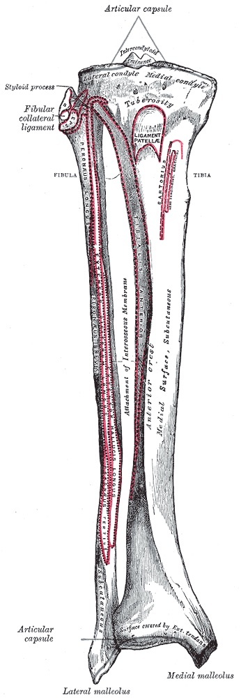
Figure 1. Bones of the right leg, anterior surface, showing the tibia, fibula and interosseous membrane with their borders and surfaces. Gray’s Anatomy (1918), Plate 258, Henry Vandyke Carter, public domain, via Wikimedia Commons.
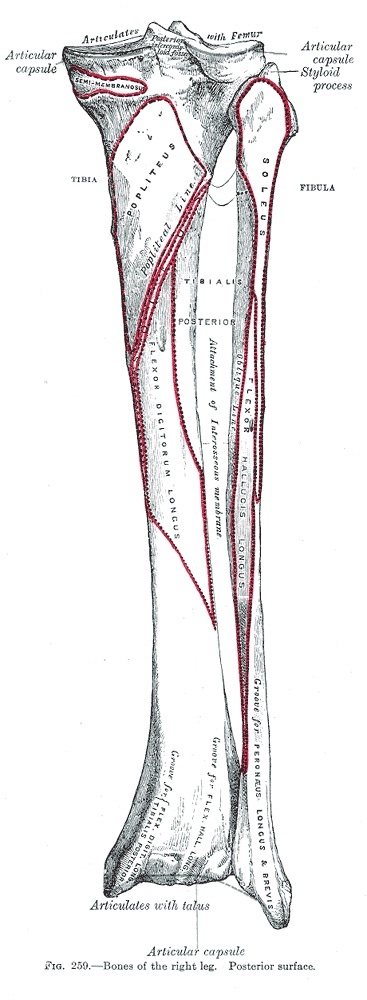
Figure 2. Bones of the right leg, posterior surface, with the popliteal and soleal lines and the attachments of the deep posterior muscles. Gray’s Anatomy (1918), Plate 259, public domain, via Wikimedia Commons.
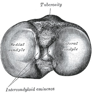
Figure 3. Upper surface of the right tibia (the tibial plateau) seen from above, with the condyles and intercondylar eminence. Gray’s Anatomy (1918), Plate 257, public domain, via Wikimedia Commons.
Part I - Applied Surgical Anatomy of the Leg
The two bones and the deep fascia
To the surgeon the tibia and fibula could hardly be more different. The tibia presents a large subcutaneous surface that gives access along its entire length, and it carries no major neurovascular structure other than its nutrient artery. The fibula is enclosed almost completely in muscle, becoming subcutaneous only at its proximal end and in its lower third where it ends as the lateral malleolus. Operating on most of it therefore means stripping muscle off bone, and it has intimate ties to the common peroneal nerve and its branches. Enclosing the calf is the deep fascia of the leg, a tough, fibrous, unyielding sheet that attaches to the bony borders where they surface. That unyielding quality is exactly why a swollen compartment cannot decompress itself.[3]
The four compartments
Four separate compartments are walled off by the deep fascia, two intermuscular septa, and the interosseous membrane. Both septa run from the deep surface of the fascia to the fibula, and together they enclose the lateral compartment. The anterior intermuscular septum divides the anterior from the lateral compartment, the posterior intermuscular septum divides the lateral from the posterior compartments, the interosseous membrane separates the deep posterior compartment from the anterior, and a fascial layer separates the superficial posterior compartment from the deep posterior one. The cardinal teaching point is one nerve per compartment, which is what makes them both anatomically distinct and surgically releasable.[4]
The anterior (extensor) compartment lies between the lateral surface of the tibia, the extensor surface of the fibula with the anterior septum, and the deep fascia, walled posteriorly by the interosseous membrane. It holds the extensor muscles of the foot and ankle (tibialis anterior, extensor hallucis longus, extensor digitorum longus, and peroneus tertius), all supplied by the deep peroneal nerve and accompanied by the anterior tibial artery; together they dorsiflex the ankle and extend the toes.[5]
The lateral (peroneal) compartment is bounded by the anterior septum in front, the posterior septum behind, and the fibula medially. It contains the peroneal muscles (peroneus longus and brevis), supplied by the superficial peroneal nerve, which evert the foot. It is the one compartment with no artery of its own: its muscles are fed by several branches of the peroneal artery.[6]
The superficial posterior (flexor) compartment holds three muscles, the gastrocnemius, soleus, and plantaris, separated from the lateral compartment by the posterior septum and from the deep posterior compartment by a fascial layer. The tibial nerve supplies it, the sural nerve is its associated cutaneous nerve, and it plantarflexes the ankle. The deep posterior (flexor) compartment holds the tibialis posterior, flexor digitorum longus, and flexor hallucis longus, and it carries the tibial nerve and the posterior tibial artery themselves. It is separated from the superficial compartment by the posterior septum and from the anterior compartment by the interosseous membrane. Its muscles plantarflex and invert the foot and flex the toes.[7]
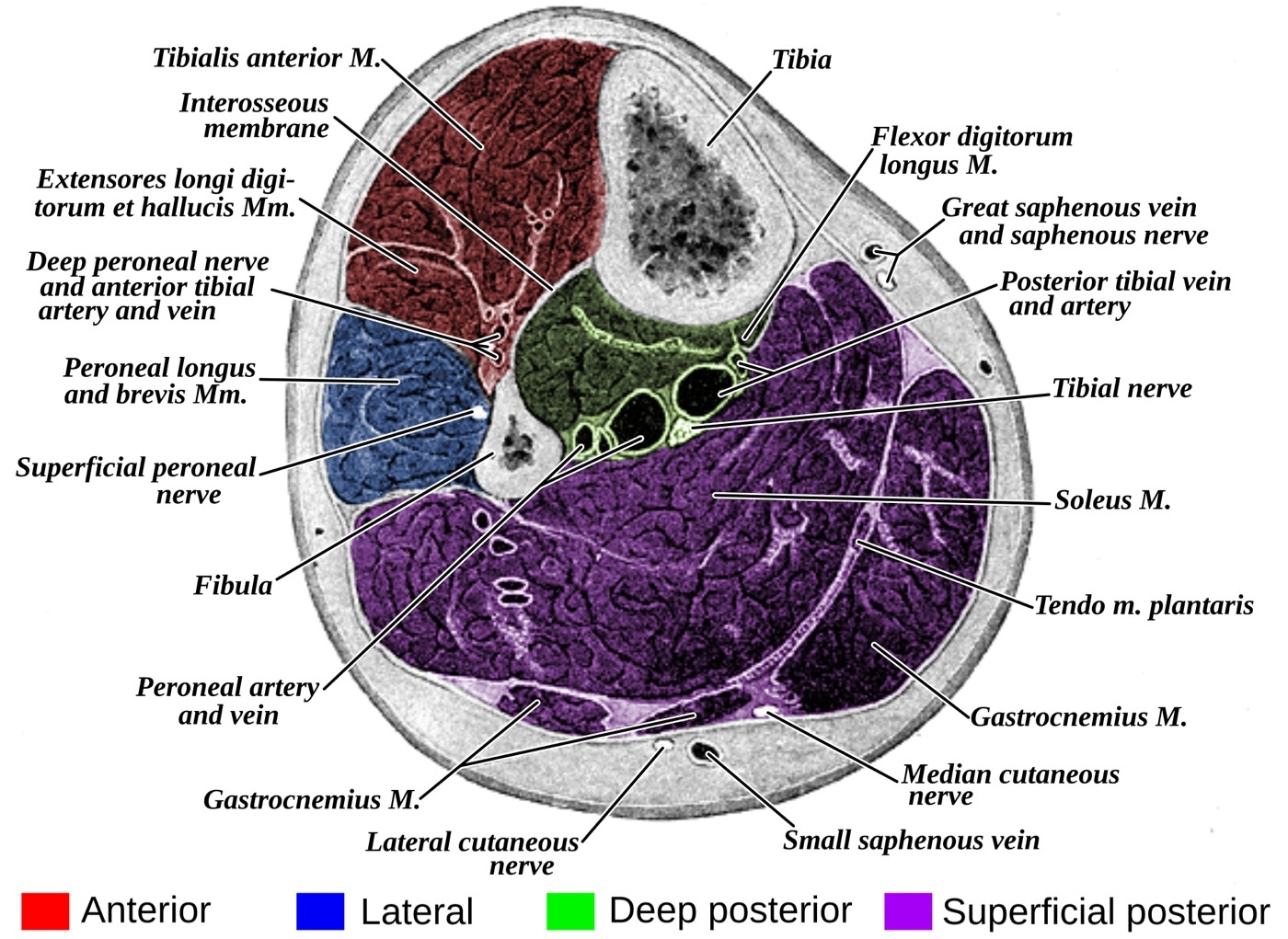
Figure 4. Transverse section through the middle of the leg showing the four fascial compartments (anterior, lateral, superficial and deep posterior, colour-coded), the tibia, fibula and interosseous membrane, the intermuscular septa and the neurovascular bundles. Gray’s Anatomy (1918), Plate 440 (colourised derivative), public domain, via Wikimedia Commons.
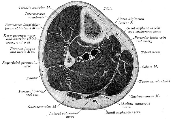
Figure 5. The same transverse section of the leg, original plate, with the muscles and neurovascular structures labelled. Gray’s Anatomy (1918), Plate 440, public domain, via Wikimedia Commons.
The neurovascular course
The fibula’s intimate relationship with the common peroneal nerve is the through-line of leg surgery. The nerve winds around the fibular neck and, by standard teaching, divides there into the superficial peroneal nerve (which descends in the lateral compartment and pierces the deep fascia in the distal third to become subcutaneous) and the deep peroneal nerve (which descends on the interosseous membrane in the anterior compartment with the anterior tibial artery). The anterior tibial artery reaches the anterior compartment by passing forward through an opening in the proximal interosseous membrane. The posterior tibial neurovascular bundle, the tibial nerve with the posterior tibial artery, runs in the deep posterior compartment, and every deep-flexor release must protect it.[8]
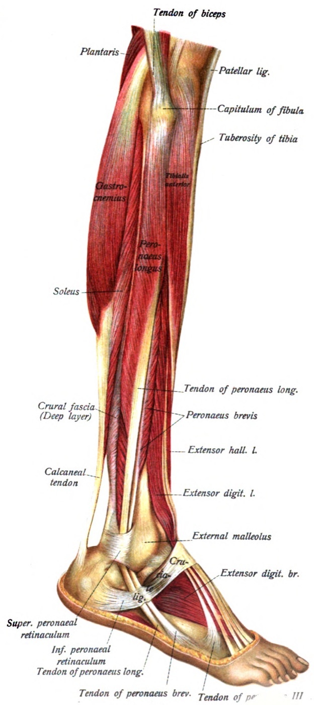
Figure 6. Lateral aspect of the leg showing the lateral (peroneal) compartment, peroneus longus and brevis with their retinacula, together with the anterior extensor muscles. Sobotta’s Atlas of Human Anatomy (1909), public domain, via Wikimedia Commons.
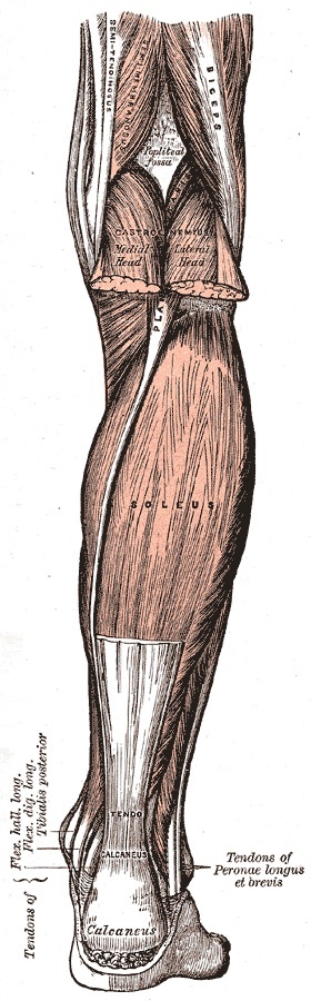
Figure 7. Superficial muscles of the posterior leg: the gastrocnemius and soleus converging on the tendo calcaneus. Gray’s Anatomy (1918), Plate 438, public domain, via Wikimedia Commons.
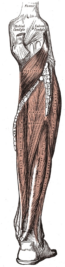
Figure 8. Deep muscles of the posterior leg: popliteus, tibialis posterior, flexor digitorum longus and flexor hallucis longus. Gray’s Anatomy (1918), Plate 439, public domain, via Wikimedia Commons.
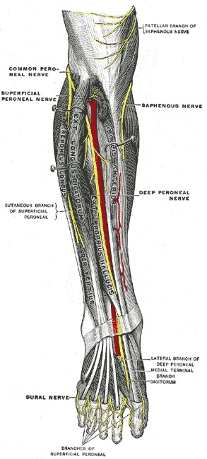
Figure 9. Deep nerves of the front of the leg: the common peroneal nerve dividing into the superficial and deep peroneal nerves, the latter accompanying the anterior tibial artery among the extensor muscles. Gray’s Anatomy (1918), Plate 835, public domain, via Wikimedia Commons.
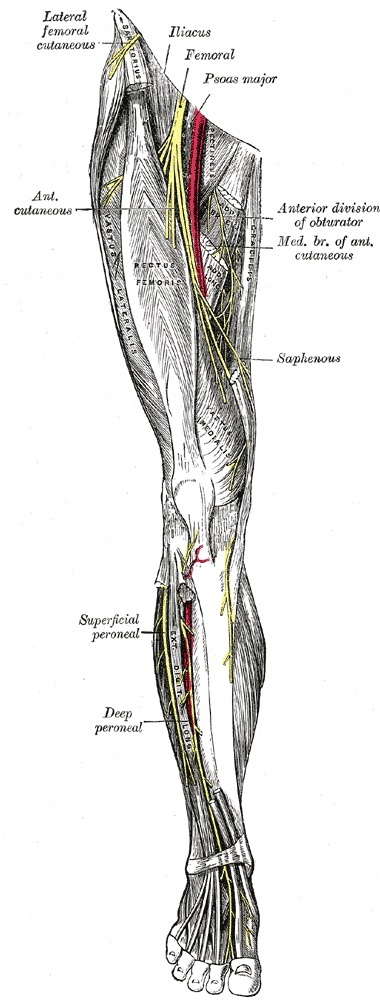
Figure 10. Nerves of the right lower limb, anterior view, showing the course of the saphenous, superficial peroneal and deep peroneal nerves. Gray’s Anatomy (1918), Plate 827, public domain, via Wikimedia Commons.
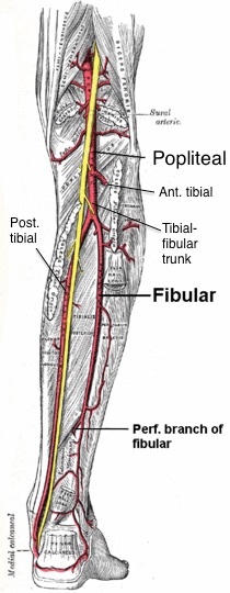
Figure 11. Arteries of the leg, posterior view: the popliteal artery dividing into the anterior tibial artery and the tibiofibular trunk, which gives the posterior tibial and fibular (peroneal) arteries. Wikimedia contributor, CC BY-SA 4.0, via Wikimedia Commons.
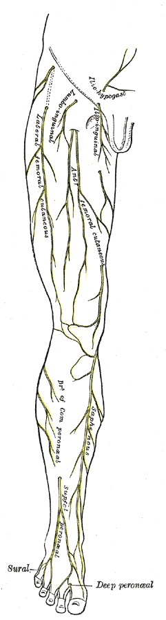
Figure 12. Cutaneous nerves of the right lower limb, anterior view: the saphenous nerve along the medial leg with the superficial peroneal and sural cutaneous territories. Gray’s Anatomy (1918), Plate 825, public domain, via Wikimedia Commons.
Part II - Approaches to the Tibial Shaft
The anterior approach to the tibia
The anterior approach is the preferred exposure of the tibia, except where the skin is scarred or carries draining sinuses. Through one incision it reaches both the medial (subcutaneous) surface and, by reflecting tibialis anterior, the lateral (extensor) surface, and the bone can be exposed along its whole length. It serves open reduction and internal fixation, bone grafting of delayed or nonunion, sequestrectomy and saucerisation in osteomyelitis, tumour excision and biopsy, and osteotomy. The incision is longitudinal, parallel to and about 1 cm lateral to the anterior border of the tibia. Because the skin here is poorly vascularised, it is safer to make a longer incision than to retract the edges forcibly.[9]
There is no internervous plane, because the subcutaneous medial surface is used directly. The dissection is epiperiosteal and leaves the extensor compartment’s nerve supply undisturbed. On the medial surface, periosteal stripping is kept to an absolute minimum, and the periosteum is never stripped from an isolated fragment, or that fragment will become avascular. On the lateral surface, tibialis anterior, the only muscle arising from the lateral surface of the tibia, is reflected laterally. The plating decision is the heart of the approach. A plate on the medial subcutaneous surface sits on the tensile side and is easy to contour, but it lies under thin skin and risks wound breakdown, so some surgeons plate the lateral surface deep to muscle instead.[10]
The dominant danger is not a nerve or vessel but the soft-tissue envelope. The skin over the lower third of the tibia is very thin and heals badly, especially in smokers and patients with chronic venous insufficiency. Longitudinal incisions heal well, but transverse and irregular wounds heal poorly, particularly in the elderly. The long saphenous vein runs up the medial side of the calf and must be protected when the medial flap is reflected, and devascularised bone, however well reduced and fixed, will not unite. The same principles drive the minimally invasive anterior approach to the distal tibia, a two-window epiperiosteal technique for percutaneous plating of multifragmentary distal metaphyseal fractures. It is used only when the soft tissues are sound (smoking and chronic venous insufficiency are relative contraindications), the plane is deliberately epiperiosteal to preserve the periosteal blood supply, and the saphenous nerve and long saphenous vein anterior to the medial malleolus are retracted and preserved.[11]
The posterolateral approach to the tibia
The posterolateral approach is a technically demanding exposure of the middle two-thirds of the tibia that exists for one purpose: to reach the bone when the anteromedial subcutaneous skin is scarred or infected and an anterior approach would not heal. Coming from behind through healthy, well-vascularised posterior muscle, it can deliver a bone graft to a nonunion under sound soft-tissue cover. The patient lies in the lateral decubitus position with the affected leg uppermost, and the incision runs along the easily palpable lateral border of the gastrocnemius, at least 10 cm long.[12]
The internervous plane lies between the peroneal muscles (superficial peroneal nerve) and the gastrocnemius-soleus and flexor group (tibial nerve). Working in the interval between the lateral gastrocnemius and soleus posteriorly and the peronei anteriorly, the surgeon detaches the soleus origin and the flexor hallucis longus from the fibula, then dissects medially across the interosseous membrane, stripping tibialis posterior to reach the posterior surface of the tibia. The protective principle is elegant. The posterior tibial artery and tibial nerve are kept safe by the bulk of tibialis posterior and flexor hallucis longus, which lie between them and the operative plane, so the dissection must stay on the interosseous membrane and never stray behind those muscles. The short saphenous vein is spared (or ligated if needed) at the skin-flap stage, and branches of the peroneal artery are ligated in the superficial plane. The approach cannot be carried into the proximal quarter of the tibia, where the popliteus and the more superficial posterior tibial vessels and tibial nerve make dissection unsafe.[13]
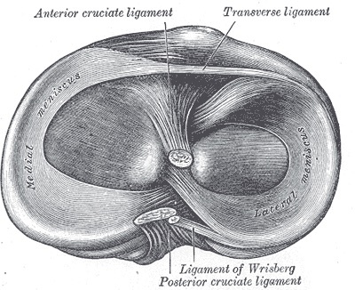
Figure 13. Head of the right tibia from above, with the menisci and the tibial attachments of the cruciate ligaments on the plateau. Gray’s Anatomy (1918), Plate 349, public domain, via Wikimedia Commons.
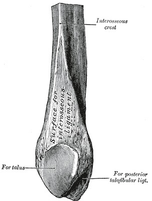
Figure 14. Lower end of the right fibula (the lateral malleolus), medial aspect, showing the articular surface for the talus and the malleolar fossa. Gray’s Anatomy (1918), Plate 262, public domain, via Wikimedia Commons.
Part III - Approaches to the Proximal Tibia and Tibial Plateau
The anterolateral approach to the lateral plateau
The anterolateral approach is the standard safe access to the lateral tibial plateau, and its workhorse indication is the open reduction and internal fixation of lateral plateau fractures. It is chosen over a direct anterior route because its skin incision does not lie directly over bone and because less retraction is needed. That matters because the proximal-tibial soft-tissue envelope is “thin and delicate,” prone to massive swelling and blistering after high-energy trauma, so definitive surgery is frequently delayed until the soft tissues recover. The knee is flexed about 60 degrees over a wedge, an inverted L-shaped incision curves over Gerdy’s tubercle, and there is no internervous plane: the dissection is epiperiosteal and spares the extensor compartment’s nerve supply.[14]
The articular surface is reached by a submeniscal arthrotomy. The capsule is opened transversely just below the lateral meniscus, and the meniscus is detached from its inferior soft-tissue attachments (the coronary/meniscotibial ligament) and lifted on stay sutures, preserving its anterior and posterior attachments for later repair. The lateral meniscus is at greatest risk during the synovial incision. The deep peroneal nerve has a variable course but normally lies well posterior to the field. A femorotibial distractor applying a varus force opens the lateral compartment for a clearer view of the articular surface.[15]
The posteromedial approach to the proximal tibia
Complex plateau fractures often carry a large posteromedial fragment, and accurate reduction of that fragment onto the shaft is frequently the first stage of bicondylar fixation. The posteromedial approach is built to address it. A buttress plate placed here, on the compression side, prevents the varus collapse that is the commonest deformity of the proximal tibia after fracture. Its soft tissues are usually spared the anterior blistering, which is part of its appeal. The patient is supine with a sandbag under the opposite hip to roll the limb into external rotation and bring the posteromedial corner forward, and the incision overlies the palpable posteromedial border.[16]
There is no internervous plane. The working interval is the epiperiosteal plane between the bone (with the pes anserinus) and the medial head of gastrocnemius, which is lifted off the bone by blunt dissection. The saphenous nerve and long saphenous vein lie just anterior to the incision and must be identified and preserved at the superficial stage. The pes anserinus can be either divided longitudinally (simple, but hard to repair beneath a plate) or reflected by detaching the sartorius, gracilis, and semitendinosus tendons. Carried proximally, the approach reaches the posteromedial corner of the knee and even the popliteal vessels for vascular surgery, and carried distally it opens the superficial and deep posterior compartments for release.[17]
The posterolateral, posterior, and minimally invasive plateau approaches
Three further plateau exposures complete the proximal-tibia repertoire. The posterolateral approach to the tibial plateau addresses depressed posterolateral articular fragments. It has no internervous plane, working between the biceps femoris and common peroneal nerve laterally and the lateral head of gastrocnemius medially, and it requires elevation of the popliteus and detachment of the soleus origin from the proximal fibula. The posterior approach to the tibial plateau is the one proximal-tibia exposure with a true internervous plane, between the semitendinosus (sciatic/tibial nerve) and the medial head of gastrocnemius (tibial nerve). That plane is safe because each muscle is innervated away from the interval. It reaches the posterior plateau but leaves a blind spot at the posterolateral corner. The minimally invasive anterolateral approach to the proximal tibia is an epiperiosteal submuscular slide for percutaneous plating that spares the deep peroneal nerve, with one hard rule: when a long plate is used, the anterior tibial artery and deep peroneal nerve lie in the path of the distal screws (around the 11th to 13th holes), so a formal open distal window is mandatory and blind stab screws are contraindicated there.[18]
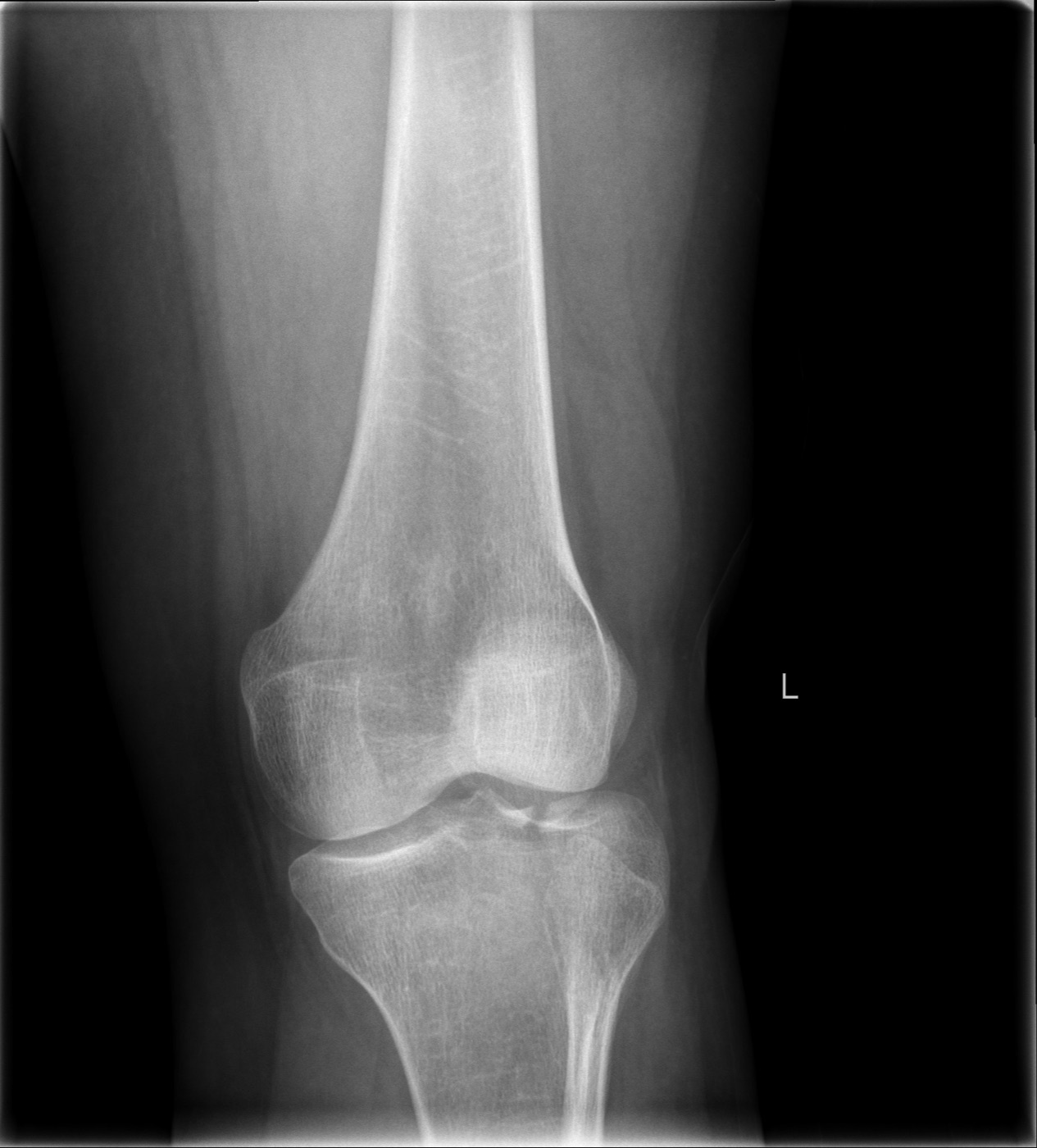
Figure 15. Anteroposterior radiograph of a lateral tibial plateau fracture with articular depression (a Schatzker-pattern injury). LottieLattes, CC BY-SA 4.0, via Wikimedia Commons.
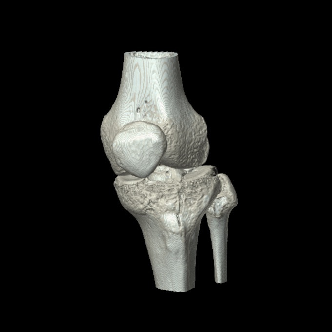
Figure 16. Three-dimensional CT reconstruction of a depressed lateral tibial plateau fracture. LottieLattes, CC BY-SA 4.0, via Wikimedia Commons.
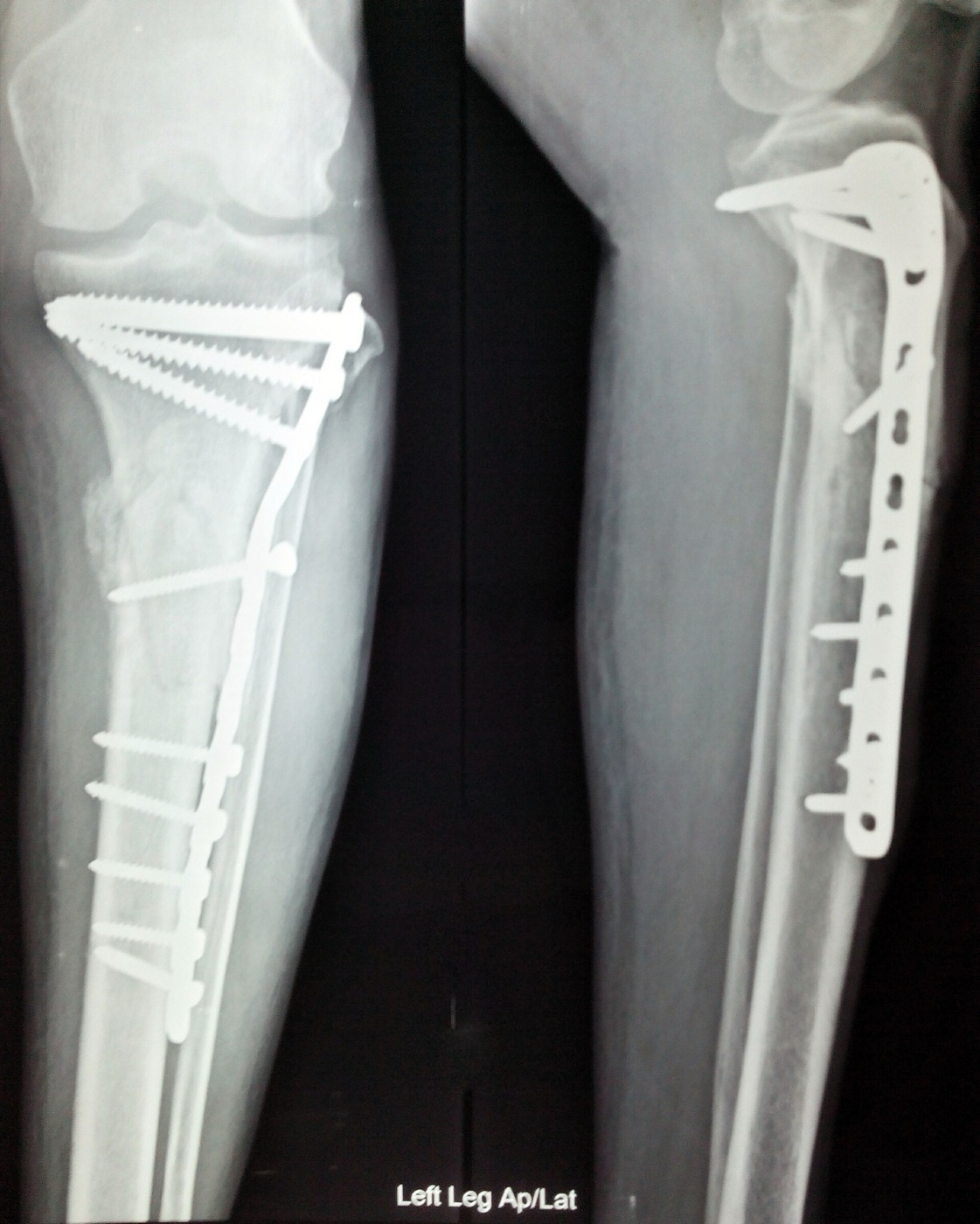
Figure 17. Proximal tibial plateau fracture after open reduction and internal fixation with a buttress plate and screws, anteroposterior and lateral views. Nizil Shah, CC BY-SA 4.0, via Wikimedia Commons.
Part IV - The Approach to the Fibula
A single plane the length of the bone
The approach to the fibula is a classic extensile exposure that offers access to all parts of the bone. The whole length can be exposed through one longitudinal incision just posterior to the fibula, though usually only a segment is needed. It serves partial resection during tibial osteotomy or nonunion surgery, decompressive fibulectomy of all four compartments, resection of tumour or osteomyelitis, fixation of fibular fractures, and the harvest of corticocancellous strut grafts and vascularised fibular grafts, which are dissected with their vascular pedicles. The patient lies in lateral decubitus with the affected side up, and the fibular head is palpable about 2 to 3 cm below the lateral femoral condyle.[19]
The internervous plane lies between the peroneal muscles (superficial peroneal nerve) and the flexor muscles (tibial nerve), operationalised as the interval between the peronei and the soleus. The muscles arising from the fibula all run distally toward the foot, so they are stripped cleanly only from distal to proximal; the interosseous membrane, whose fibres run obliquely upward, is stripped the opposite way, proximal to distal. Throughout the deep dissection the rule is to stay subperiosteal, close to the bone, to spare the peroneal vessels (whose terminal branches lie deep to the lateral malleolus) and the deep posterior neurovascular structures.[20]
The common peroneal nerve - the danger of the approach
The common peroneal nerve is the danger that defines fibular surgery. It is subcutaneous over the fibular neck, can be rolled under the fingers there, and can be cut at the very first stroke if the skin incision is too bold. It is at risk again when the deep fascia is incised. The key to preserving it is to identify it proximally where it lies on the posterior border of the biceps femoris, then trace it safely through the peroneal muscle mass and around the neck before any bone work, mobilising it by cutting the overlying peroneus longus fibres and lifting it gently forward, preserving every branch, and avoiding retraction if at all possible. A more distal hazard is the dorsal cutaneous branch of the superficial peroneal nerve at the junction of the middle and distal thirds, whose injury numbs the dorsum of the foot.[21]
How much fibula can be removed is governed by the joints at each end. By standard teaching, the proximal fibula carries the lateral collateral ligament and the biceps insertion and the common peroneal nerve at its neck, so resecting it risks posterolateral knee instability and demands reconstruction. The distal fibula forms the lateral wall of the ankle mortise and is anchored by the syndesmosis, so its loss destabilises the ankle. The middle shaft is the expendable safe segment harvested for grafting, sparing roughly the proximal and distal quarters.[22]
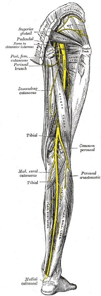
Figure 18. Nerves of the right lower limb, posterior view: the sciatic nerve dividing into the tibial and common peroneal nerves at the popliteal fossa, the common peroneal nerve coursing toward the fibula, and the medial sural cutaneous nerve contributing to the sural nerve. Gray’s Anatomy (1918), Plate 832, public domain, via Wikimedia Commons.
Part V - Acute Compartment Syndrome and Fasciotomy
Principles
The unyielding deep fascia that encloses the calf is what turns rising intracompartmental pressure into a surgical emergency, because a swollen compartment cannot expand and its perfusion fails. Hoppenfeld states two principles plainly: the diagnosis is still mainly clinical, and routine decompression of all four compartments is the current gold standard in the absence of strong evidence that a compartment is uninvolved. The classic clinical signs, by standard teaching, are pain out of proportion and pain on passive stretch, the earliest and most reliable, with paraesthesia, pallor, pulselessness, and paralysis appearing late.[23]
The two-incision fasciotomy
Hoppenfeld describes the double-incision technique, which decompresses all four compartments through an anterolateral and a posteromedial incision, each running from the level of the tibial tubercle to about 6 cm above the ankle. The anterolateral incision releases the anterior and lateral compartments through two separate fascial incisions. The posteromedial incision releases the superficial posterior compartment by incising the fascia over the soleus, and then the deep posterior compartment by lifting the soleus off the intermuscular septum and dividing that septum under direct vision. Both fascial releases must run the full length of the skin incision. (By standard teaching, a single-incision perifibular technique also exists, releasing all four compartments through one lateral incision around the fibula, but it is not described here.)[24]
The dangers of fasciotomy are as much conceptual as anatomical. The structure the extract names is the posterior neurovascular bundle, the tibial nerve and posterior tibial artery, which lies just beneath the deep-flexor septum and is injured if that septum is divided carelessly. A tourniquet is clearly contraindicated, since it would deepen the ischaemia of already-compromised tissue. The cardinal warning is that the main danger is inadequate decompression: “a compartment syndrome is not an indication for minimally invasive surgery.” By standard teaching, the superficial peroneal nerve is at risk at the anterolateral incision and the saphenous nerve and long saphenous vein at the posteromedial incision.[25]
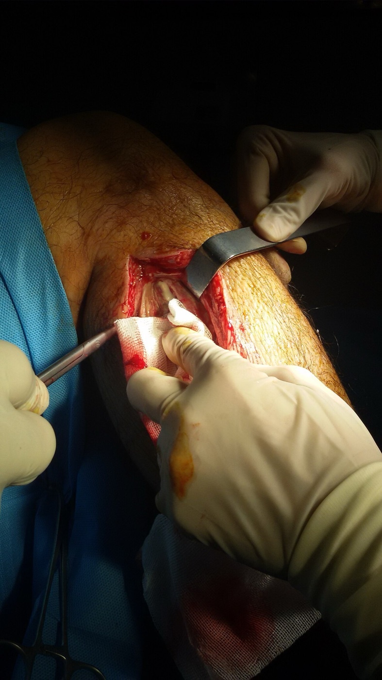
Figure 19. Intra-operative fasciotomy of the leg for compartment syndrome, decompressing a fascial compartment through a long longitudinal incision. آرمین, CC0, via Wikimedia Commons.
Part VI - Intramedullary Tibial Nailing
The approach and the entry point
Most tibial shaft fractures treated operatively are treated by intramedullary nailing, and the minimally invasive approach exists to introduce the nail. By standard teaching the nail is favoured because it is a load-sharing implant placed by a closed technique that preserves the fracture haematoma. The source notes that tibial nails are angled at their upper end to allow an anterior entry and are straight in the AP plane. The patient is positioned either on a traction table (most common, with better fracture control and easier distal locking) or with the free leg flexed over the table edge (allowing the greater flexion that eases insertion). A support behind the distal thigh must never sit in the popliteal fossa, where it would compress the popliteal vein and raise the risk of deep vein thrombosis. A 5 cm incision is made over the medial border of the patellar tendon, which is retracted laterally.[26]
The entry point is the most testable fact of the operation. On the AP view it lies in the axis of the medullary canal. On the lateral view it lies at the junction of the anterior and superior surfaces of the proximal tibia (the anterior edge of the articular surface), classically described as just medial to the lateral tibial spine. It is extrasynovial, sitting anterior to the tibial insertion of the anterior cruciate ligament and lateral to the anterior horn of the medial meniscus, and it must be confirmed on both AP and lateral imaging before entry. A wrong entry point malreduces the fracture. Too medial gives valgus and too lateral gives varus in proximal fractures, too posterior damages the ACL insertion and the anterior horn of the medial meniscus, and too anterior splinters the anterior cortex on insertion. By standard teaching, proximal-third fractures classically malreduce into valgus and apex-anterior (procurvatum) deformity, which is the rationale for semi-extended and suprapatellar techniques not described here.[27]
Anterior knee pain and the other hazards
The infrapatellar trajectory makes anterior knee pain the signature problem of tibial nailing. The infrapatellar branch of the saphenous nerve is frequently damaged, and patients should be warned that an area of numbness is likely. If the knee is not flexed beyond 90 degrees, the nail presses on the anterior patella hard enough to cause a patellofemoral compression lesion or even transient patellar subluxation with cartilage damage, which is why many surgeons prefer the free-leg position for its greater flexion. By standard teaching, anterior knee pain is the commonest complication of antegrade infrapatellar nailing, and the source supplies the anatomical substrate rather than the epidemiology. Reaming carries a risk of thermal necrosis, mitigated by sharp reamers, and by standard teaching the popliteal artery behind the proximal tibia is at risk from over-aggressive reaming or posteriorly directed drilling.[28]
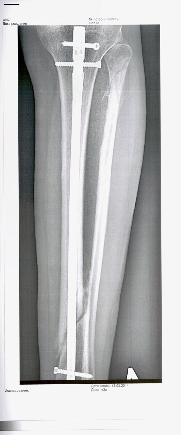
Figure 20. Tibial shaft fracture stabilised with a locked intramedullary nail and interlocking screws, anteroposterior view; the fibula is intact alongside. NiaPol, CC BY 4.0, via Wikimedia Commons.
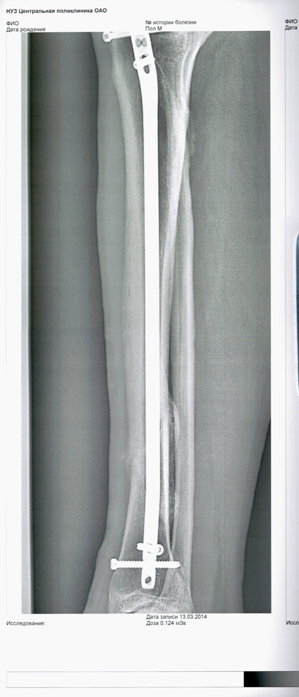
Figure 21. The same locked intramedullary tibial nail, lateral view. NiaPol, CC BY 4.0, via Wikimedia Commons.
References
-
The contrasting anatomy of the two bones, the tibia’s large subcutaneous surface accessible along its whole length, and the fibula being almost completely enclosed in muscle with close ties to the common peroneal nerve, are from Hoppenfeld, Surgical Exposures in Orthopaedics: The Anatomic Approach, 5th ed. (2016), Chapter 11 (The Tibia & Fibula), applied anatomy of the leg (Hoppenfeld p.1074). General teaching that supplements the source is flagged in-line as standard teaching and carries no page citation.
-
The recurring danger structures are drawn together from the chapter: the common peroneal nerve subcutaneous over the fibular neck, cut “if the skin incision is too bold” (Hoppenfeld p.1068); and the posterior neurovascular bundle lying just beneath the deep-flexor intermuscular septum during fasciotomy (Hoppenfeld p.1081). The clinical consequence of common peroneal nerve injury (footdrop) is standard teaching.
-
The tibia’s large subcutaneous surface and absence of major neurovascular structures on it (only the nutrient artery), the fibula being almost completely muscle-enclosed and surfacing only proximally and in the lower third at the lateral malleolus, the fibula’s close ties to the common peroneal nerve, and the deep fascia of the leg as “a tough, fibrous, unyielding structure” attached to the bony borders where they are subcutaneous are from Hoppenfeld’s applied anatomy of the leg (Hoppenfeld pp.1074-1075). The deduction that the unyielding fascia is the mechanical basis of compartment syndrome is standard teaching.
-
The four compartments, the two intermuscular septa passing from the deep fascia to the fibula and enclosing the lateral compartment, the anterior septum (anterior vs lateral), the posterior septum (lateral vs posterior, and superficial vs deep posterior), the interosseous membrane (deep posterior vs anterior), and the fascial layer between superficial and deep posterior compartments are from Hoppenfeld’s applied anatomy of the leg (Hoppenfeld pp.1075-1077).
-
The anterior (extensor) compartment’s boundaries (lateral surface of the tibia medially, extensor surface of the fibula and anterior intermuscular septum laterally, deep fascia, interosseous membrane posteriorly), its content of “the extensor muscles of the foot and ankle,” its deep peroneal nerve supply and anterior tibial artery are from Hoppenfeld (Hoppenfeld p.1075, p.1077). The individual muscle names (tibialis anterior, extensor hallucis longus, extensor digitorum longus, peroneus tertius) and the dorsiflexion/toe-extension actions are standard teaching; the extract names only “the extensor muscles of the foot and ankle.”
-
The lateral (peroneal) compartment’s boundaries (anterior septum anteriorly, posterior septum posteriorly, fibula medially), its content of “the peroneal muscles, which evert the foot,” its superficial peroneal nerve supply, and the fact that no artery runs in it (its muscles supplied “from several branches of the peroneal artery”) are from Hoppenfeld (Hoppenfeld pp.1075-1076). The individual muscle names (peroneus longus and brevis) are standard teaching; the extract says only “the peroneal muscles.”
-
The superficial posterior compartment containing gastrocnemius, soleus, and plantaris and its boundaries, and the deep posterior compartment containing tibialis posterior, flexor digitorum longus, and flexor hallucis longus plus the tibial nerve and posterior tibial artery, with its boundaries (posterior septum from the superficial compartment, interosseous membrane from the anterior compartment), are from Hoppenfeld (Hoppenfeld pp.1076-1077). The tibial-nerve supply and sural cutaneous nerve of the superficial compartment, the popliteus also belonging to the deep posterior group, the peroneal artery’s origin within the deep posterior compartment, and the plantarflexion/inversion/toe-flexion actions are standard teaching; the extract states the tibial nerve and posterior tibial artery for the deep posterior compartment but does not name the nerve of the superficial compartment.
-
That the fibula has close ties to the common peroneal nerve, that the deep peroneal nerve supplies and runs in the anterior compartment with the anterior tibial artery, that the superficial peroneal nerve supplies the lateral compartment, and that the posterior tibial neurovascular bundle (tibial nerve + posterior tibial artery) lies in the deep posterior compartment are from Hoppenfeld (Hoppenfeld pp.1074-1077, p.1081). The common peroneal nerve dividing at the fibular neck into superficial and deep peroneal nerves, the superficial peroneal nerve piercing the deep fascia in the distal third, and the anterior tibial artery passing through an opening in the proximal interosseous membrane are standard teaching, consistent with but not spelled out in the extract.
-
The anterior approach giving “safe, easy access to the medial (subcutaneous) and lateral (extensor) surfaces of the tibia,” being the preferred approach except when the skin is scarred or has draining sinuses, exposing the whole length of the bone, its indications, and the longitudinal incision ~1 cm lateral to the anterior border with the warning that a longer incision is safer than forcible retraction (poor skin vascularity) are from Hoppenfeld’s anterior approach to the tibia (Hoppenfeld pp.1049-1050).
-
The absence of an internervous plane (subcutaneous surface used directly; epiperiosteal dissection sparing the extensor compartment’s nerve supply), the rule of minimal periosteal stripping and never stripping an isolated fragment (which would become avascular), tibialis anterior as the only muscle arising from the lateral tibial surface (reflected laterally to expose it), and the medial-subcutaneous (tensile, easy to contour, but wound-breakdown risk) versus lateral (deep to muscle, soft-tissue-safer) plating trade-off are from Hoppenfeld’s anterior approach to the tibia (Hoppenfeld p.1049, p.1051).
-
The thin lower-third skin healing badly in smokers/venous insufficiency, longitudinal incisions healing well versus poorly healing transverse/irregular wounds (especially in the elderly), the long saphenous vein vulnerable during superficial dissection (“preserved for future vascular procedures, if at all possible”), and “devascularized bone… will not unite” are from Hoppenfeld (Hoppenfeld p.1053). The minimally invasive anterior approach to the distal tibia (two windows for multifragmentary distal metaphyseal fractures, used only on good soft tissues with smoking/venous insufficiency as relative contraindications, an epiperiosteal not subperiosteal plane preserving periosteal blood supply, and the saphenous nerve and long saphenous vein anterior to the medial malleolus retracted and preserved) is from Hoppenfeld (Hoppenfeld pp.1055-1059).
-
The posterolateral approach exposing the middle two-thirds of the tibia, used when the subcutaneous (anteromedial) skin is badly scarred or infected, described as “a technically demanding operation,” its uses (internal fixation, bone grafting of delayed/nonunion, exposure of the middle of the posterior fibula), the lateral decubitus position with the affected leg uppermost, and the incision over the lateral border of the gastrocnemius (minimum 10 cm) are from Hoppenfeld’s posterolateral approach to the tibia (Hoppenfeld pp.1060-1062). That the rationale is to bring healthy posterior muscle over the graft is standard teaching; the extract states the indication and the exposed posterior tibial surface.
-
The internervous plane between the peroneal muscles (superficial peroneal nerve) and the gastrocnemius/soleus/flexor hallucis longus group (tibial nerve), the deep dissection detaching the soleus and flexor hallucis longus origins from the fibula and crossing the interosseous membrane (stripping tibialis posterior) to reach the posterior tibial surface, the posterior tibial artery and tibial nerve protected by the bulk of tibialis posterior and flexor hallucis longus (stay on the interosseous membrane, never behind those muscles), the short saphenous vein at the skin-flap stage (ligate if needed) and peroneal artery branches ligated in the superficial plane, and the inability to extend into the proximal quarter (popliteus + superficial posterior tibial vessels/tibial nerve) are from Hoppenfeld’s posterolateral approach to the tibia (Hoppenfeld pp.1062-1066). The sural nerve travelling with the short saphenous vein is standard teaching.
-
The anterolateral approach offering “safe access to the lateral tibial plateau,” its indications (ORIF of lateral plateau fractures, bone grafting, osteomyelitis, tumour excision/biopsy, graft harvest), preference over the direct anterior approach (incision not over bone, less retraction), the thin/delicate proximal-tibia soft tissues prone to swelling and blistering with frequent delay of definitive surgery, the knee flexed ~60° over a wedge, the inverted L-shaped incision over Gerdy’s tubercle, and the absence of an internervous plane (epiperiosteal, sparing the extensor compartment’s nerve supply) are from Hoppenfeld’s anterolateral approach to the lateral tibial plateau (Hoppenfeld pp.1015-1018). The Schatzker I-III association is standard teaching.
-
The submeniscal arthrotomy (transverse capsulotomy just below the lateral meniscus, detaching the meniscus from its inferior attachments on stay sutures while preserving anterior and posterior attachments), the lateral meniscus at most risk “during the incision of the knee joint synovium,” the deep branch of the peroneal nerve having a variable course but normally well posterior to the dissection, and the femorotibial distractor applying a varus force to open the lateral compartment are from Hoppenfeld’s anterolateral approach to the lateral tibial plateau (Hoppenfeld pp.1018-1020). The “soft tissue attachments inferiorly” correspond to the coronary/meniscotibial ligament (standard teaching).
-
The posteromedial approach addressing the large posteromedial fragment of complex plateau fractures (reduction onto the shaft often the first stage of bicondylar fixation), the posteromedial buttress plate on the compression side preventing varus (“the most common deformity of the proximal tibia after fracture”), the usually-spared posteromedial soft tissues, its indications (Schatzker IV medial and V-VI bicondylar fractures, upper tibial osteotomy, abscess drainage, tumour biopsy), and the supine position with a contralateral-hip sandbag rolling the limb ~20° into external rotation are from Hoppenfeld’s posteromedial approach to the proximal tibia (Hoppenfeld pp.1021-1023).
-
The absence of an internervous plane (the epiperiosteal plane between bone and the medial head of gastrocnemius), the saphenous nerve and vein just anterior to the approach (identify and preserve), the two pes anserinus options (longitudinal division, simple but hard to repair under a plate, versus reflection of the sartorius/gracilis/semitendinosus tendons), the medial head of gastrocnemius freed by blunt dissection, and the extensile options (proximally to the posteromedial corner and popliteal vessels; distally to the superficial and deep posterior compartments) are from Hoppenfeld’s posteromedial approach to the proximal tibia (Hoppenfeld pp.1023-1026).
-
The posterolateral approach to the tibial plateau (for depressed posterolateral fragments, no internervous plane, between biceps femoris/common peroneal nerve laterally and the lateral head of gastrocnemius medially, elevating popliteus and detaching the soleus origin from the proximal fibula), the posterior approach to the tibial plateau as the one with a true internervous plane between semitendinosus and the medial head of gastrocnemius (safe because each is innervated away from the interval) with a posterolateral-corner blind spot, and the minimally invasive anterolateral approach to the proximal tibia (epiperiosteal/submuscular, sparing the deep peroneal nerve, with the anterior tibial artery and deep peroneal nerve in the path of distal screws around the 11th-13th holes mandating a formal open distal window) are from Hoppenfeld’s posterolateral, posterior, and minimally invasive plateau approaches (Hoppenfeld pp.1029-1048).
-
The approach to the fibula as a “classic extensile exposure” offering “access to all parts of the fibula” with the whole length exposable through one incision just posterior to the bone, its uses (partial resection for tibial osteotomy/nonunion, decompressive fibulectomy of all four compartments, tumour and osteomyelitis resection, ORIF of fibular fractures, corticocancellous strut and vascularised fibular grafts dissected with their pedicles), the lateral decubitus position, and the fibular head palpable 2-3 cm below the lateral femoral condyle are from Hoppenfeld’s approach to the fibula (Hoppenfeld pp.1067-1068).
-
The internervous plane between the peroneal muscles (superficial peroneal nerve) and the flexor muscles (tibial nerve), operationalised between the peronei and soleus, the principle that fibula-origin muscles run distally and are stripped distal-to-proximal while the interosseous membrane (oblique upward fibres) is stripped proximal-to-distal, and the rule to stay subperiosteal/close to bone to avoid the peroneal vessels (terminal branches deep to the lateral malleolus) and deep neurovascular structures are from Hoppenfeld’s approach to the fibula (Hoppenfeld p.1069, pp.1072-1073).
-
The common peroneal nerve as the chief danger (subcutaneous over the fibular neck, “rolled underneath the fingers,” cut “if the skin incision is too bold,” and at risk when incising the deep fascia), the rule that “the key to preserving the nerve is to identify it proximally as it lies on the posterior border of the biceps femoris” and trace it through the peroneal muscle mass before bone work (mobilising it by cutting the overlying peroneus longus fibres, preserving all branches, avoiding retraction if possible), and the dorsal cutaneous branch of the superficial peroneal nerve at the middle/distal-third junction (injury numbs the dorsum of the foot) are from Hoppenfeld’s approach to the fibula (Hoppenfeld pp.1068-1070). The clinical picture of a complete common peroneal palsy (footdrop) is standard teaching.
-
That the whole fibula is exposable and removable for decompression, tumour, or graft is from Hoppenfeld (Hoppenfeld p.1067, p.1068, p.1072); the lower fourth being subcutaneous is from Hoppenfeld (p.1068). The safe-resection limits - preserving the proximal fibula/head (lateral collateral ligament, biceps, common peroneal nerve; otherwise posterolateral knee instability) and the distal fibula/lateral malleolus and syndesmosis (otherwise ankle instability), and harvesting only the expendable middle shaft - are standard teaching and are not quantified in the extract.
-
That the pathophysiology and diagnosis are “beyond the scope of this book” but “diagnosis is still mainly clinical” and “routine decompression of all four compartments is the current gold standard in treatment in the absence of strong evidence that a compartment is not involved” are from Hoppenfeld’s compartment-syndrome section (Hoppenfeld p.1077). The unyielding deep fascia as the mechanical basis of the syndrome (Hoppenfeld p.1075) supports the pathophysiology. The classic “P” signs (pain out of proportion, pain on passive stretch, paraesthesia, pallor, pulselessness, paralysis) are standard teaching.
-
The double-incision fasciotomy - the anterolateral incision (tibial tubercle to 6 cm above the ankle) releasing the anterior and lateral compartments through two separate fascial incisions, and the posteromedial incision (same length) releasing the superficial posterior compartment over the soleus and then the deep posterior compartment by lifting the soleus off the intermuscular septum and dividing it under direct vision, with the rule that the fascial incision must extend the full length of the skin incision - is from Hoppenfeld (Hoppenfeld pp.1078-1081). The single-incision perifibular technique is standard teaching, not in the extract.
-
The posterior neurovascular bundle (tibial nerve + posterior tibial artery) at risk if the deep-flexor fascia is divided carelessly (lying “just beneath” the septum), the tourniquet “clearly contraindicated,” and the principles that “the main danger of the approach is inadequate decompression” and “a compartment syndrome is not an indication for minimally invasive surgery” are from Hoppenfeld (Hoppenfeld p.1081). The superficial peroneal nerve at the anterolateral incision and the saphenous nerve and long saphenous vein at the posteromedial incision are standard teaching.
-
That most operatively treated tibial shaft fractures are treated by intramedullary nailing and the minimally invasive approach is for nail insertion, the nail design (angled at the upper end for an anterior route, straight in the AP plane), the two positions (traction table, most common, with better control and easier distal locking; free leg flexed over the table end for greater flexion), the warning never to place the thigh support in the popliteal fossa (popliteal vein compression, deep vein thrombosis risk), and the 5 cm incision over the medial border of the patellar tendon (retracted laterally) are from Hoppenfeld’s minimally invasive approach for tibial nailing (Hoppenfeld pp.1082-1086). The load-sharing/haematoma-preserving rationale is standard teaching.
-
The entry point - in the axis of the medullary canal on the AP view, at the junction of the anterior and superior surfaces of the proximal tibia on the lateral view, extrasynovial, anterior to the tibial ACL insertion and lateral to the anterior horn of the medial meniscus, and confirmed on both AP and lateral imaging before entry
- and the wrong-entry malreductions (too medial → valgus, too lateral → varus in proximal fractures; too posterior → ACL insertion and anterior horn of medial meniscus; too anterior → anterior-cortex splintering) are from Hoppenfeld (Hoppenfeld p.1086, pp.1089-1091). The classic “just medial to the lateral tibial spine” landmark, and the proximal-third valgus + apex-anterior (procurvatum) malreduction pattern with the semi-extended/suprapatellar rationale, are standard teaching.
-
The infrapatellar branch of the saphenous nerve frequently damaged (warn the patient of likely numbness), the nail pressing on the anterior patella if the knee is not flexed beyond 90° causing a patellofemoral compression lesion or transient patellar subluxation with cartilage damage (driving the preference for the free-leg position), and thermal necrosis during reaming mitigated by sharp reamers are from Hoppenfeld (Hoppenfeld p.1088, pp.1082-1083, p.1091). Anterior knee pain as the commonest complication of antegrade nailing, and popliteal-artery risk during reaming/posterior drilling, are standard teaching; the extract’s only posterior-vessel caution is popliteal-vein compression by a mal-placed thigh rest.
-
The four compartments and their nerves (anterior-deep peroneal, lateral-superficial peroneal, posterior compartments-tibial), with the deep posterior compartment carrying the tibial nerve and posterior tibial artery, are from Hoppenfeld’s applied anatomy of the leg (Hoppenfeld pp.1075-1077); the tibial-nerve supply of the superficial posterior compartment is standard teaching.
-
The anterior tibial artery in the anterior compartment, the posterior tibial artery in the deep posterior compartment, and the lateral compartment having no artery (supplied by branches of the peroneal artery) are from Hoppenfeld (Hoppenfeld pp.1075-1077). The peroneal artery’s origin in the deep posterior compartment is standard teaching.
-
The absence of an internervous plane (subcutaneous surface used directly, epiperiosteal dissection), the soft-tissue/wound-breakdown danger over the thin tibial skin, and the medial-versus-lateral plating trade-off are from Hoppenfeld’s anterior approach to the tibia (Hoppenfeld p.1049, p.1051, p.1053).
-
The posterolateral plane between the peroneal muscles (superficial peroneal nerve) and the gastrocnemius/soleus/flexor group (tibial nerve), the indication (scarred/infected anteromedial skin), and the protection of the posterior tibial bundle by tibialis posterior and flexor hallucis longus (stay on the interosseous membrane) are from Hoppenfeld’s posterolateral approach to the tibia (Hoppenfeld pp.1062-1066). The “healthy muscle over the graft” rationale is standard teaching.
-
The common peroneal nerve as the chief danger of the fibular approach and the rule to identify it proximally on the posterior border of biceps femoris and trace it before bone work are from Hoppenfeld’s approach to the fibula (Hoppenfeld pp.1068-1070).
-
The distal-to-proximal stripping of fibula-origin muscles and the proximal-to-distal stripping of the interosseous membrane (because of its oblique upward fibres), with subperiosteal technique to protect the neurovascular structures, are from Hoppenfeld’s approach to the fibula (Hoppenfeld p.1069, p.1073).
-
The double-incision fasciotomy with the anterolateral incision releasing the anterior and lateral compartments and the posteromedial incision releasing the superficial and deep posterior compartments (lifting soleus off the septum and dividing it under direct vision, protecting the posterior neurovascular bundle), each fascial cut running the full skin length, is from Hoppenfeld (Hoppenfeld pp.1078-1081).
-
That diagnosis is mainly clinical, four-compartment decompression is the gold standard, the tourniquet is clearly contraindicated, and the main danger is inadequate decompression (“a compartment syndrome is not an indication for minimally invasive surgery”) are from Hoppenfeld (Hoppenfeld p.1077, p.1081).
-
The nail entry point (axis of the medullary canal on AP; junction of anterior and superior surfaces on lateral; extrasynovial, anterior to the ACL insertion and lateral to the anterior horn of the medial meniscus; confirmed on both views) is from Hoppenfeld’s tibial nailing approach (Hoppenfeld p.1086, p.1089).
-
The wrong-entry malreductions (too medial → valgus, too lateral → varus; too posterior → ACL insertion and anterior horn of medial meniscus; too anterior → anterior-cortex splintering) are from Hoppenfeld (Hoppenfeld pp.1089-1091). The valgus + apex-anterior (procurvatum) proximal-third pattern and the semi-extended/suprapatellar rationale are standard teaching.
-
The infrapatellar branch of the saphenous nerve frequently damaged and the patellofemoral compression/subluxation from insufficient flexion (mitigated by the free-leg position’s greater flexion) are from Hoppenfeld (Hoppenfeld p.1088, p.1091). Anterior knee pain as the commonest complication and the suprapatellar mitigation are standard teaching.
-
That the whole fibula is exposable and the lower fourth is subcutaneous is from Hoppenfeld (Hoppenfeld p.1068); the safe-resection limits (preserve the proximal head/neck for the lateral collateral ligament, biceps, and common peroneal nerve, and the distal malleolus/syndesmosis for the ankle mortise, harvesting only the middle shaft) are standard teaching and are not quantified in the extract.