Contents
- Orientation
- Part I - Applied Surgical Anatomy of the Hip
- Part II - Blood Supply of the Femoral Head and Acetabulum
- Part III - Anterior (Smith-Petersen) Approach and the Direct Anterior Approach
- Part IV - Anterolateral (Watson-Jones) Approach
- Part V - Lateral (Hardinge) Approach
- Part VI - Posterior (Moore / Southern) Approach
- Part VII - Medial (Ludloff) Approach
- References
Orientation
The hip can be reached from every side, and each route is defined by the muscle layer it crosses and the nerve or vessel that guards it. Hoppenfeld describes five classic exposures: the anterior (Smith-Petersen) approach and its minimally invasive variant, the anterolateral (Watson-Jones) approach, the lateral (Hardinge) approach, the posterior (Moore or Southern) approach, and the medial (Ludloff) approach. Beneath all of them sits the one fact that dominates hip surgery: the blood supply of the femoral head is precarious. It is carried mostly by the retinacular branches of the medial femoral circumflex artery, and losing it causes avascular necrosis. For that reason this summary treats the femoral head blood supply as a topic in its own right, as the konspekt requires, before working through the approaches.[1]
Each approach has its own characteristic danger. The anterior and direct anterior approaches threaten the lateral femoral cutaneous nerve; the anterolateral and lateral approaches threaten the superior gluteal nerve (and with it the hip abductors); the posterior approach threatens the sciatic nerve; the medial approach threatens the medial femoral circumflex artery and the obturator nerve. The internervous plane is the other recurring theme. It is a true plane in the anterior and direct-anterior approaches and, deeply, in the medial approach, whereas the anterolateral, lateral, and posterior approaches have no true internervous plane and instead split a muscle distal to its nerve supply.[2]
Part I - Applied Surgical Anatomy of the Hip
The muscle layers and the “pelvic deltoid”
Posteriorly the hip is covered by two muscle sheaths with the sciatic nerve running between them. The outer layer is formed by the gluteus maximus and, in continuity through the fascia lata and iliotibial tract, the tensor fasciae latae (the “pelvic deltoid” that covers the hip as the deltoid covers the shoulder); the inner layer is the short external rotators (piriformis, superior gemellus, obturator internus, inferior gemellus, quadratus femoris). Anterolaterally the relationship that matters is between the tensor fasciae latae (superficial, anterior, from the anterior outer lip of the iliac crest) and the gluteus medius (deeper, from the outer ilium between the gluteal lines). The fascia lata splits to enclose the tensor and the gluteus maximus but only covers the gluteus medius.[3]
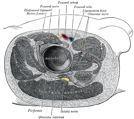
Figure 1. Transverse section through the right hip: the femoral neurovascular bundle anteriorly and the sciatic nerve posteriorly, with the two muscle layers, gluteus maximus and the short external rotators (piriformis, obturator internus). Gray’s Anatomy plate 344 (public domain), via Wikimedia Commons.
The capsule, the femoral triangle, and the bony landmarks
The hip joint capsule is reached only by parting the muscles that cross it, every one of which sends fibres into it (the iliopsoas contributes the iliocapsularis). Anterior to the joint, with the psoas interposed, lies the femoral neurovascular bundle in the femoral triangle. From medial to lateral these are the femoral Vein, Artery, and Nerve (“VAN”), the femoral nerve being the most lateral and therefore the structure most at risk from anterior retractors. The bony landmarks that orient every approach are the anterior superior iliac spine, the iliac crest, the greater trochanter (whose posterior aspect is the most palpable and muscle-free), the vastus lateralis ridge, and the pubic tubercle.[4]
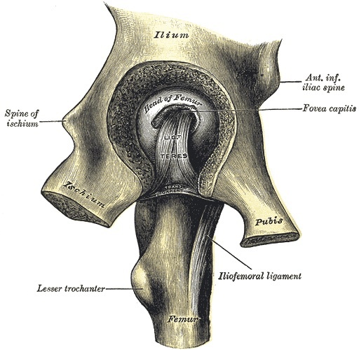
Figure 2. The osseous hip joint: the femoral head with the fovea for the ligamentum teres seated in the acetabulum, with the iliofemoral ligament and lesser trochanter. Gray’s Anatomy plate 341 (public domain), via Wikimedia Commons.
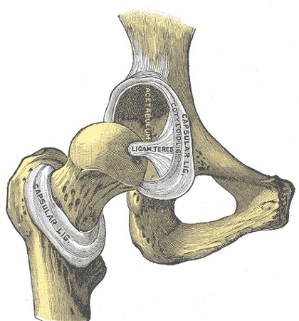
Figure 3. Hip joint, anterior view with the capsule removed, exposing the femoral head, acetabulum and ligamentum teres. Gray’s Anatomy plate 342 (public domain), via Wikimedia Commons.
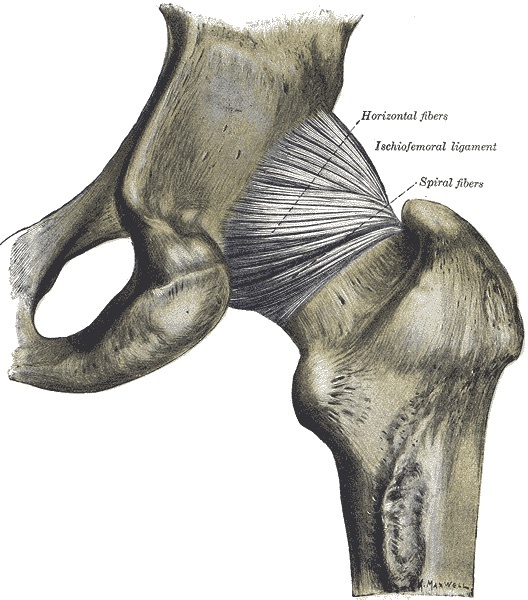
Figure 4. Posterior aspect of the hip joint showing the ischiofemoral ligament (horizontal and spiral fibres) reinforcing the capsule. Gray’s Anatomy plate 340 (public domain), via Wikimedia Commons.
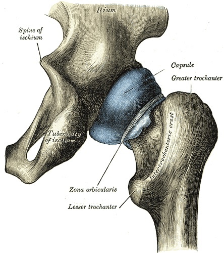
Figure 5. Distended hip-joint capsule, posterior view, demonstrating the zona orbicularis and its relation to the trochanters. Gray’s Anatomy plate 343 (public domain), via Wikimedia Commons.
Part II - Blood Supply of the Femoral Head and Acetabulum
The adult femoral head is supplied overwhelmingly by the medial femoral circumflex artery (MFCA), a branch of the profunda femoris. The MFCA runs posteriorly, deep to the quadratus femoris and between the obturator externus and the gemelli/obturator internus, and its deep branch reaches the posterosuperior femoral neck where it becomes the lateral epiphyseal (superior retinacular) vessels. Hoppenfeld’s text anchors this clinically. The quadratus femoris carries “troublesome vessels that arise from the medial circumflex artery”, so it is normally left alone (or divided only about 1 cm from its insertion), and the short external rotators in the posterior approach are detached about 1 cm from the femur; both manoeuvres preserve the MFCA. The detailed retinacular anatomy below is standard teaching, flagged as such.[5]
The supporting network is conventionally described as follows (standard teaching). At the base of the femoral neck an extracapsular arterial ring is formed mainly by the MFCA posteriorly and the lateral femoral circumflex artery anteriorly, with contributions from the superior and inferior gluteal arteries. From this ring the retinacular (ascending cervical) vessels, the arteries of Weitbrecht, run up the neck beneath the synovial reflections of the capsule to a subsynovial ring and enter the head near the articular margin. The lateral femoral circumflex artery supplies a smaller anterior and inferior share (Hoppenfeld notes its ascending branch crossing the anterior approach and its transverse branch cut in the lateral approach). The artery of the ligamentum teres (the foveolar artery, usually from the obturator artery) supplies only a small region around the fovea; it matters in children but cannot sustain the head alone in adults. A trochanteric anastomosis of the gluteal and circumflex arteries provides collateral flow.[6]
The clinical consequence ties the whole topic together. Because the head is fed mainly by retinacular vessels running along the neck, a displaced femoral neck fracture tears these vessels and produces avascular necrosis (and may also tamponade the intracapsular vessels), with the risk rising as displacement increases. This is why displaced neck fractures in the elderly are treated by arthroplasty rather than fixation. For the same reason a posterior dislocation or a poorly executed posterior approach endangers the MFCA behind the neck, and the Ganz surgical-dislocation technique is designed specifically to protect its deep branch. The acetabulum is supplied by the acetabular branch of the obturator artery (entering the acetabular notch with the ligamentum teres) together with gluteal and circumflex contributions.[7]
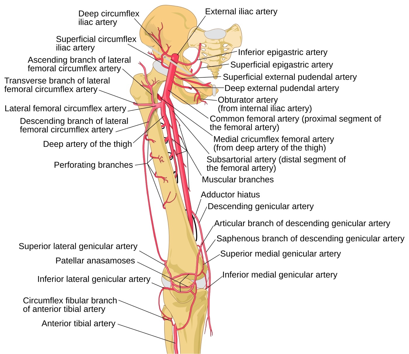
Figure 6. Arterial supply around the proximal femur: the medial and lateral femoral circumflex arteries (from the profunda femoris) forming the extracapsular ring that is the dominant blood supply to the femoral head and neck. Mcstrother, CC BY 3.0, via Wikimedia Commons.
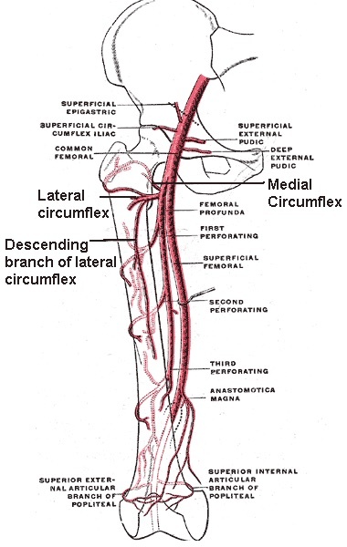
Figure 7. The circumflex femoral arteries on the proximal femur: the medial and lateral femoral circumflex arteries, the principal contributors to the retinacular supply of the femoral head. Mikael Häggström (public domain), via Wikimedia Commons.
Part III - Anterior (Smith-Petersen) Approach and the Direct Anterior Approach
The classic Smith-Petersen approach
The anterior approach gives safe access to the hip and ilium, though it exposes the acetabulum less completely than other routes unless muscle is extensively stripped. Its uses are open reduction of a congenital hip dislocation (when the head lies anterosuperior to the true acetabulum), synovial biopsy, intra-articular fusion, total hip replacement, hemiarthroplasty, tumour excision, and pelvic osteotomy. The patient is supine. The incision follows the anterior half of the iliac crest to the anterior superior iliac spine, then turns vertically down toward the lateral patella. It uses a single internervous plane at two levels: superficially between the sartorius (femoral nerve) and the tensor fasciae latae (superior gluteal nerve), and deeply between the rectus femoris (femoral nerve) and the gluteus medius (superior gluteal nerve).[8]
The superficial interval is best found 4-5 cm below the anterior superior iliac spine, and the deep fascia is incised on the medial side of the tensor fasciae latae. That keeps the dissection within the tensor’s sheath and protects the lateral femoral cutaneous nerve, which runs over the sartorius fascia. The ascending branch of the lateral femoral circumflex artery crosses the interval and is ligated. Deep, the rectus femoris is detached from both heads (the direct head from the anterior inferior iliac spine, the reflected head from the rim of the acetabulum and the anterior capsule), the iliocapsularis fibres of the iliopsoas are released from the capsule, and the capsule is opened longitudinally or as a T, the hip dislocating on external rotation.[9]
The central danger is the lateral femoral cutaneous nerve, which crosses the field about 2.5 cm below the anterior superior iliac spine, usually passing over the sartorius. It is very variable, often with three or more branches, and injury gives a painful neuroma and numbness over the lateral thigh (meralgia paraesthetica). The femoral nerve sits well medial to the rectus femoris in the femoral triangle and is safe unless the surgeon strays far out of plane; the femoral pulse, which lies well medial, is the landmark that confirms the plane. The approach extends proximally onto the ilium (for pelvic osteotomy, with care for the sciatic nerve at the notch) and distally between the vastus lateralis and rectus femoris to the whole femoral shaft.[10]
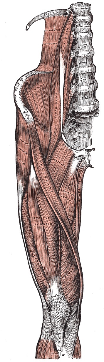
Figure 8. Muscles of the iliac and anterior femoral regions: sartorius, tensor fasciae latae, rectus femoris and iliopsoas, defining the intervals of the anterior (Smith-Petersen) and anterolateral (Watson-Jones) approaches. Gray’s Anatomy plate 430 (public domain), via Wikimedia Commons.
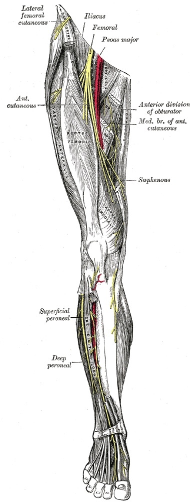
Figure 9. Anterior nerves of the lower limb: the lateral femoral cutaneous nerve crossing near the anterior superior iliac spine, where it is vulnerable in the anterior approach, with the femoral and obturator nerves. Gray’s Anatomy plate 827 (public domain), via Wikimedia Commons.
The minimally invasive (direct) anterior approach
The minimally invasive anterior approach (the Hueter interval, the modern direct anterior approach) uses the same true internervous plane (superficially sartorius versus tensor fasciae latae, deeply rectus femoris versus gluteus medius and minimus) through a short incision, and is used almost exclusively for total hip arthroplasty. Because exposure is limited, an image intensifier checks the femoral neck osteotomy and component position, which the supine position makes easy. The dangers match the open anterior approach: the lateral femoral cutaneous nerve (protected by staying in the tensor’s fascial sheath), the lateral femoral circumflex vessels, and the femoral nerve. If the surgeon loses the plane, the operation is converted to the classic anterior approach rather than risking a vital structure.[11]
Part IV - Anterolateral (Watson-Jones) Approach
The anterolateral approach, popularised by Watson-Jones, is a common approach for joint replacement. It combines good acetabular exposure with safe femoral reaming, and is used for total hip replacement, hemiarthroplasty, ORIF of femoral neck fractures, and synovial or femoral-neck biopsy. The patient is supine at the edge of the table, tilted away so the buttock falls back (remember the tilt when orienting the acetabular component). It exploits the interval between the tensor fasciae latae and the gluteus medius, but this is not a true internervous plane, because both muscles are supplied by the superior gluteal nerve. It is safe only because the nerve enters the tensor near the iliac crest, so the interval must not be developed proximally to the muscles’ origins.[12]
To expose the joint the abductor mechanism is neutralised so the femur can fall back and adduct for reaming, either by a trochanteric osteotomy (reflecting the gluteus medius and minimus on the trochanter) or by detaching the anterior part of the gluteus medius and the whole gluteus minimus. The dangers are the femoral nerve (the most lateral structure of the bundle and the one closest to the field, usually a compression neurapraxia from over-medial retraction), the superior gluteal nerve (which limits proximal splitting), and the femoral vessels and profunda femoris (pierced by retractors that stray off the bone into the iliopsoas, sometimes with occult retroperitoneal bleeding; the anterior retractor sits at 1 o’clock for a right hip and 11 o’clock for a left). One particular hazard is femoral shaft fracture during forced external rotation at dislocation or adduction for reaming, especially if the fascia lata was incised too far anteriorly, so an adequate capsulotomy is done first and the fascia lata is incised at the posterior border of the trochanter.[13]
Part V - Lateral (Hardinge) Approach
The lateral, direct lateral, or transgluteal approach (Hardinge) gives excellent exposure for joint replacement while avoiding a trochanteric osteotomy and preserving the bulk of the gluteus medius, which allows early mobilisation. It is a common approach for total hip replacement, hemiarthroplasty, and femoral neck fixation. The patient is supine with the trochanter at the table edge. The incision is centred on the tip of the greater trochanter, beginning 5 cm above it and running about 8 cm down the shaft. There is no true internervous plane: the gluteus medius is split in the line of its fibres distal to its nerve supply, and the vastus lateralis is split lateral to its femoral supply, the two joined into a single anterior myofascial sleeve (the anterior third of the gluteus medius with the gluteus minimus and the anterior vastus lateralis) reflected off the trochanter to expose the anterior capsule.[14]
The critical danger is the superior gluteal nerve, which runs between the gluteus medius and minimus about 3-5 cm above the upper border of the trochanter. The operative rule is therefore not to split the gluteus medius more than 3 cm above the trochanter, with a stay suture placed at the apex of the split so it cannot propagate. Injury denervates the abductors and produces a Trendelenburg gait, which is why the approach cannot be usefully extended proximally. Anteriorly the femoral nerve and vessels are at risk from retractors that leave the bone, and the transverse branch of the lateral femoral circumflex artery is cauterised as the vastus lateralis is mobilised. The approach extends distally by splitting the vastus lateralis to the whole femur.[15]
Part VI - Posterior (Moore / Southern) Approach
The posterior approach is the most common and practical exposure of the hip. Popularised by Moore and often called the Southern approach, it is quick, needs only one assistant, spares the abductor mechanism, and gives excellent femoral-shaft access for revision arthroplasty. Its uses are hemiarthroplasty, total and revision hip replacement, ORIF of posterior acetabular fractures, dependent drainage of sepsis, removal of loose bodies, pedicle bone grafting, and open reduction of posterior dislocations. Its one drawback is that dividing the posterior capsule predisposes to posterior dislocation (on flexion, adduction, and internal rotation), with a higher dislocation rate than anterior approaches in elderly bedridden patients. The patient is in the lateral position. A curved incision is centred on the posterior greater trochanter, the proximal limb in the line of the gluteus maximus fibres (pointing toward the posterior superior iliac spine). There is no true internervous plane: the gluteus maximus is split in the line of its fibres, which does not denervate it because its supply enters well medially.[16]
The gluteus maximus is split bluntly (its gluteal-artery branches ramify like spokes and bleed), revealing the short external rotators with the sciatic nerve lying on them in fat. The nerve is palpated but not dissected out, the hip is internally rotated to draw the field away from it, and the piriformis and obturator internus are tagged. The rotators are then detached about 1 cm from the femur and reflected back over the sciatic nerve to shield it. The quadratus femoris is left alone (its medial femoral circumflex vessels supply the head), the capsule is opened, and the hip is dislocated by internal rotation. The central danger is the sciatic nerve, injured by the posterior retractor blade or by failure to protect it during reduction of the prosthetic head. It may divide into tibial and peroneal components within the pelvis (so a “small-looking” nerve means the other branch must be found), the peroneal division being the more vulnerable by standard teaching, and a postoperative common peroneal palsy is localised by an EMG of the short head of biceps femoris. The inferior gluteal artery, if torn, retracts into the pelvis and is controlled by turning the patient supine and ligating the internal iliac artery.[17]
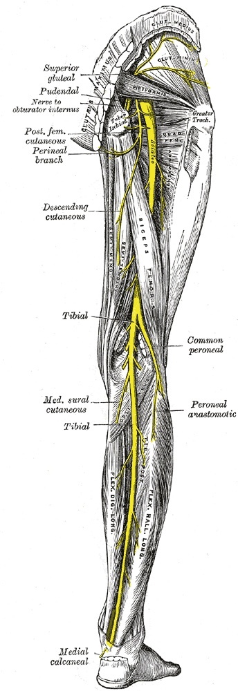
Figure 10. Posterior nerves of the lower limb: the sciatic nerve emerging below piriformis and the superior gluteal nerve, the structures at risk in the posterior (Moore) and lateral (Hardinge) approaches respectively. Gray’s Anatomy plate 832 (public domain), via Wikimedia Commons.
Part VII - Medial (Ludloff) Approach
The medial approach, attributed to Ludloff, reaches the hip from the inferomedial side through the adductor compartment. It is principally a children’s approach for open reduction of a congenital (developmental) hip dislocation, because it gives an excellent view of the psoas tendon that blocks reduction. Its other uses are biopsy of inferomedial femoral-neck and proximal-shaft lesions, psoas release, and obturator neurectomy. The patient is supine with the hip flexed, abducted, and externally rotated, the sole of the foot against the opposite knee. The incision runs down over the adductor longus from 3 cm below the pubic tubercle. The plane is staged: superficially between the adductor longus and gracilis (both anterior-obturator-supplied, so not a true internervous plane but safe, the nerves entering proximally), and deeply between the adductor brevis (anterior division of the obturator nerve) and the adductor magnus (posterior division, plus an ischial part from the tibial nerve), a true internervous plane leading to the lesser trochanter and psoas tendon.[18]
The adductor brevis is the key landmark. It is sandwiched between the two divisions of the obturator nerve, the anterior division on its anterior surface and the posterior division behind it on the adductor magnus, an arrangement that makes this the approach for obturator neurectomy in adductor spasticity. The critical danger is the medial femoral circumflex artery, which passes around the medial side of the distal psoas tendon. It is at risk, especially in children, if the psoas is detached without first isolating the tendon and cutting it under direct vision, and its injury causes avascular necrosis of the femoral head. The obturator nerve divisions are themselves protected (or deliberately cut for spasticity), the posterior division being preserved where possible to keep the adductor magnus working. The approach is rarely extended.[19]
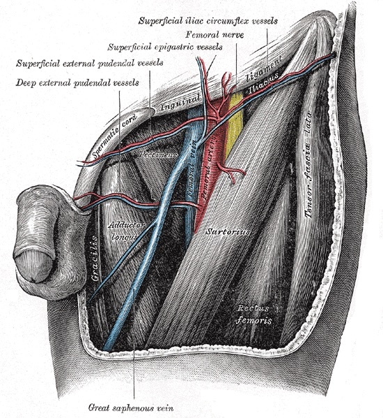
Figure 11. The femoral triangle: the femoral nerve, artery and vein bounded by sartorius and adductor longus, with the adductor and gracilis muscles relevant to the medial (Ludloff) approach. Gray’s Anatomy plate 549 (public domain), via Wikimedia Commons.
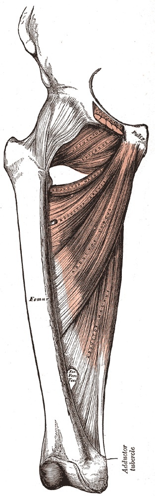
Figure 12. Deep muscles of the medial thigh: adductor brevis and magnus and obturator externus, the planes traversed in the medial (Ludloff) approach. Gray’s Anatomy plate 433 (public domain), via Wikimedia Commons.
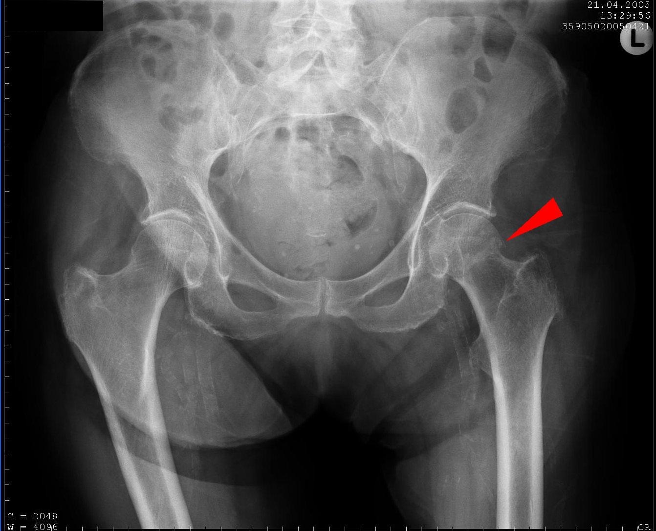
Figure 13. Non-displaced subcapital femoral neck fracture (arrow) on an AP pelvic radiograph, the injury that tears the retinacular vessels and risks avascular necrosis. Hellerhoff, CC BY-SA 3.0, via Wikimedia Commons.
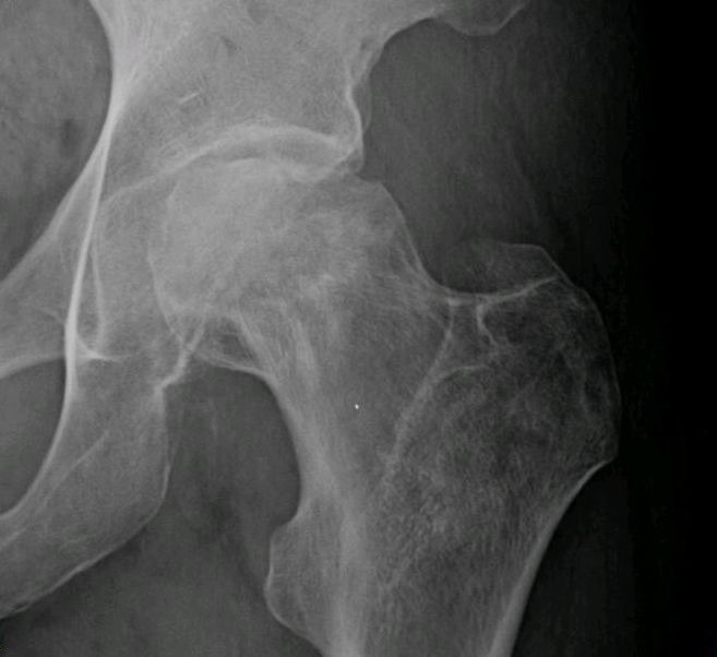
Figure 14. Avascular necrosis of the femoral head: radiograph showing subchondral collapse. Jmarchn, CC BY-SA 3.0, via Wikimedia Commons.
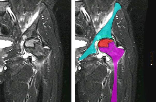
Figure 15. Avascular necrosis of the femoral head on coronal MRI (necrotic segment highlighted). Jmarchn, CC BY-SA 3.0, via Wikimedia Commons.
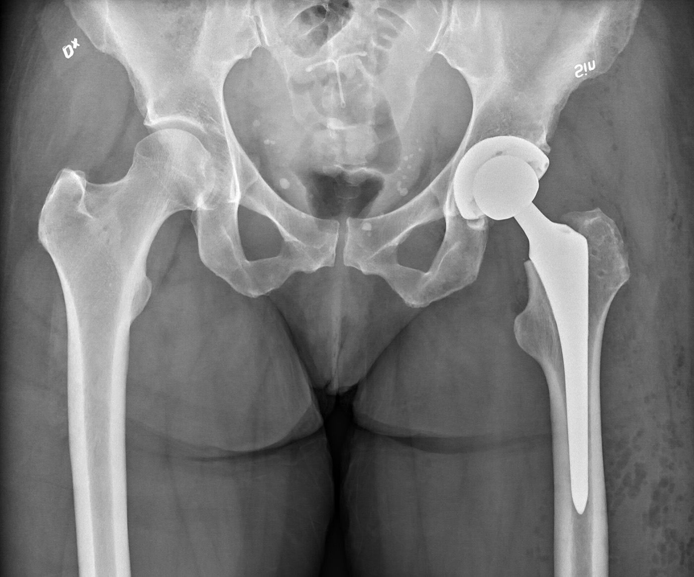
Figure 16. Total hip replacement: AP pelvic radiograph of a left total hip arthroplasty. Mikael Häggström, CC0, via Wikimedia Commons.
References
-
Hoppenfeld ch.8 describes the anterior (Smith-Petersen) approach (p.733) and its minimally invasive variant (p.754), the anterolateral (Watson-Jones) approach (p.763), the lateral (Hardinge) approach (p.780), the posterior (Moore/Southern) approach (p.800), and the medial (Ludloff) approach (p.820). The femoral head’s dependence on the medial femoral circumflex artery and the resulting risk of avascular necrosis is the cross-cutting theme; the detailed retinacular supply is largely standard teaching (the extract gives the circumflex branches and the MFCA’s relation to quadratus femoris).
-
Characteristic dangers: lateral femoral cutaneous nerve (anterior/direct anterior), superior gluteal nerve and the abductors (anterolateral/lateral), sciatic nerve (posterior), and the medial femoral circumflex artery and obturator nerve (medial); true internervous planes exist in the anterior, direct-anterior, and (deep) medial approaches, while the anterolateral, lateral, and posterior approaches split a muscle distal to its nerve supply (Hoppenfeld p.735, p.766, p.781, p.801, p.821-823).
-
Posteriorly two layers with the sciatic nerve between them: outer = gluteus maximus + (via the iliotibial tract) tensor fasciae latae (the “pelvic deltoid”), inner = the short external rotators (piriformis, superior gemellus, obturator internus, inferior gemellus, quadratus femoris) (Hoppenfeld p.811); anterolaterally the tensor fasciae latae is superficial/anterior and the gluteus medius deeper, the fascia lata enclosing the tensor and gluteus maximus but only covering the gluteus medius (Hoppenfeld p.789-790).
-
The capsule is reached by parting the muscles crossing the hip, all of which send fibres into it (iliopsoas → iliocapsularis) (Hoppenfeld p.798-799, p.742); anterior to the joint, with psoas interposed, the femoral vein, artery, and nerve lie medial-to-lateral (“VAN”), the femoral nerve most lateral and most at risk from anterior retractors (Hoppenfeld p.799); landmarks = ASIS, iliac crest, greater trochanter (posterior aspect most palpable), vastus lateralis ridge, pubic tubercle (Hoppenfeld p.781, p.791-793, p.801, p.814).
-
The MFCA (a branch of the profunda femoris) is the dominant adult femoral-head supply, running deep to quadratus femoris and between obturator externus and the gemelli/obturator internus, its deep branch becoming the lateral epiphyseal/superior retinacular vessels at the posterosuperior neck (standard teaching); Hoppenfeld grounds this in the quadratus femoris carrying “troublesome vessels that arise from the medial circumflex artery” (left alone or divided ~1 cm from insertion, p.804, p.810) and the short external rotators detached ~1 cm from the femur in the posterior approach (p.804).
-
Standard teaching: an extracapsular arterial ring at the neck base (MFCA posteriorly, LFCA anteriorly, with superior/inferior gluteal contributions) gives the retinacular (ascending cervical) vessels of Weitbrecht, which ascend the neck beneath the synovial reflections to a subsynovial ring and enter the head near the articular margin; the LFCA supplies the anterior/inferior head (Hoppenfeld documents its ascending branch crossing the anterior approach, p.795-796, and its transverse branch cut in the lateral approach, p.788); the artery of the ligamentum teres (foveolar, usually from the obturator artery) supplies a small foveal region (important in children, insufficient in adults); a trochanteric anastomosis of the gluteal and circumflex arteries gives collateral flow.
-
Standard teaching: a displaced femoral neck fracture tears the retinacular vessels → avascular necrosis (and may tamponade the intracapsular hematoma), the risk rising with displacement, which is why displaced elderly neck fractures are treated by arthroplasty (the indications Hoppenfeld lists, p.800; he corroborates the vascular logic by noting the quadratus femoris pedicle’s use for neck-fracture nonunion, p.810); a posterior dislocation or posterior approach endangers the MFCA behind the neck, and the Ganz surgical dislocation protects its deep branch; the acetabulum is supplied by the acetabular branch of the obturator artery (via the acetabular notch with the ligamentum teres) plus gluteal and circumflex contributions.
-
The anterior (Smith-Petersen) approach gives safe access to the hip and ilium but exposes the acetabulum less completely unless muscle is stripped; uses = open reduction of congenital dislocation (head anterosuperior), synovial biopsy, intra-articular fusion, THA, hemiarthroplasty, tumour excision, pelvic osteotomy; supine, incision along the anterior iliac crest to the ASIS then vertically toward the lateral patella; one internervous plane at two levels: superficial sartorius (femoral nerve)/tensor fasciae latae (superior gluteal nerve), deep rectus femoris (femoral nerve)/gluteus medius (superior gluteal nerve) (Hoppenfeld p.733-735).
-
Find the superficial interval 4-5 cm below the ASIS and incise the deep fascia on the medial side of the tensor fasciae latae (staying in the tensor’s sheath protects the lateral femoral cutaneous nerve, which runs over the sartorius fascia); ligate the ascending branch of the lateral femoral circumflex artery crossing the interval; deep, detach the rectus femoris from its direct head (anterior inferior iliac spine) and reflected head (acetabular rim and anterior capsule), release the iliocapsularis from the capsule, and open the capsule longitudinally or as a T, dislocating on external rotation (Hoppenfeld p.736, p.741-742).
-
The central danger is the lateral femoral cutaneous nerve, crossing ~2.5 cm below the ASIS usually over the sartorius, very variable (often ≥3 branches), injury giving a painful neuroma and lateral-thigh numbness (meralgia paraesthetica, standard-teaching term); the femoral nerve is well medial to the rectus femoris and safe unless far out of plane, the medial femoral pulse confirming the plane; the approach extends proximally onto the ilium (sciatic-nerve care at the notch) and distally in the vastus lateralis/rectus femoris interval to the whole femur (Hoppenfeld p.742-744, p.752-753).
-
The minimally invasive (direct) anterior approach uses the same true internervous plane (sartorius/tensor fasciae latae superficially, rectus femoris/gluteus medius and minimus deeply) through a short incision, almost exclusively for THA, with an image intensifier to check the neck osteotomy and component position; dangers = the lateral femoral cutaneous nerve (stay in the tensor’s sheath), the lateral femoral circumflex vessels, and the femoral nerve; convert to the classic approach if the plane is lost (Hoppenfeld p.754-763). The “Hueter interval” / “direct anterior approach” eponyms are standard teaching.
-
The anterolateral (Watson-Jones) approach combines good acetabular exposure with safe femoral reaming; uses = THA, hemiarthroplasty, ORIF of femoral neck fractures, synovial/femoral-neck biopsy; supine at the table edge, tilted away (remember the tilt when orienting the acetabular component); it uses the tensor fasciae latae/gluteus medius interval, which is NOT a true internervous plane (both are superior-gluteal-supplied), safe only if not developed proximally to the iliac origins (the superior gluteal nerve enters the tensor near the crest) (Hoppenfeld p.763-766).
-
The abductor mechanism is neutralised by a trochanteric osteotomy or by detaching the anterior gluteus medius and the whole gluteus minimus; dangers = the femoral nerve (most lateral, compression neurapraxia from over-medial retraction), the superior gluteal nerve (limits proximal splitting), and the femoral vessels/profunda femoris (retractors straying off bone into iliopsoas, with possible occult retroperitoneal bleeding; anterior retractor at 1 o’clock for a right hip, 11 o’clock for a left); femoral shaft fracture occurs on forced external rotation at dislocation or adduction for reaming, especially if the fascia lata was incised too anteriorly, so do an adequate capsulotomy first and incise the fascia lata at the posterior trochanter (Hoppenfeld p.763-778).
-
The lateral/direct lateral/transgluteal (Hardinge) approach gives excellent exposure for joint replacement, avoids trochanteric osteotomy, preserves gluteus medius bulk and allows early mobilisation; uses = THA, hemiarthroplasty, femoral neck fixation; supine with the trochanter at the table edge, incision centred on the trochanter tip from 5 cm above to ~8 cm down the shaft; no true internervous plane (gluteus medius split distal to its nerve supply, vastus lateralis split lateral to its femoral supply), the two joined into a single anterior myofascial sleeve (anterior gluteus medius + gluteus minimus + anterior vastus lateralis) reflected off the trochanter (Hoppenfeld p.780-782). The Hardinge eponym is standard teaching.
-
The critical danger is the superior gluteal nerve, between the gluteus medius and minimus ~3-5 cm above the upper border of the trochanter, so do not split the gluteus medius more than 3 cm above the trochanter and place a stay suture at the apex; injury denervates the abductors → Trendelenburg gait, so the approach cannot be extended proximally; anteriorly the femoral nerve and vessels are at risk from retractors off the bone, and the transverse branch of the lateral femoral circumflex artery is cauterised; extend distally by splitting the vastus lateralis (Hoppenfeld p.782-783, p.788, p.798).
-
The posterior (Moore/Southern) approach is the most common hip exposure: quick, one assistant, spares the abductors, excellent femoral-shaft access for revision; uses = hemiarthroplasty, THA/revision, ORIF of posterior acetabular fractures, drainage of sepsis, loose-body removal, pedicle bone grafting, open reduction of posterior dislocations; dividing the posterior capsule predisposes to posterior dislocation (flexion, adduction, internal rotation), with a higher dislocation rate in elderly bedridden patients; lateral position, curved incision centred on the posterior trochanter with the proximal limb in the gluteus maximus fibres (toward the PSIS, standard teaching); no true internervous plane (gluteus maximus split, supply enters medially) (Hoppenfeld p.800-801).
-
Split the gluteus maximus bluntly (its gluteal-artery branches ramify like spokes and bleed), revealing the short external rotators with the sciatic nerve on them in fat; palpate but do not dissect out the nerve; internally rotate the hip to draw the field away; tag the piriformis and obturator internus and detach the rotators ~1 cm from the femur, reflecting them over the sciatic nerve; leave the quadratus femoris alone (its medial femoral circumflex vessels supply the head); open the capsule and dislocate by internal rotation; the central danger is the sciatic nerve (posterior retractor blade; protect during prosthetic-head reduction; may divide into tibial+peroneal in the pelvis so find the other branch; peroneal division more vulnerable; common peroneal palsy localised by EMG of the short head of biceps); the inferior gluteal artery, if torn, retracts into the pelvis (turn supine, ligate the internal iliac artery) (Hoppenfeld p.801-809, p.818-819).
-
The medial (Ludloff) approach reaches the hip inferomedially through the adductor compartment, principally a children’s approach for open reduction of congenital/developmental hip dislocation (excellent psoas-tendon exposure), also for inferomedial femoral-neck/proximal-shaft biopsy, psoas release, and obturator neurectomy; supine, hip flexed/abducted/externally rotated with the foot against the opposite knee, incision over the adductor longus from 3 cm below the pubic tubercle; staged plane: superficial adductor longus/gracilis (both anterior-obturator-supplied, not a true internervous plane but safe) and deep adductor brevis (anterior division)/adductor magnus (posterior division + ischial part from the tibial nerve), a true internervous plane to the lesser trochanter and psoas tendon (Hoppenfeld p.820-823, p.829).
-
The adductor brevis is sandwiched between the two obturator divisions (anterior on its surface, posterior behind it on the adductor magnus), making this the approach for obturator neurectomy in adductor spasticity; the critical danger is the medial femoral circumflex artery passing around the medial side of the distal psoas tendon, at risk especially in children if the psoas is detached without isolating and cutting the tendon under direct vision, its injury causing femoral-head avascular necrosis (Hoppenfeld p.823, p.826, p.831; the explicit AVN consequence and the profunda femoris parentage are standard teaching); preserve the posterior obturator division to keep the adductor magnus working; the approach is rarely extended (p.823, p.826).
-
The adult head is supplied mainly by the MFCA (deep branch → lateral epiphyseal/superior retinacular vessels along the posterosuperior neck), with lesser LFCA and foveolar (ligamentum teres) supply; a displaced neck fracture tears the retinacular vessels → AVN, the risk rising with displacement, hence arthroplasty for displaced elderly fractures (standard teaching; Hoppenfeld grounds the MFCA in its relation to quadratus femoris, p.804, p.810).
-
Anterior approach: superficial plane sartorius (femoral nerve)/tensor fasciae latae (superior gluteal nerve), deep plane rectus femoris (femoral nerve)/gluteus medius (superior gluteal nerve) (Hoppenfeld p.735).
-
Find the interval 4-5 cm below the ASIS and incise the deep fascia on the medial side of the tensor fasciae latae, staying in its sheath (the nerve runs over the sartorius fascia); the nerve crosses ~2.5 cm below the ASIS, usually over the sartorius, very variable; injury → meralgia paraesthetica (Hoppenfeld p.736, p.741, p.743-744).
-
The Watson-Jones interval (tensor fasciae latae/gluteus medius) is not internervous because both are superior-gluteal-supplied; it is safe only if not developed proximally to the iliac origins, since the superior gluteal nerve enters the tensor near the crest (Hoppenfeld p.766).
-
The superior gluteal nerve runs between gluteus medius and minimus ~3-5 cm above the trochanter; do not split the gluteus medius more than 3 cm above the trochanter and place a stay suture at the apex; injury → abductor weakness/Trendelenburg gait, so no proximal extension (Hoppenfeld p.782-783).
-
The posterior (Moore/Southern) approach spares the abductors and gives excellent femoral-shaft access for revision; its drawback is posterior dislocation (flexion, adduction, internal rotation) from dividing the posterior capsule, with a higher dislocation rate, especially in the elderly bedridden (Hoppenfeld p.800).
-
Palpate but do not dissect out the sciatic nerve; internally rotate the hip; tag the piriformis and obturator internus and detach the rotators ~1 cm from the femur, reflecting them over the nerve; keep retractors on the cut rotators and protect the nerve during prosthetic-head reduction (Hoppenfeld p.803-804, p.808, p.818).
-
The short external rotators are detached ~1 cm from the femur and the quadratus femoris is left alone (it carries the medial circumflex vessels) to protect the medial femoral circumflex artery, the dominant femoral-head supply, and avoid AVN (Hoppenfeld p.804, p.810; the dominant-supply/AVN rationale is standard teaching).
-
A post-posterior-approach common peroneal palsy is localised by EMG of the short head of biceps femoris (the only thigh muscle supplied by the peroneal division): denervated → pelvis/hip lesion, spared → fibular-neck lesion (Hoppenfeld p.819).
-
Medial approach: superficial adductor longus/gracilis (both anterior-obturator, not a true internervous plane but safe) and deep adductor brevis (anterior division)/adductor magnus (posterior division + ischial part from the tibial nerve), a true internervous plane to the lesser trochanter and psoas tendon (Hoppenfeld p.821-823).
-
The medial femoral circumflex artery passes around the medial side of the distal psoas tendon and is at risk, especially in children, if the psoas is detached without isolating and cutting the tendon under direct vision; injury causes femoral-head AVN (Hoppenfeld p.826, p.828; the dominant-supply/AVN statements are standard teaching).
-
True internervous planes: anterior and direct-anterior (femoral vs superior gluteal) and the deep medial (adductor brevis vs adductor magnus); no true internervous plane: Watson-Jones (two superior-gluteal muscles), Hardinge (split gluteus medius and vastus lateralis distal to their supplies), and posterior (split gluteus maximus, nerve entering medially) (Hoppenfeld p.735, p.766, p.781, p.801, p.823).