Contents
- Orientation
- Part I - Applied Surgical Anatomy of the Hand and Wrist
- Skin, creases, and the cardinal incision rule
- The palmar aponeurosis and the digital neurovascular bundle
- The carpal tunnel
- The median nerve and its two variable branches
- Guyon’s canal and the ulnar nerve
- The intrinsic muscles and their nerve supply
- The palmar arches and the dorsal extensor compartments
- The extensor expansion
- Part II - Dorsal Approach to the Wrist
- Part III - Volar Approaches to the Wrist
- Part IV - Approaches to the Flexor Tendons and Digits
- Part V - Approaches to the Scaphoid
- Part VI - Drainage of Hand Infections
- Part VII - Z-Plasty
- References
Orientation
Nowhere else in the body are anatomy, incision design, and outcome bound together so tightly. A scar placed across a flexion crease at a right angle contracts the finger; a flexor tendon repaired in the wrong zone fails; a carpal tunnel released without respect for the recurrent motor branch leaves the thumb without opposition. Hoppenfeld’s chapter on the wrist and hand is therefore as much about where NOT to cut as where to cut. This summary works through the dorsal and volar approaches to the wrist, the approaches to the flexor tendons and the digits, the two approaches to the scaphoid, the drainage of the hand’s characteristic deep infections, and finally the Z-plasty, the geometric trick that lengthens a contracted scar.[1]
Three structures dominate the dangers. On the volar side it is the median nerve and its two unpredictable branches: the recurrent (thenar) motor branch, protected by releasing the carpal tunnel on the ulnar side of the nerve, and the palmar cutaneous branch, protected by keeping the incision ulnar to palmaris longus and angling it across the wrist. On the radial and dorsal side it is the superficial radial nerve and the posterior interosseous nerve’s territory, with the radial artery in the anatomical snuffbox lying directly on the scaphoid. In the fingers it is the digital neurovascular bundles, bracketed by Cleland’s and Grayson’s ligaments, that govern where every incision may safely run.[2]
One theme worth fixing at the outset: the hand resists exposure because its tendons are confined in tight fibro-osseous tunnels and its skin is inelastic and tethered. The flexor tendons run through the carpal tunnel and then the digital fibrous sheath with its pulleys; the extensors run through six dorsal compartments. Open one of these tunnels and the price is potential adhesion, so the recurring discipline is to preserve sheaths and pulleys (A2 and A4 above all), raise thick skin flaps, and keep dissection in bloodless internervous-equivalent planes.[3]
Part I - Applied Surgical Anatomy of the Hand and Wrist
Skin, creases, and the cardinal incision rule
Palmar skin is tough and almost immobile, tethered to the palmar aponeurosis by fibrous bands that divide the subcutaneous fat into pressure-resistant loculi; it is so inelastic that even small defects need V-Y advancement or grafting. The governing surgical rule follows from the flexion creases: to avoid flexion contractures, a flexure crease must never be crossed at 90°. Cutting within a crease avoids contracture but heals poorly, which is why so many hand incisions parallel the creases or zig-zag across them. The dorsal skin is the opposite, thin and mobile with poor blood supply, carrying the venous return of the hand.[4]
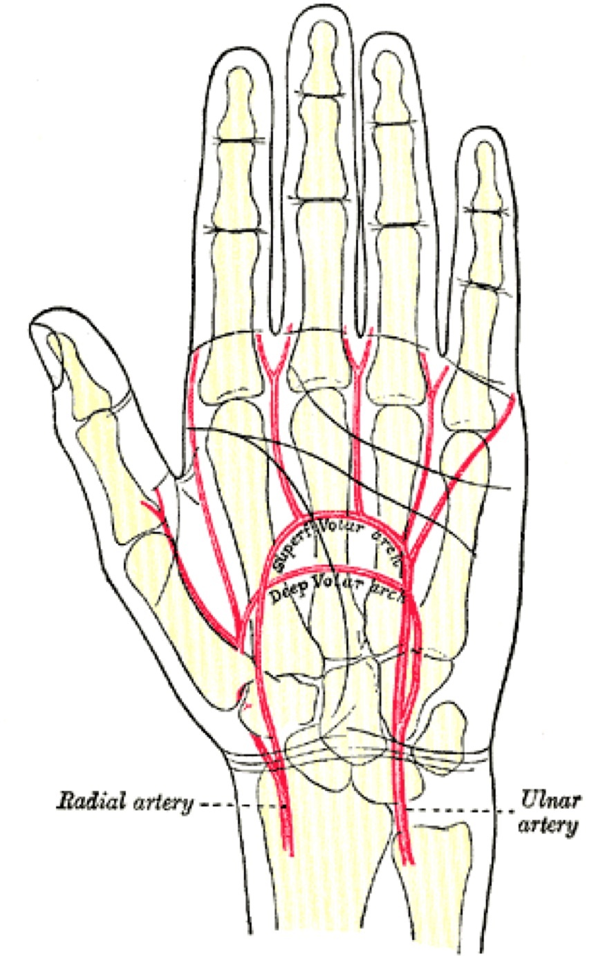
Figure 1. Surface markings and flexion creases of the palm and wrist, the landmarks that govern incision placement (a flexion crease must not be crossed at a right angle). Gray’s Anatomy plate 1237 (public domain), via Wikimedia Commons.
The palmar aponeurosis and the digital neurovascular bundle
The palmar aponeurosis is continuous with the palmaris longus tendon and fans into four digital bands; the palmar nerves and vessels lie immediately deep to it, in contact with its deep surface, which is why blunt dissection in the line of its fibres is used to reach the deep palm. In Dupuytren contracture this fascia thickens and encloses the digital nerves. In the finger the digital neurovascular bundle is bracketed by two ligaments: Grayson’s ligament lies volar to the bundle (named by Hoppenfeld, and the layer opened to expose the bundle) and Cleland’s ligament lies dorsal to it; together they tether the bundle during flexion. The digital nerves lie lateral to the flexor tendons, with the artery on the dorsal side of the nerve in the digit.[5]
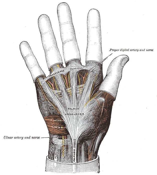
Figure 2. The palmar aponeurosis, continuous with the palmaris longus tendon; the digital nerves and vessels lie immediately deep to it. Gray’s Anatomy plate 425 (public domain), via Wikimedia Commons.
The carpal tunnel
The carpal tunnel is a fibro-osseous canal whose floor is the concave volar surface of the carpal bones and whose roof is the flexor retinaculum (transverse carpal ligament). The retinaculum has four palpable bony attachments: the scaphoid tubercle and the ridge of the trapezium radially, and the pisiform and the hook of the hamate ulnarly. The tunnel transmits nine flexor tendons (four superficialis, four profundus, and flexor pollicis longus) plus the median nerve. Within the tunnel the median nerve is the most superficial and radial structure, lying superficial to the profundus and flexor pollicis longus tendons, with the superficialis tendons ulnar to it; the superficialis tendons to the middle and ring fingers lie superficial to those of the index and little fingers.[6]
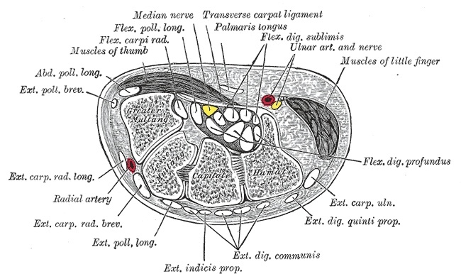
Figure 3. Transverse section through the carpal tunnel: the carpal bones forming the floor, the transverse carpal ligament the roof, and the flexor tendons with the median nerve within. Gray’s Anatomy plate 422 (public domain), via Wikimedia Commons.
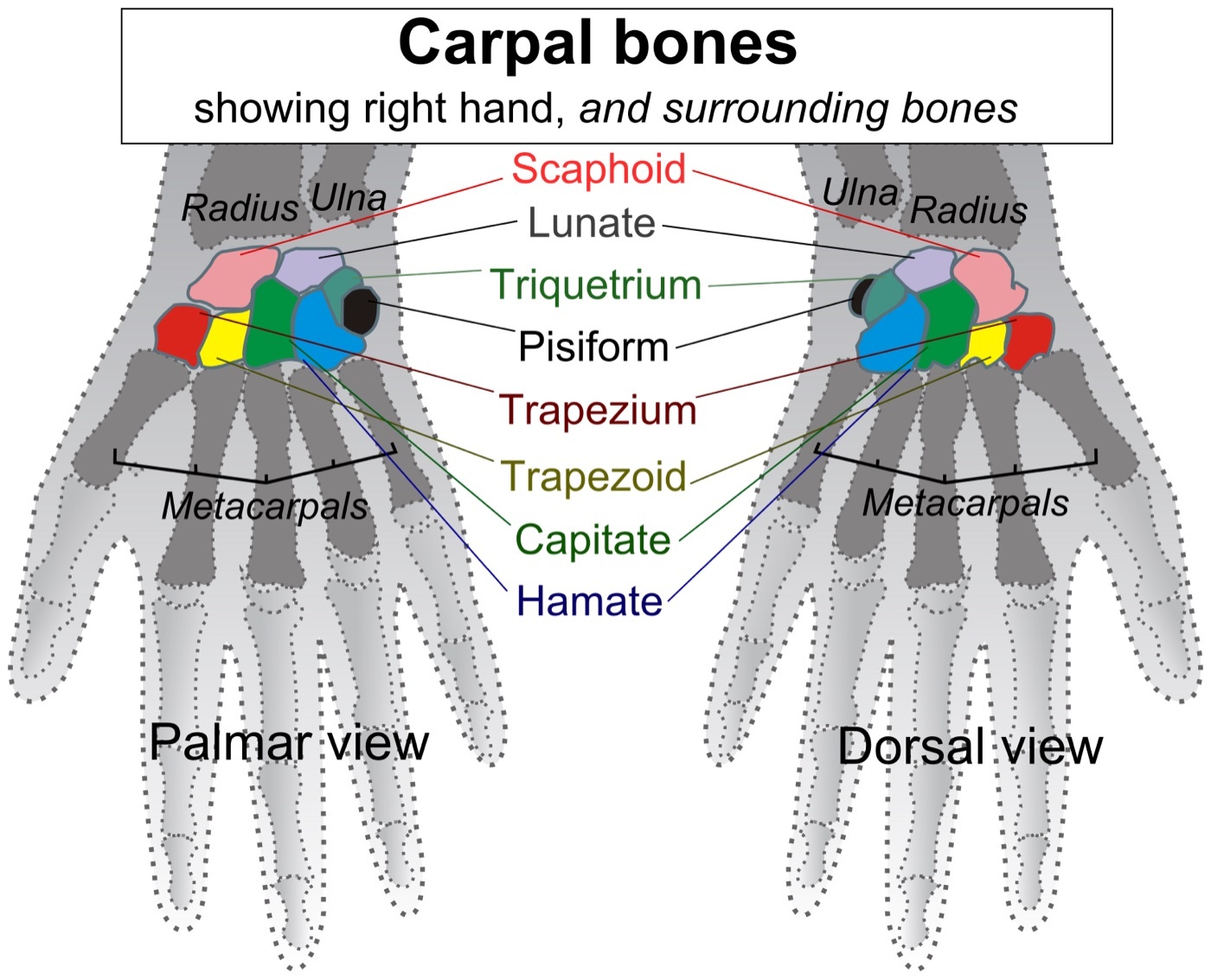
Figure 4. The carpal bones of the wrist, labelled. Mikael Häggström, CC BY-SA 3.0, via Wikimedia Commons.
The median nerve and its two variable branches
Two branches of the median nerve govern volar wrist surgery, and both are notoriously variable. The palmar cutaneous branch arises about 5 cm proximal to the wrist, runs along the ulnar side of the flexor carpi radialis, and crosses superficial to the retinaculum to supply the thenar skin; it is protected by keeping incisions ulnar to palmaris longus and angled across the wrist rather than transverse. The recurrent (thenar) motor branch usually arises from the anterolateral median nerve as it leaves the tunnel and curves into the thenar muscles between abductor pollicis brevis and flexor pollicis brevis. Its course has eight described variations; the classic (≈50%) is extraligamentous, ≈30% subligamentous, and ≈20% transligamentous (piercing the retinaculum). The safe rule is to divide the transverse carpal ligament on the ulnar side of the median nerve, where the motor branch will be spared unless it lies on that same side.[7]
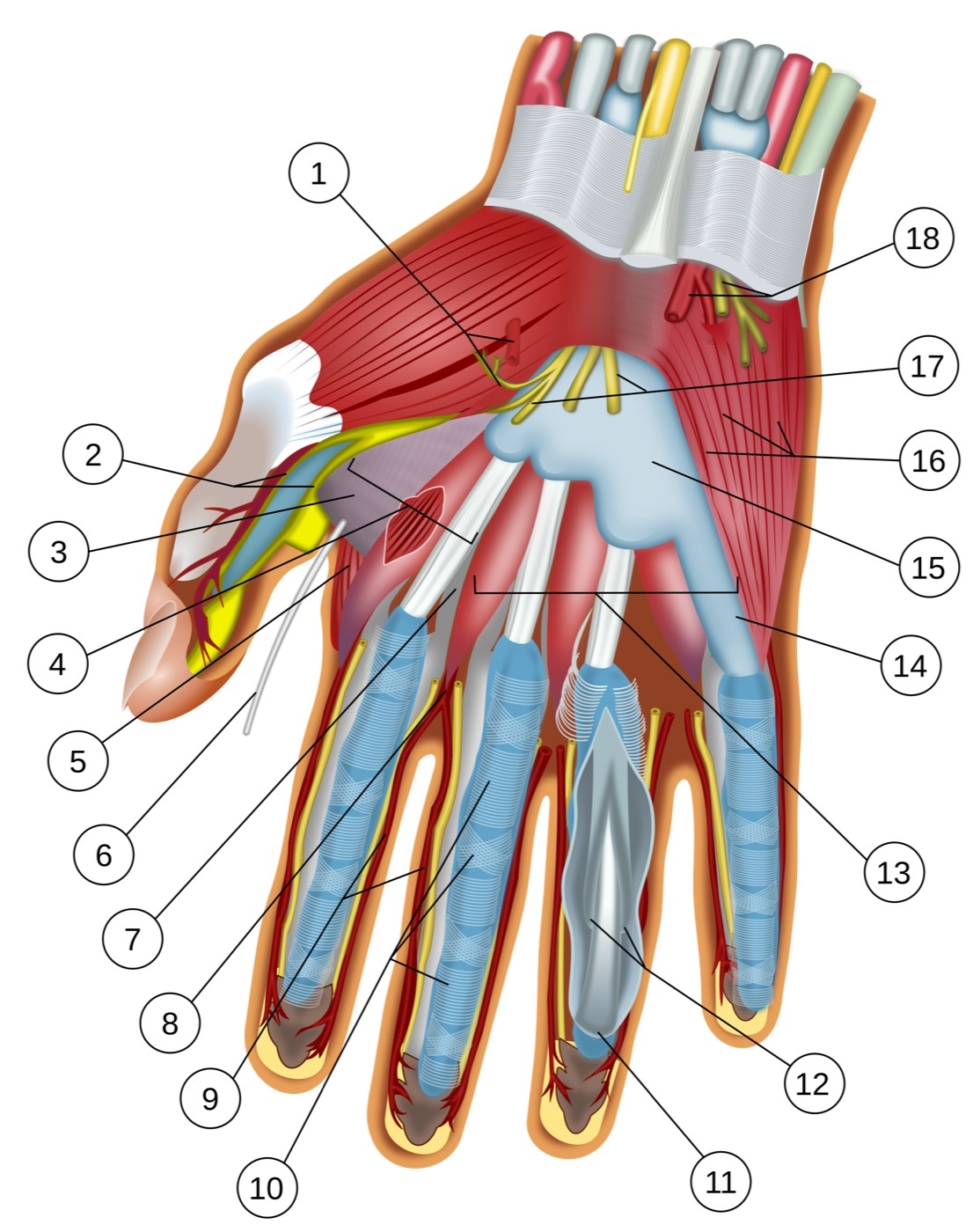
Figure 5. Deep palmar dissection of the wrist and hand showing the flexor tendons, the median and ulnar nerves, and the palmar arches. Wilfredor, released under CC0, via Wikimedia Commons.
Guyon’s canal and the ulnar nerve
The ulnar nerve does not pass through the carpal tunnel; it crosses over the flexor retinaculum in its own fibro-osseous tunnel, the canal of Guyon. The canal has four boundaries: its floor is the transverse carpal ligament, its roof the volar carpal ligament (a condensation of forearm fascia and flexor carpi ulnaris expansions), its medial wall the pisiform, and its lateral wall the hook of the hamate. Inside, from lateral to medial, lie the artery, then the nerve, then the flexor carpi ulnaris tendon (the “ANT” mnemonic). Around the pisiform the ulnar nerve divides into a superficial branch (sensory to the ulnar one and a half digits, plus motor to palmaris brevis) and a deep motor branch (to all the intrinsics except the thenar muscles and the radial two lumbricals). The deep branch lies on the hook of the hamate, where it can be injured by a hook fracture.[8]
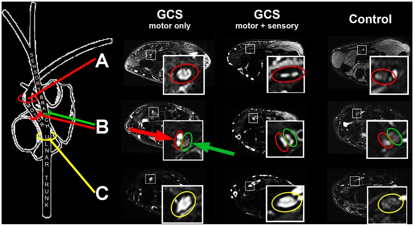
Figure 6. Magnetic resonance neurography of the ulnar nerve and its branches in the canal of Guyon. Kollmer et al., CC BY 4.0, via Wikimedia Commons.
The intrinsic muscles and their nerve supply
The thenar eminence has three muscles (abductor pollicis brevis, flexor pollicis brevis, opponens pollicis), all median-supplied through the recurrent motor branch, except that the deep head of flexor pollicis brevis takes an ulnar supply, which is why a complete median palsy does not fully flatten the thenar eminence. The hypothenar eminence has three ulnar-supplied muscles, with palmaris brevis the only muscle of the superficial ulnar branch. The lumbricals arise from the flexor digitorum profundus tendons and are split-supplied: the radial two by the median nerve, the ulnar two by the ulnar nerve. The interossei (dorsal abduct, “DAB”; palmar adduct, “PAD”) and the adductor pollicis are all supplied by the deep branch of the ulnar nerve. Lumbricals and interossei flex the metacarpophalangeal joints while extending the interphalangeal joints; their loss produces clawing.[9]
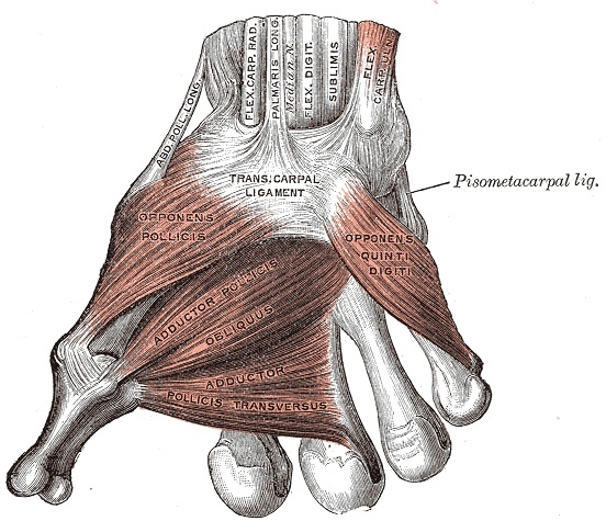
Figure 7. The deep muscles of the palm: the thenar muscles, the adductor pollicis and the interossei. Gray’s Anatomy plate 426 (public domain), via Wikimedia Commons.
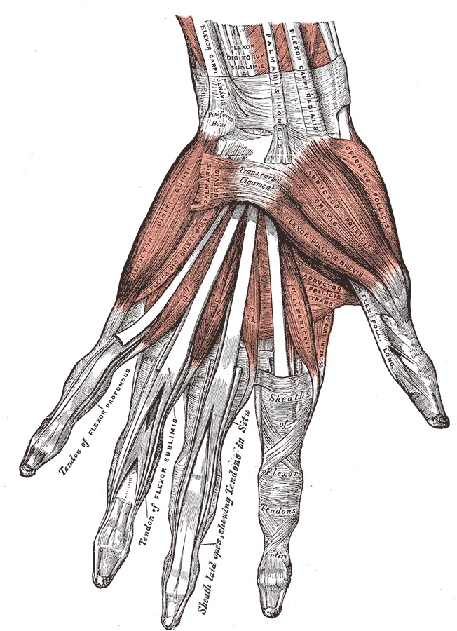
Figure 8. The muscles, tendons and synovial sheaths of the palm. Gray’s Anatomy plate 427 (public domain), via Wikimedia Commons.
The palmar arches and the dorsal extensor compartments
The superficial palmar arch, formed mainly by the ulnar artery (and often incomplete when the radial contribution is absent), lies superficial to the digital nerves in the palm and gives the common digital arteries; the deep palmar arch, the terminal radial artery joined by the deep ulnar branch, lies deep to the flexor tendons with the deep motor branch of the ulnar nerve. On the dorsum, twelve extensor tendons cross the wrist in six compartments beneath the extensor retinaculum (which prevents bowstringing): (1) abductor pollicis longus and extensor pollicis brevis (the de Quervain compartment); (2) extensor carpi radialis longus and brevis, radial to Lister’s tubercle; (3) extensor pollicis longus, which angles around the ulnar side of Lister’s tubercle; (4) extensor digitorum communis and extensor indicis; (5) extensor digiti minimi over the distal radioulnar joint; and (6) extensor carpi ulnaris at the ulnar styloid.[10]
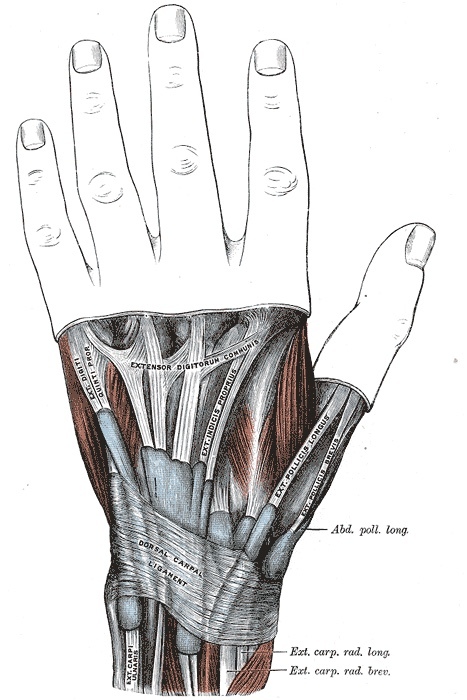
Figure 9. The extensor tendons of the wrist crossing beneath the extensor retinaculum in their dorsal compartments. Gray’s Anatomy plate 424 (public domain), via Wikimedia Commons.
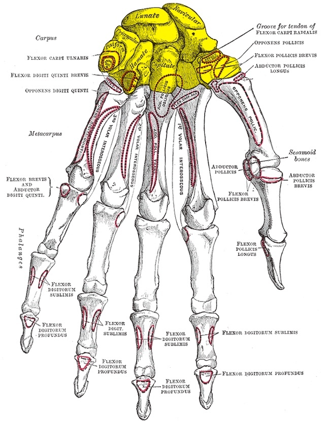
Figure 10. The bones of the wrist and hand, dorsal aspect: the carpal bones, metacarpals and phalanges. Gray’s Anatomy plate 219 (public domain), via Wikimedia Commons.
The extensor expansion
Over the dorsum of the finger the extensor tendon broadens into the extensor expansion (hood). It divides into a central slip, inserting into the base of the middle phalanx, and two marginal (lateral) slips, which receive the lumbrical and interosseous tendons and converge to insert into the base of the distal phalanx. The hood is anchored to the volar plate of the metacarpophalangeal joint by the sagittal bands, and the marginal slips are joined over the middle phalanx by the triangular ligament. Disruption of the central slip and triangular ligament lets the lateral slips slip volar to the proximal interphalangeal axis, producing the flexed proximal interphalangeal joint of a boutonnière deformity; loss of the terminal tendon at the distal phalanx produces a mallet finger.[11]
Part II - Dorsal Approach to the Wrist
The dorsal approach gives excellent access to all six extensor compartments, the dorsal carpus, and the distal radius. It is used for rheumatoid synovectomy and extensor repair, wrist arthrodesis, excision of distal radial tumours, ORIF of certain distal radial and carpal injuries (dorsal lip fractures, transscaphoid perilunate dislocations), and proximal row carpectomy; dorsal plating of the distal radius, however, irritates the overlying extensor tendons, so volar approaches are now often preferred for plate fixation. The patient is supine with the forearm pronated and a tourniquet; the incision is an 8 cm longitudinal line midway between the radial and ulnar styloids over Lister’s tubercle. The dorsal skin is loose enough that this is one of the rare incisions that may cross a major skin crease at a right angle without causing contracture.[12]
There is no true internervous plane, because the extensors all share the posterior interosseous nerve; the intermuscular plane is safe because that nerve enters them proximally, near the elbow. Lister’s tubercle is the key landmark, separating the second compartment (radial) from the third (extensor pollicis longus, ulnar). The deep dissection depends on the target: a rheumatoid synovectomy deroofs each compartment sequentially and tucks the preserved retinaculum beneath the extensor tendons to protect them; exposure of the intermediate column opens the third compartment and retracts the extensor pollicis longus radially on a vascular loop; exposure of the radial column opens the first compartment; and full wrist exposure for arthrodesis opens the fourth compartment, retracts its tendons, and incises the dorsal capsule (the dorsal radiocarpal ligament) longitudinally.[13]
The dangers are cutaneous nerves, the extensor pollicis longus, and the radial artery. The superficial radial nerve (emerging from under brachioradialis, most often over the second compartment) and the dorsal cutaneous branch of the ulnar nerve are protected by taking the incision down to the retinaculum before raising flaps, so they stay in the fat. The extensor pollicis longus is the tendon at risk: it can be crushed and devascularised between Lister’s tubercle and the base of the third metacarpal, causing delayed rupture even after an undisplaced distal radial fracture, and it abrades on dorsal plates. The radial artery crosses the lateral wrist and is safe if dissection stays subperiosteal. The approach cannot be extended proximally (no internervous plane) but reaches the distal half of the radius by retracting the abductor pollicis longus and extensor pollicis brevis.[14]
Part III - Volar Approaches to the Wrist
Volar (FCR) approach to the distal radius
This is the workhorse approach for volar locking-plate fixation of distal radial fractures, whose popularity has surged with locking plates; it is also used for nonunion grafting, osteotomy, and radial styloid excision. The patient is supine with the forearm supinated and a tourniquet; the incision is a 5-7 cm longitudinal line over the flexor carpi radialis tendon ending just above the wrist crease. The internervous plane is between the flexor carpi radialis (median nerve) and the brachioradialis (radial nerve), but the working interval is developed between the flexor carpi radialis tendon and the radial artery, through the FCR sheath, down onto the pronator quadratus; choosing this plane (rather than the brachioradialis-radial-artery plane of the forearm Henry approach) means the radial artery need not be retracted to reach the central radius.[15]
The pronator quadratus is the deep cover over the bone and may be detached as a flap (and repaired over the plate), tunnelled under (minimally invasive), or simply divided. Its subperiosteal reflection is what protects the median nerve and the ulnar-side flexor structures. The dangers are the radial artery (at the lateral wound edge, identified by its venae comitantes under tourniquet) and the median nerve (on the ulnar side, safe as long as the surgeon stays radial to the flexor carpi radialis and keeps the pronator quadratus sleeve). The approach extends proximally along the FCR/brachioradialis plane and distally into the wrist joint toward the scaphoid tubercle.[16]
Volar approach to the carpal tunnel
Carpal tunnel decompression is one of the commonest hand operations, and its whole technique is dictated by the two variable median-nerve branches, which are so unpredictable that blind division is condemned: the retinaculum must be divided full-length under direct vision. The incision begins just ulnar to the thenar crease (but never in it, for skin healing), stays ulnar to the palmaris longus tendon (protecting the palmar cutaneous branch), and curves to the ulnar side as it approaches the wrist so the flexion crease is not crossed transversely. The median nerve is found between palmaris longus and flexor carpi radialis (closer to palmaris longus), a flat dissector is passed beneath the retinaculum, and the transverse carpal ligament is divided on the ulnar side of the nerve, cutting onto the dissector, to protect the recurrent motor branch.[17]
The dangers are the recurrent motor branch (protected by the ulnar-side division; the transligamentous variant may itself need release), the palmar cutaneous branch (protected by the ulnar-angled incision), and, distally, the superficial palmar arch, which lies at the level of the outstretched thumb tip and is the reason not to push a closed instrument too far distally. The approach extends proximally up the forearm to expose the median nerve (which adheres to the deep surface of the flexor digitorum superficialis, so reflecting that muscle carries the nerve with it) and distally as a volar zigzag into the digits.[18]
Volar approach to the ulnar nerve (Guyon’s canal)
This approach decompresses the canal of Guyon and explores the ulnar nerve at the wrist; it is freely extensile up the forearm. The patient is supine and supinated with a tourniquet; the incision follows the radial border of the hypothenar eminence and crosses the wrist obliquely at about 60° onto the distal forearm. The flexor carpi ulnaris tendon is the landmark: incising the fascia on its radial border and retracting it ulnarward exposes the ulnar nerve and artery, which are then traced distally by dividing the volar carpal ligament, the roof of the canal. The nerve is at risk at two moments, when the flexor carpi ulnaris fascia is incised and when the volar carpal ligament is divided; the palmar cutaneous branch of the ulnar nerve is at risk superficially. Proximal extension uses the flexor carpi ulnaris (ulnar nerve) / flexor digitorum superficialis (median nerve) plane up to the elbow.[19]
Part IV - Approaches to the Flexor Tendons and Digits
Incision planning: the Bruner zigzag and the midlateral approach
Two incision designs reach the finger flexor apparatus, and both exist to obey the no-90°-crease rule. The volar zigzag (Bruner) incision runs diagonally across the finger between the three flexion creases, with the zigzag limbs angled at about 90° to each other (sharper angles necrose the corners) and never carried too far dorsally, where the neurovascular bundle would be endangered; it gives the best exposure of the tendons and both neurovascular bundles and extends freely into the palm. The midlateral (midaxial) incision runs along the line joining the dorsal ends of the flexion creases, the bloodless plane dorsal to the neurovascular bundle (which stays in the volar flap), on the watershed between dorsal and palmar digital nerve territories; it is harder to extend into the palm and gives less exposure.[20]
The flexor sheath, the pulleys, and Camper’s chiasm
Within the digit the flexor digitorum superficialis and profundus run in a common fibro-osseous sheath thickened into pulleys: four annular pulleys (A1 over the metacarpophalangeal joint, A2 over the proximal phalanx, A3 over the proximal interphalangeal joint, A4 over the middle phalanx) and three cruciate pulleys (C1-C3). The cruciate pulleys are not critical, but A2 and A4 must be preserved to prevent bowstringing; A1 is the pulley divided in trigger-finger release. To reach the volar plate of the proximal interphalangeal joint, the C1, A3, and C2 pulleys are divided and reconstructed, sparing A2 and A4. The superficialis tendon splits and spirals around the profundus to reunite deep to it (Camper’s chiasm) before inserting on the middle phalanx, while the profundus passes through to the distal phalanx; the tendons are nourished by the vincula and their volar surfaces are relatively avascular, so core sutures are placed volarly.[21]
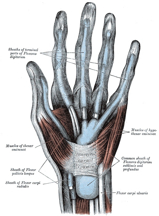
Figure 11. The synovial sheaths of the flexor tendons of the fingers and the radial and ulnar bursae, within the fibrous flexor sheath whose thickenings form the pulleys. Gray’s Anatomy plate 423 (public domain), via Wikimedia Commons.
The flexor zones (Milford) and zone II
The flexor tendons are divided into five zones from distal to proximal. Zone I is distal to the superficialis insertion (profundus alone). Zone II, from the distal palmar crease to the middle of the middle phalanx, is where both tendons run together in the tight fibro-osseous sheath and must glide over each other; adhesions here cripple the finger, which is why Bunnell nicknamed it “no-man’s land,” and repairs here have the worst prognosis. Zone III is the palm (the lumbrical origin). Zone IV is the carpal tunnel (eight tendons in the common sheath). Zone V is the distal forearm, where repairs do best. The prognosis hierarchy runs from best in zone V to worst in zone II.[22]
The volar approach to the flexor tendons exposes the sheath best and reaches both neurovascular bundles; skin flaps are raised thick and not widely mobilised until the sheath is reached, and Grayson’s ligament is opened to expose the bundle. The midlateral approach keeps the bundle in the volar flap and is used for phalangeal fractures and sheath access. The dorsal approach to the phalanges and interphalangeal joints is the route for fracture fixation, since the only structure between skin and bone is the extensor mechanism: the metacarpophalangeal joint is entered by splitting the extensor or the hood, the proximal interphalangeal joint between the central slip and a lateral band, and the distal interphalangeal joint through the terminal tendon and triangular ligament.[23]
Part V - Approaches to the Scaphoid
The scaphoid is approached volarly or dorsolaterally, and the choice turns on its retrograde blood supply: the dominant vessels (dorsal-ridge branches of the radial artery) enter the bone distally and flow proximally, so the proximal pole is supplied last and is the commonest site of nonunion and avascular necrosis. The volar approach is based on the scaphoid tubercle and extended proximally between the flexor carpi radialis and the radial artery; it has no true internervous plane (only the flexor carpi radialis, median-supplied, is mobilised), spares the dorsal proximal blood supply and the superficial radial nerve, and leaves a cosmetic scar, but it endangers the radial artery and its superficial palmar branch. The distal two-thirds are exposed by an oblique capsulotomy, the proximal pole by forced dorsiflexion; the palmar (radioscaphocapitate) ligament complex must be preserved to stabilise the proximal pole.[24]
The dorsolateral approach uses the anatomical snuffbox, whose floor is the scaphoid with the radial artery running directly across it. The patient is pronated; an S-shaped incision is centred over the snuffbox, and the plane is developed between the extensor pollicis longus and the extensor pollicis brevis (both posterior-interosseous-supplied, so no denervation). The dangers are the superficial radial nerve (which crosses directly beneath the incision in two or more variable branches and must be sought and preserved), the radial artery on the scaphoid, and the dorsal carpal branch supplying the bone, so dorsal-ridge soft tissue is preserved. A longitudinal capsulotomy exposes the radioscaphoid joint, and ulnar deviation delivers the whole proximal pole. Screw fixation often combines the volar and dorsolateral approaches; the snuffbox’s anatomical boundaries are the extensor pollicis longus dorsally and the abductor pollicis longus and extensor pollicis brevis volarly.[25]
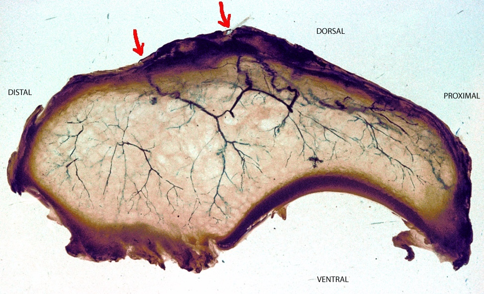
Figure 12. The retrograde blood supply of the scaphoid, entering distally so that the proximal pole is perfused last and is prone to avascular necrosis. Nelsonmleite, CC BY-SA 3.0, via Wikimedia Commons.
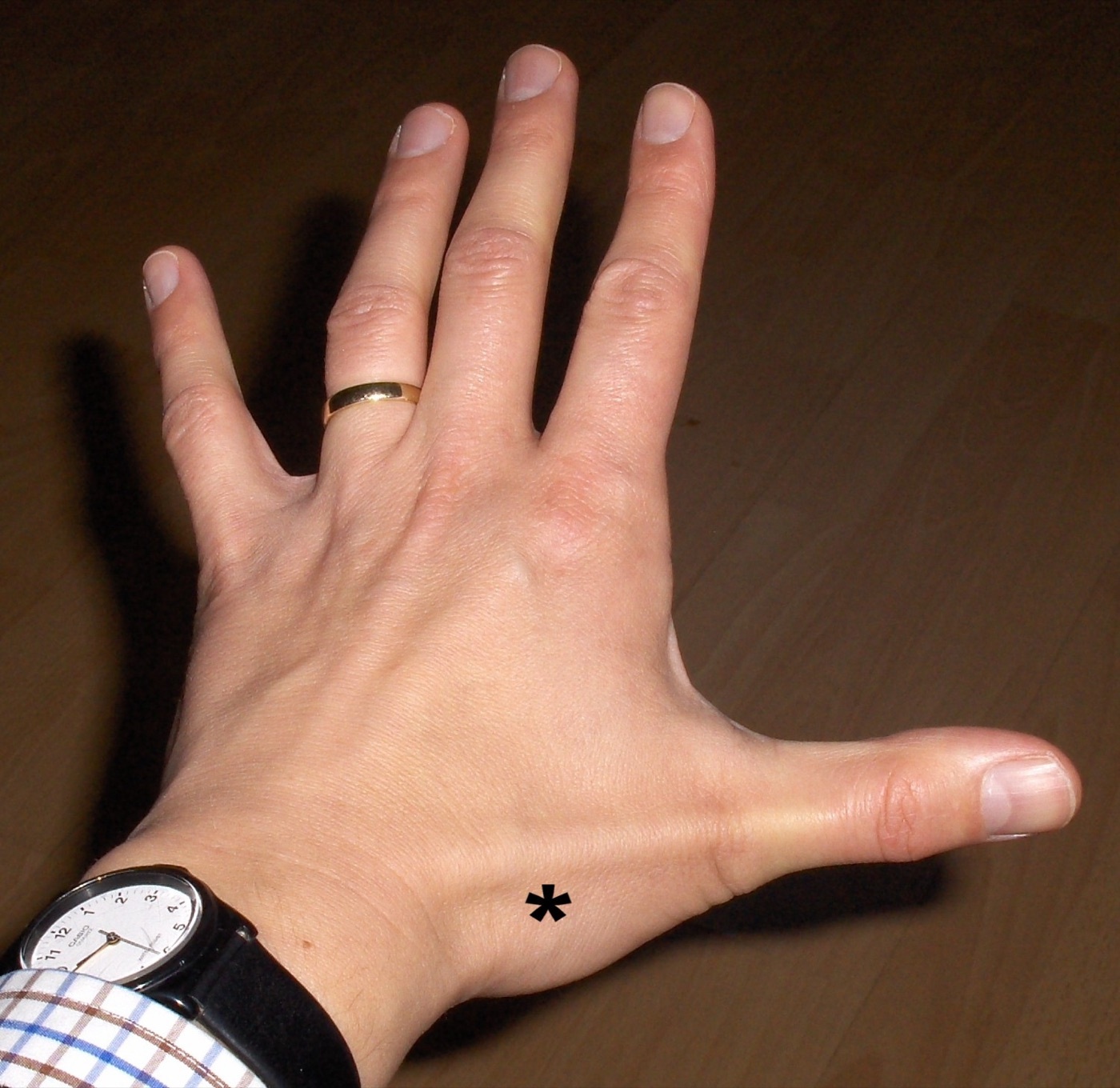
Figure 13. The anatomical snuffbox, whose floor is the scaphoid crossed by the radial artery, bounded by the extensor pollicis longus and the abductor pollicis longus / extensor pollicis brevis tendons. James Heilman MD, CC BY-SA 4.0, via Wikimedia Commons.
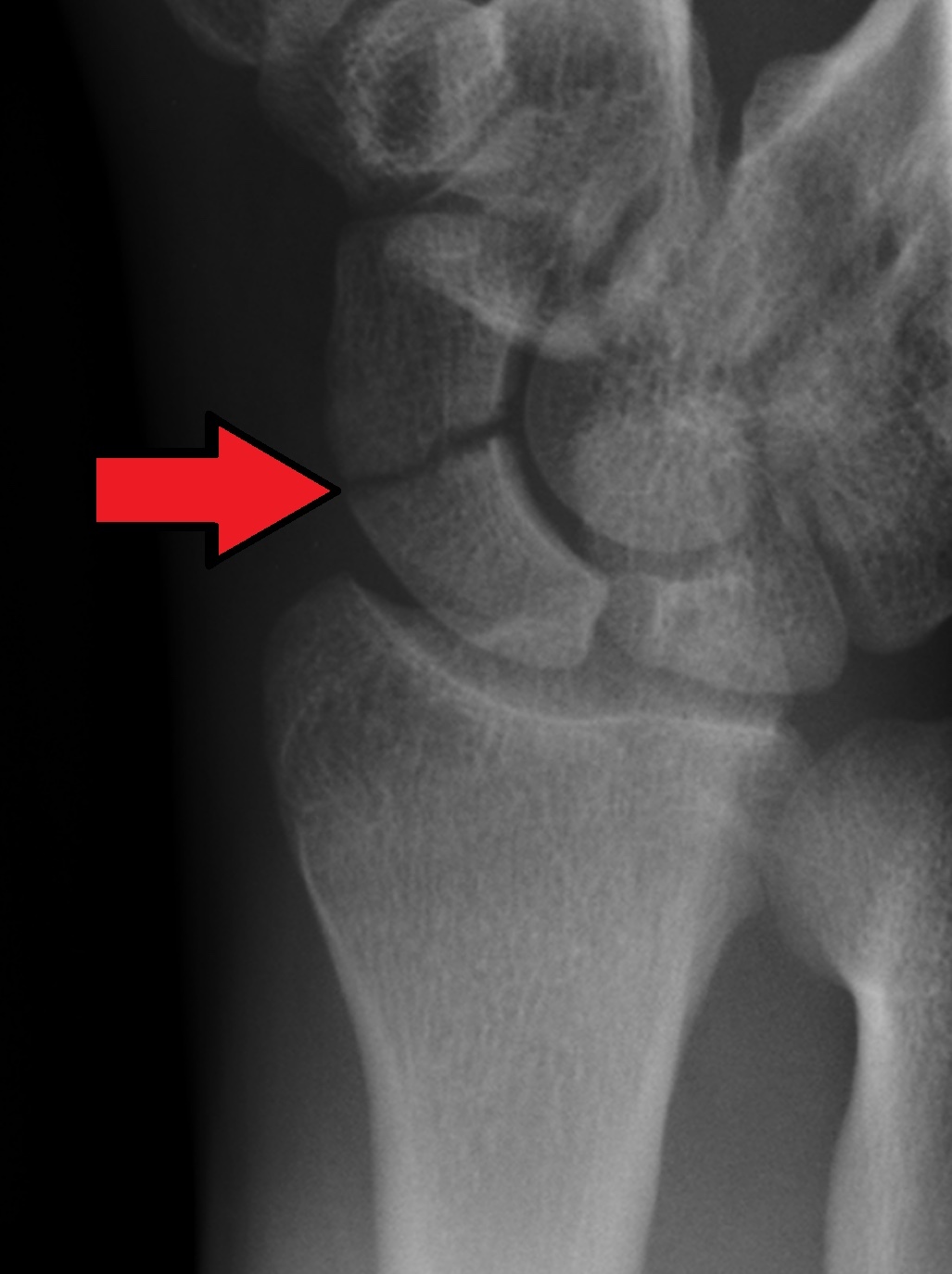
Figure 14. Radiograph of a scaphoid waist fracture. Gilo1969, CC BY 3.0, via Wikimedia Commons.
Part VI - Drainage of Hand Infections
Principles and Kanavel’s signs
Hand infections turn on accurate localisation and the timing of surgery: drainage too early may incise cellulitis and spread the infection, too late lets pus damage tendons. Pus is hard to detect (fat is itself fluctuant at body temperature), but inability to sleep for pain and pain on passive extension of a digit are reliable warnings; the latter is one of Kanavel’s four cardinal signs of flexor tenosynovitis (the others being fusiform swelling of the digit, tenderness along the sheath, and the finger held flexed). Operative principles are fixed: a general anaesthetic or distal block (never local infiltration into the infected area), a tourniquet without exsanguination by bandage, perfect lighting with identification of every neurovascular bundle, wounds left open, and immobilisation in the safe position (metacarpophalangeal joints at 80°, interphalangeal joints at 10°) where the collateral ligaments are at maximum length.[26]
Paronychia and felon
Paronychia is an infection of the nail fold, usually staphylococcal and often from a torn hangnail cuticle; it is drained by a short longitudinal incision at the corner of the nail fold, removing part of the nail if pus is subungual, protecting the nail bed. The felon is a closed pulp-space infection in which fibrous septa tether the distal phalanx to the skin and loculate the pus, risking osteomyelitis of the distal phalanx. It is drained through a unilateral high lateral incision (not a midline volar scar, which is painful, and not a through-and-through “fish-mouth” incision, which devascularises the pulp), kept dorsal to the digital nerve and not extended within 1 cm of the distal interphalangeal crease (to spare the flexor sheath), dividing the septa to open all loculi.[27]
Web space, flexor sheath, and deep palmar spaces
A web-space (collar-button) abscess points dorsally because the dorsal web skin is thinner, spreads the fingers apart, and can track up the lumbrical canal into the palm; it is drained by a volar incision (longitudinal or a transverse one just proximal to the web), protecting the digital nerves. Flexor tenosynovitis is an emergency drained through a proximal transverse incision at the A1 pulley plus, if turbid fluid is found, a distal midlateral incision for through-and-through irrigation, both kept off the digital bundles. The deep palmar space lies between the flexor tendons and the interossei/adductor, divided by an oblique septum running from the middle-finger flexor sheath to the third metacarpal into a radial thenar space (infection disables the thumb and index) and an ulnar midpalmar space (infection disables the middle and ring fingers); each is drained directly, dissecting out the digital nerves and (for the thenar space) the recurrent motor branch before incising the aponeurosis.[28]
The radial bursa is the synovial sheath of the flexor pollicis longus and the ulnar bursa the common flexor sheath (continuous with the little-finger sheath and enveloping the other finger flexors through the carpal tunnel); both run to the volar wrist just proximal to the retinaculum. Each is drained with a distal digital midlateral incision and a proximal wrist incision, using a probe to define the proximal extent; if the probe lies in the carpal tunnel, the retinaculum is divided protecting the median nerve and its branches, and if in the forearm (ulnar bursa), the ulnar nerve and artery. Because the two bursae often communicate proximal to the wrist, infection can cross between thumb and little finger as a horseshoe abscess.[29]
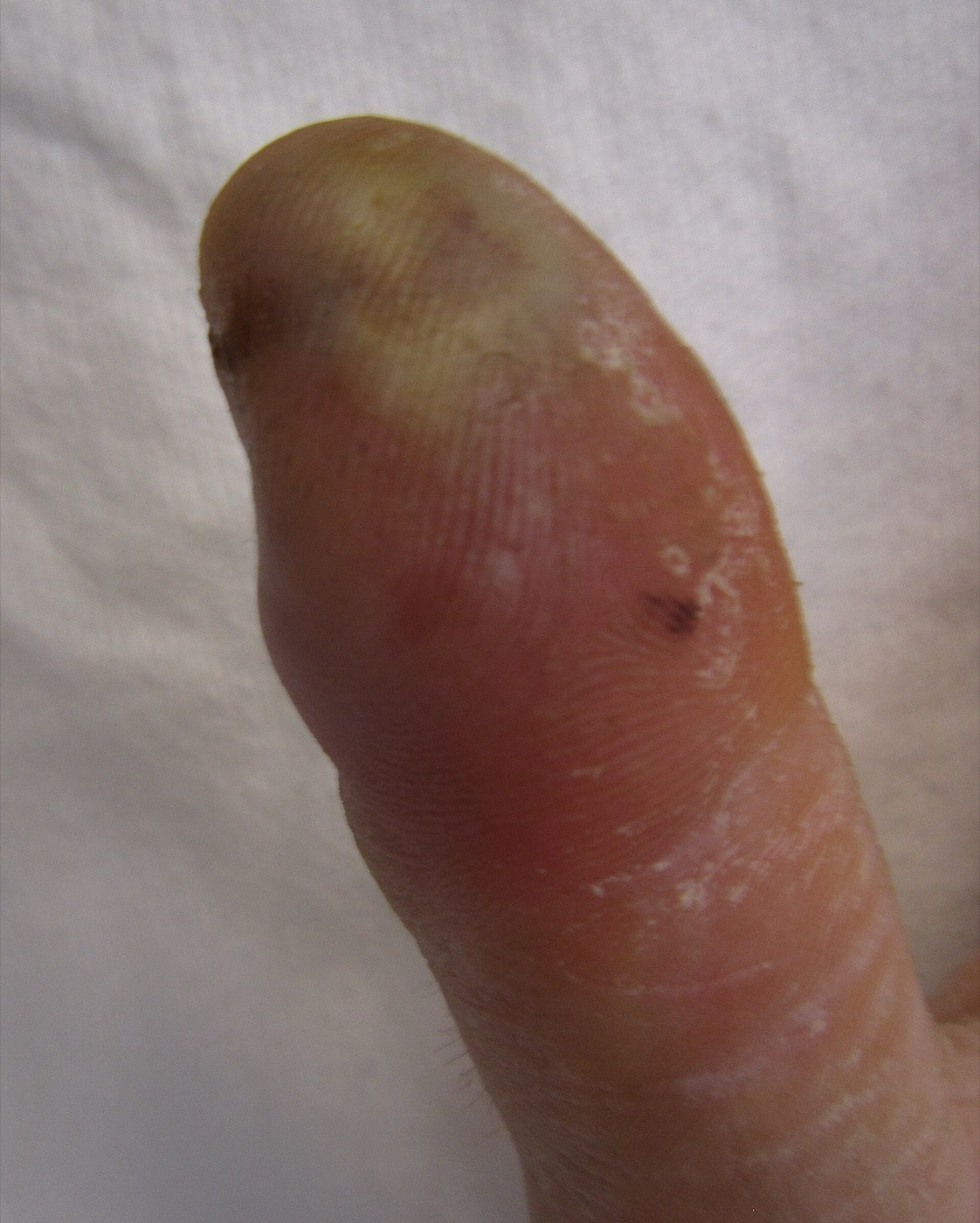
Figure 15. A felon: a closed, septate pulp-space infection of the thumb. James Heilman MD, CC BY-SA 3.0, via Wikimedia Commons.
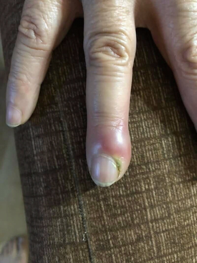
Figure 16. Acute paronychia: infection of the nail fold. Chris Craig (public domain), via Wikimedia Commons.
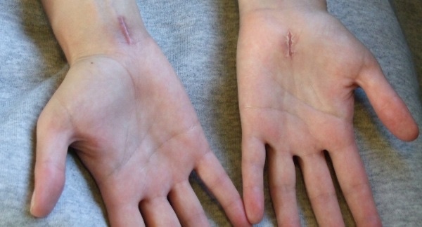
Figure 17. Healed open carpal-tunnel-release scars in the palm, placed ulnar to the thenar crease. HenrykGerlach, CC BY-SA 3.0, via Wikimedia Commons.
Part VII - Z-Plasty
The Z-plasty is the geometric operation that lengthens a contracted scar, and it is the operative corollary of the cardinal hand rule that a scar must not cross a flexion crease at a right angle: such a scar contracts, and the Z-plasty re-angles it. Two triangular flaps are raised on either side of the contracture, with the central limb laid along the line of the scar and two lateral limbs of equal length drawn off each end to form a “Z”; transposing (swapping) the two flaps reorients the central limb by about 90°, gaining length along the original line of tension at the cost of width. The theoretical gain depends on the limb angle: about 25% at 30°, 50% at 45%, 75% at the classic 60°, and 100% at 75°, with 60° the usual compromise because wider angles exceed the available skin laxity.[30]
Long, linear contractures are better treated by several smaller Z-plasties in series than by one large one, since this recruits less adjacent skin per flap and keeps the flaps viable. In the hand the Z-plasty is used for digital flexion contractures (converting a longitudinal scar crossing the creases into a broken zig-zag line) and for first web-space (thumb-index) adduction contractures, where a Z-plasty (or a four-flap or five-flap “jumping-man” variant) deepens and widens the web to restore thumb abduction and opposition; it is also used in releasing Dupuytren and burn contractures.[31]
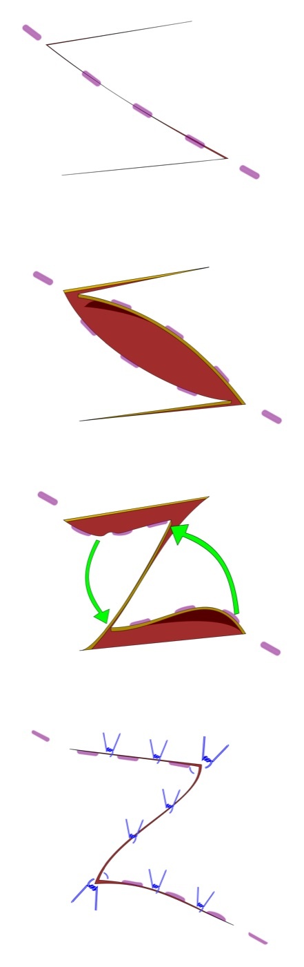
Figure 18. The Z-plasty in four stages: the Z incision design, the two triangular flaps raised, their transposition (arrows), and the closed result with the central limb re-oriented and lengthened. Evan Mason, CC BY-SA 4.0, via Wikimedia Commons.
References
-
Hoppenfeld ch.5 covers the dorsal approach to the wrist (p.356), the volar approaches to the distal radius, carpal tunnel, and ulnar nerve (p.373, p.379, p.391), the flexor-tendon and digit approaches (p.411-426), the scaphoid approaches (p.432, p.437), the drainage of hand infections (p.442-475), and the anatomy of the hand (p.476). The cardinal incision rule, that a flexion crease must not be crossed at 90°, recurs throughout (Hoppenfeld p.418, p.477).
-
The recurrent motor branch and palmar cutaneous branch of the median nerve are the two variable structures that govern carpal tunnel surgery (Hoppenfeld p.379, p.409); release on the ulnar side of the nerve protects the motor branch (p.381, p.387). The superficial radial nerve and the radial artery on the scaphoid are the key dangers of the dorsolateral scaphoid approach (p.437-438). The digital neurovascular bundles, held by Grayson’s (volar) and Cleland’s (dorsal) ligaments, govern digital incisions (p.414; Cleland’s is standard teaching).
-
The flexor tendons run in the carpal tunnel (a fibro-osseous canal) and the digital fibrous sheath with annular (A1-A4) and cruciate (C1-C3) pulleys, A2 and A4 being critical to prevent bowstringing (Hoppenfeld p.428-429); the extensors run in six dorsal compartments under the extensor retinaculum (p.368). Adhesions follow when these sheaths are opened, so sheaths/pulleys are preserved and thick flaps raised (p.414-415).
-
Palmar skin is tough and immobile, tethered to the palmar aponeurosis (fat in pressure-resistant loculi), often needing V-Y flaps or grafting for closure; “to avoid flexion contractures, the flexure creases should not be crossed at 90 degrees”; a triangular flap apex should exceed 60° and distally based palmar flaps are avoided; dorsal skin is thin and mobile with poorer blood supply and carries venous return (Hoppenfeld p.476-477, p.481).
-
The palmar aponeurosis is continuous with palmaris longus, fans into four bands, and the palmar nerves and vessels lie immediately deep to it; Dupuytren contracture thickens it around the digital nerves (Hoppenfeld p.477); Grayson’s ligament lies volar to the digital neurovascular bundle and must be opened to expose it (p.414); the digital nerves lie lateral to the tendons (p.416). Cleland’s ligament (dorsal to the bundle) and the “Grayson = ground / Cleland = ceiling” relationship are standard teaching.
-
The carpal tunnel floor is the concave volar carpal bones, roof the flexor retinaculum/transverse carpal ligament; four attachments = scaphoid tubercle and trapezial ridge (radial), pisiform and hook of hamate (ulnar); contents = nine flexor tendons (4 FDS, 4 FDP, FPL) + the median nerve; the median nerve is most superficial/radial, superficial to FDP and FPL with the FDS tendons ulnar to it; FDS to middle and ring fingers lies superficial to index and little (Hoppenfeld p.397, p.409-410).
-
The palmar cutaneous branch arises ~5 cm proximal to the wrist, runs along the ulnar side of FCR, and crosses superficial to the retinaculum (four variations), protected by keeping incisions ulnar to palmaris longus and angled (Hoppenfeld p.399-402); the recurrent motor branch usually arises anterolaterally as the nerve exits the tunnel, entering the thenar muscles between APB and FPB, with eight variations (classic ≈50%, ~30% from within the tunnel, ~20% transligamentous), the danger minimised by dividing the retinaculum on the ulnar side of the nerve (Hoppenfeld p.409-410). The extra/sub/transligamentous terms are standard teaching.
-
The ulnar nerve crosses over the flexor retinaculum in the canal of Guyon (floor = transverse carpal ligament, roof = volar carpal ligament, medial wall = pisiform, lateral wall = hook of hamate); contents lateral to medial = artery, nerve, FCU tendon (“ANT”); around the pisiform it divides into a superficial branch (sensory ulnar 1.5 digits + palmaris brevis) and a deep motor branch (all intrinsics except the thenar muscles and radial two lumbricals); the deep branch lies on the hook of the hamate (Hoppenfeld p.397, p.400, p.408).
-
Thenar muscles (APB, FPB, opponens pollicis) are median-supplied via the recurrent branch, but the deep head of FPB is ulnar-supplied (so median palsy spares part of the eminence); hypothenar muscles are ulnar-supplied, palmaris brevis being the only superficial-ulnar-branch muscle; lumbricals are split (radial two median, ulnar two ulnar); interossei (PAD/DAB) and adductor pollicis are deep-ulnar-supplied; lumbricals/interossei flex MCP and extend IP joints, loss gives clawing (Hoppenfeld p.478-482). The LOAF mnemonic for median intrinsics is standard teaching.
-
The superficial palmar arch (mainly ulnar artery, often incomplete) lies superficial to the palmar digital nerves and gives the common digital arteries; the deep palmar arch (terminal radial + deep ulnar branch) lies deep to the flexor tendons with the deep ulnar motor branch (Hoppenfeld p.478-480). Twelve extensor tendons cross in six compartments under the extensor retinaculum: 1 = APL/EPB (de Quervain), 2 = ECRL/ECRB (radial to Lister), 3 = EPL (ulnar to Lister, angling ~45°), 4 = EDC/EIP, 5 = EDM (over the DRUJ), 6 = ECU (at the ulnar styloid) (Hoppenfeld p.368-370).
-
The extensor expansion divides into a central slip (to the base of the middle phalanx) and two marginal slips (receiving lumbrical/interosseous tendons, inserting into the base of the distal phalanx); the hood is anchored to the MCP volar plate (sagittal bands), the marginal slips joined over the middle phalanx by the triangular ligament; central-slip + triangular-ligament disruption gives a boutonnière deformity (Hoppenfeld p.481-482). Mallet finger (terminal tendon disruption) and the term “sagittal bands” are standard teaching.
-
The dorsal approach exposes all six extensor compartments, the dorsal carpus, and the distal radius; uses = RA synovectomy/extensor repair, wrist fusion, tumour excision, ORIF of dorsal lip fractures and transscaphoid perilunate dislocations, proximal row carpectomy; dorsal plates irritate extensor tendons so volar approaches are often preferred; position supine, forearm pronated, tourniquet; 8 cm longitudinal incision midway between the styloids over Lister’s tubercle; the loose dorsal skin tolerates crossing a crease at a right angle (Hoppenfeld p.356-357, p.368).
-
No true internervous plane (all extensors share the posterior interosseous nerve, which enters them proximally, so the intermuscular plane is safe); Lister’s tubercle separates the second (radial) from the third (EPL, ulnar) compartment; synovectomy deroofs each compartment sequentially and tucks the retinaculum beneath the tendons; the intermediate-column exposure opens the third compartment and retracts EPL radially on a vascular loop; the radial-column exposure opens the first compartment; full wrist exposure opens the fourth compartment and incises the dorsal capsule (dorsal radiocarpal ligament) longitudinally (Hoppenfeld p.357-360).
-
Dangers: the superficial radial nerve (from under brachioradialis, commonest over the second compartment) and the dorsal cutaneous branch of the ulnar nerve, protected by taking the incision to the retinaculum before raising flaps; the EPL, crushed/devascularised between Lister’s tubercle and the third metacarpal base (delayed rupture even after undisplaced fractures) and abraded by dorsal plates; the radial artery on the lateral wrist (safe if subperiosteal); no proximal extension (no internervous plane), but the distal half of the radius is reached by retracting APL and EPB (Hoppenfeld p.365-366, p.368, p.370). The snuffbox course of the radial artery and formal EPL transposition are standard teaching.
-
The volar FCR approach is the workhorse for volar locking-plate fixation of distal radial fractures (and nonunion grafting, osteotomy, radial styloid excision); supine, forearm supinated, tourniquet; 5-7 cm incision over the FCR tendon ending just above the wrist crease; internervous plane = FCR (median nerve) vs brachioradialis (radial nerve), the working interval developed between the FCR tendon and the radial artery via the FCR sheath onto the pronator quadratus, so the radial artery is not retracted for central access (Hoppenfeld p.373-374).
-
The pronator quadratus is the deep cover, detached as a flap (and repaired over the plate), tunnelled under, or divided; its subperiosteal reflection protects the median nerve and ulnar-side flexors; dangers = the radial artery (lateral wound edge, found by its venae comitantes under tourniquet) and the median nerve (ulnar side, safe if you stay radial to FCR and keep the PQ sleeve); extends proximally along the FCR/brachioradialis plane and distally into the wrist toward the scaphoid tubercle (Hoppenfeld p.378-379). Placing volar plates proximal to the watershed line to avoid FPL rupture is standard teaching.
-
Carpal tunnel decompression is governed by the two variable median branches, so blind division is condemned and the retinaculum is divided full-length under direct vision; the incision begins just ulnar to the thenar crease (not in it), stays ulnar to palmaris longus (protecting the palmar cutaneous branch), and curves ulnarward at the wrist so the crease is not crossed transversely; find the median nerve between palmaris longus and FCR (closer to PL), pass a flat dissector beneath the retinaculum, and divide the transverse carpal ligament on the ulnar side of the nerve, cutting onto the dissector, to protect the recurrent motor branch (Hoppenfeld p.379-381).
-
Dangers: the recurrent motor branch (protected by ulnar-side division; the transligamentous variant may need its own release), the palmar cutaneous branch (protected by the ulnar-angled incision), and distally the superficial palmar arch at the level of the outstretched thumb tip (do not push a closed instrument too far distally); the approach extends proximally to the forearm median nerve (which adheres to the deep surface of FDS) and distally as a volar zigzag (Hoppenfeld p.386-390). Kaplan’s cardinal line approximating the arch is standard teaching.
-
The volar ulnar approach decompresses Guyon’s canal and explores the ulnar nerve, freely extensile up the forearm; supine, supinated, tourniquet; incision along the radial border of the hypothenar eminence crossing the wrist obliquely at ~60°; the FCU tendon is the landmark, its radial-border fascia incised and the muscle retracted ulnarward to expose the ulnar nerve and artery, traced distally by dividing the volar carpal ligament (the canal roof); the nerve is at risk when the FCU fascia is incised and when the volar carpal ligament is divided, the palmar cutaneous branch at risk superficially; proximal extension uses the FCU (ulnar) / FDS (median) plane to the elbow (Hoppenfeld p.391-395).
-
Two digital incisions both obey the no-90°-crease rule: the volar zigzag (Bruner) runs diagonally between the three flexion creases, limbs at ~90° (sharper angles necrose corners), not carried too far dorsally (neurovascular bundle), giving the best exposure and extending into the palm; the midlateral (midaxial) incision joins the dorsal ends of the flexion creases, in the bloodless plane dorsal to the neurovascular bundle (kept in the volar flap), on the dorsal/palmar nerve watershed, harder to extend and giving less exposure (Hoppenfeld p.411-421). The eponyms “Bruner” and the Cleland-ligament plane are standard teaching.
-
In the digit FDS and FDP run in a common fibro-osseous sheath with four annular pulleys (A1 over the MCP joint, A2 over the proximal phalanx, A3 over the PIP joint, A4 over the middle phalanx) and three cruciate pulleys (C1-C3); the cruciates are non-critical but A2 and A4 must be preserved to prevent bowstringing, A1 is divided in trigger-finger release; for PIP volar-plate access divide C1/A3/C2 and reconstruct, sparing A2/A4; FDS splits and spirals around FDP, reuniting deep to it (Camper’s chiasm) to insert on the middle phalanx while FDP passes to the distal phalanx; the vincula supply the tendons and the volar surfaces are relatively avascular, so core sutures go volarly (Hoppenfeld p.414, p.428-431). The A5 pulley and the “Camper’s chiasm” eponym are standard teaching.
-
The flexor tendons are divided into five Milford zones: zone I distal to the FDS insertion (FDP alone), zone II (“no-man’s land,” Bunnell) from the distal palmar crease to the mid-middle-phalanx where both tendons run in the tight sheath and adhesions cripple the finger (worst prognosis), zone III the palm/lumbrical origin, zone IV the carpal tunnel (eight tendons in the common sheath), zone V the distal forearm (best repairs); prognosis runs best (V) to worst (II) (Hoppenfeld p.427-430). Roman-numeral zone notation is standard teaching.
-
The volar flexor-tendon approach exposes the sheath best and both neurovascular bundles, raising thick flaps not widely mobilised until the sheath is reached and opening Grayson’s ligament; the midlateral approach keeps the bundle in the volar flap (phalangeal fractures, sheath access); the dorsal approach to the phalanges/IP joints is the route for fracture fixation (only the extensor mechanism overlies bone), entering the MCP joint by splitting the extensor/hood, the PIP joint between the central slip and a lateral band, and the DIP joint through the terminal tendon and triangular ligament (Hoppenfeld p.411-425).
-
The scaphoid’s retrograde blood supply (dorsal-ridge branches enter distally and flow proximally) makes the proximal pole the commonest site of nonunion and avascular necrosis; the volar approach is based on the scaphoid tubercle and extended proximally between FCR and the radial artery, with no true internervous plane (only FCR mobilised), sparing the dorsal proximal supply and the superficial radial nerve and leaving a cosmetic scar but endangering the radial artery and its superficial palmar branch; the distal two-thirds are exposed by oblique capsulotomy, the proximal pole by forced dorsiflexion, preserving the palmar ligament complex (Hoppenfeld p.432-437). The retrograde-supply/AVN mechanism, the radioscaphocapitate name, and proximal-pole AVN are standard teaching.
-
The dorsolateral approach uses the anatomical snuffbox (floor = scaphoid, with the radial artery across it); pronated, S-shaped incision over the snuffbox, plane between EPL and EPB (both PIN-supplied, no denervation); dangers = the superficial radial nerve (crossing directly beneath the incision in two or more variable branches, sought and preserved), the radial artery on the scaphoid, and the dorsal carpal branch supplying the bone (preserve dorsal-ridge soft tissue); longitudinal capsulotomy exposes the radioscaphoid joint, ulnar deviation delivers the proximal pole; screw fixation often combines both approaches (Hoppenfeld p.437-441). The full snuffbox boundaries (APL volarly, trapezium in the floor) and snuffbox tenderness as a fracture sign are standard teaching.
-
Hand-infection surgery turns on localisation and timing (too early spreads cellulitis, too late damages tendons); pus is hard to detect, but sleeplessness from pain and pain on passive extension warn of it, the latter being one of Kanavel’s four signs of flexor tenosynovitis (with fusiform swelling, tenderness along the sheath, and the finger held flexed); operative rules = general/distal-block anaesthesia (never local infiltration), tourniquet without bandage exsanguination, perfect lighting and identification of neurovascular bundles, wounds left open, immobilisation in the safe position (MCP 80°, IP 10°, collaterals at maximum length) (Hoppenfeld p.443-444).
-
Paronychia is a nail-fold infection (usually staphylococcal, from a torn cuticle), drained by a short longitudinal incision at the corner of the nail fold (removing part of the nail if subungual), protecting the nail bed; the felon is a closed, septate, loculated pulp-space infection risking distal-phalanx osteomyelitis, drained through a unilateral high lateral incision (not midline volar, not through-and-through), kept dorsal to the digital nerve and short of 1 cm from the DIP crease, dividing all septa (Hoppenfeld p.445-449). The “fish-mouth” eponym is standard teaching.
-
A web-space (collar-button) abscess points dorsally (thinner dorsal skin), spreads the fingers, and can track up the lumbrical canal, drained by a volar incision protecting the digital nerves; flexor tenosynovitis is an emergency drained through a proximal A1-pulley incision plus a distal midlateral incision for through-and-through irrigation, off the bundles; the deep palmar space (between flexor tendons and interossei/adductor) is split by an oblique septum from the middle-finger sheath to the third metacarpal into a radial thenar space (disables thumb/index) and an ulnar midpalmar space (disables middle/ring), each drained directly after dissecting out the digital nerves and, for the thenar space, the recurrent motor branch (Hoppenfeld p.449-469). The “collar-button” eponym is standard teaching.
-
The radial bursa is the FPL synovial sheath and the ulnar bursa the common flexor sheath (continuous with the little-finger sheath and enveloping the other flexors through the carpal tunnel), both running to the volar wrist just proximal to the retinaculum; each is drained with a distal digital midlateral incision and a proximal wrist incision, a probe defining the proximal extent (protecting the median nerve/branches if in the carpal tunnel, the ulnar nerve and artery if in the forearm) (Hoppenfeld p.469-475). The horseshoe abscess (radial-ulnar bursa communication, via Parona’s space) is standard teaching.
-
The extract does not describe Z-plasty; the standard teaching is that a Z-plasty transposes two equal-limbed triangular flaps to lengthen a contracted scar along its central limb (re-angling a scar that crosses a flexion crease, the corollary of Hoppenfeld’s no-90° rule, p.477); the central limb lies along the scar, the lateral limbs are equal, and transposition reorients the central limb ~90°; theoretical gain ≈25% (30°), 50% (45°), 75% (60°, the classic), 100% (75°), with 60° the usual compromise (all standard teaching).
-
Standard teaching: long contractures are treated by serial small Z-plasties rather than one large one (less skin recruitment per flap, better viability); hand uses include digital flexion contractures (breaking up a longitudinal scar) and first web-space adduction contractures (Z-plasty or four-/five-flap “jumping-man” variants to deepen the web and restore thumb abduction/opposition), and Dupuytren/burn-contracture release; this is the operative application of the no-90°-crease principle (Hoppenfeld p.477).
-
“To avoid flexion contractures, the flexure creases should not be crossed at 90 degrees”; incisions parallel or zig-zag across creases (Hoppenfeld p.418, p.477). The Z-plasty correction is standard teaching.
-
Carpal tunnel contents = nine flexor tendons (4 FDS, 4 FDP, FPL) + the median nerve; floor = carpal bones, roof = transverse carpal ligament; attachments = scaphoid tubercle and trapezial ridge (radial), pisiform and hook of hamate (ulnar) (Hoppenfeld p.397, p.409-410).
-
Divide the transverse carpal ligament on the ulnar side of the median nerve onto a flat dissector, full-length under direct vision (sparing the recurrent motor branch unless it lies ulnar; the transligamentous variant may need release); keep the incision ulnar to palmaris longus and angled to protect the palmar cutaneous branch (Hoppenfeld p.381, p.386-387).
-
Guyon’s canal: floor = transverse carpal ligament, roof = volar carpal ligament, medial wall = pisiform, lateral wall = hook of hamate; contents = ulnar artery and nerve (with the FCU tendon, “ANT” lateral to medial), the nerve dividing into superficial and deep branches (Hoppenfeld p.400, p.408).
-
1 = APL/EPB, 2 = ECRL/ECRB (radial to Lister), 3 = EPL (ulnar to Lister, angling around it), 4 = EDC/EIP, 5 = EDM (over the DRUJ), 6 = ECU (at the ulnar styloid) (Hoppenfeld p.368-370).
-
The EPL angles around Lister’s tubercle and is crushed between it and the third metacarpal base in hyperextension; an undisplaced distal radial fracture can devascularise the intact tendon, causing delayed rupture (Hoppenfeld p.368).
-
Plane = FCR (median nerve) vs brachioradialis (radial nerve), the interval developed between the FCR tendon and the radial artery onto the pronator quadratus; the median nerve (ulnar side) is protected by staying radial to FCR and keeping the PQ sleeve; the radial artery is the lateral danger (Hoppenfeld p.374, p.378-379).
-
Four annular pulleys (A1 MCP, A2 proximal phalanx, A3 PIP, A4 middle phalanx) and three cruciate (C1-C3); cruciates non-critical, A2 and A4 must be preserved (bowstringing), A1 divided in trigger-finger release; for PIP volar-plate access divide C1/A3/C2 and reconstruct, sparing A2/A4 (Hoppenfeld p.414, p.429).
-
Zone II (distal palmar crease to mid-middle-phalanx) is where FDS and FDP run together in the tight sheath and must glide; adhesions cripple the finger, giving the worst repair prognosis (“no-man’s land,” Bunnell) (Hoppenfeld p.428-430).
-
The scaphoid’s dominant supply enters distally (dorsal-ridge branches) and flows retrograde, so the proximal pole is perfused last and prone to avascular necrosis/nonunion; the volar approach spares the dorsal proximal supply and the dorsolateral approach preserves the dorsal-ridge soft tissue and dorsal carpal branch (Hoppenfeld p.432, p.437, p.439; the retrograde mechanism is standard teaching, and the proximal pole is “the site of most cases of nonunion,” p.437).
-
Kanavel’s four signs = finger held flexed, fusiform swelling, tenderness along the sheath, pain on passive extension (cardinal); drainage via a proximal A1-pulley transverse incision plus a distal midlateral incision for through-and-through irrigation, off the bundles, wound left open (Hoppenfeld p.443-444, p.455-457).
-
Standard teaching: a Z-plasty transposes two equal-limbed triangular flaps to lengthen a scar along its central limb (re-oriented ~90°); 60° gives ~75% gain (30/45/75° give ~25/50/100%); hand uses = digital flexion contractures and first web-space contractures (four-/five-flap variants), with serial Z-plasties for long contractures; this applies Hoppenfeld’s no-90°-crease principle (p.477).