Contents
- Orientation
- Part I - Applied Surgical Anatomy of the Forearm
- Part II - Anterior (Henry) Approach to the Radius
- Part III - Exposure of the Shaft of the Ulna
- Part IV - Posterior (Thompson) Approach to the Radius
- Part V - Forearm Fasciotomy for Compartment Syndrome
- References
Orientation
The forearm is two parallel bones joined by an interosseous membrane, and almost everything that makes its surgery interesting comes down to one nerve: the posterior interosseous nerve (PIN). This is the nerve that winds around the neck of the radius inside the supinator, close to the bone and sometimes touching it, and every approach to the proximal radius is built to find it and protect it. Hoppenfeld describes three approaches to the forearm bones, each giving complete access to its bone: the anterior (Henry) approach to the radius, the posterior (Thompson) approach to the radius, and the direct approach to the subcutaneous border of the ulna. To these he adds the fasciotomy approaches for compartment syndrome, in which the forearm’s compartments are released to prevent a Volkmann contracture.[1]
This summary opens with the applied anatomy of the forearm, because the approaches make sense only once the layered flexor and extensor muscle groups and the courses of the median, ulnar, and radial nerves are fixed in the mind. It then works through the approaches in turn: the anterior (Henry) approach to the radius, the direct approach to the ulna, the posterior (Thompson) approach to the radius, and finally forearm fasciotomy. For every bony approach the two highest-yield items are the internervous plane and the dangers, both flagged throughout.[2]
One theme deserves stating once, because this is where examiners trip candidates. The proximal radius is exposed by rotating the forearm so that the supinator’s insertion, which lies on the anterior aspect of the radius, is brought into view while the PIN is carried out of the operative field. In Hoppenfeld’s fifth edition this is done by full supination in the deep step of both the anterior (Henry) and the posterior (Thompson) approaches, because supination delivers that anterior supinator insertion to the surgeon. The familiar examination shorthand “supinate for Henry, pronate for Thompson” captures the broad idea of rotating the forearm to swing the nerve away, but it is a simplification that this edition does not follow for the Thompson deep step. What you want to remember is the anatomy, not the mnemonic.[3]
Part I - Applied Surgical Anatomy of the Forearm
The two compartments and their layers
The forearm is organised into an anterior (volar, flexor) compartment and a posterior (dorsal, extensor) compartment, with a lateral group, the mobile wad, straddling the two. The flexor side is built in three layers: a superficial group from the common flexor origin (pronator teres, flexor carpi radialis, palmaris longus, flexor carpi ulnaris), an intermediate layer (flexor digitorum superficialis), and a deep layer (flexor digitorum profundus, flexor pollicis longus, pronator quadratus). The extensor side has twelve muscles in three groups: the mobile wad of three (brachioradialis, extensor carpi radialis longus and brevis) arising from the lateral supracondylar ridge and epicondyle, the four superficial extensors from the common extensor origin (anconeus, extensor carpi ulnaris, extensor digiti minimi, extensor digitorum communis), and five deep muscles (the three thumb muscles abductor pollicis longus, extensor pollicis brevis and longus, together with supinator and extensor indicis).[4]
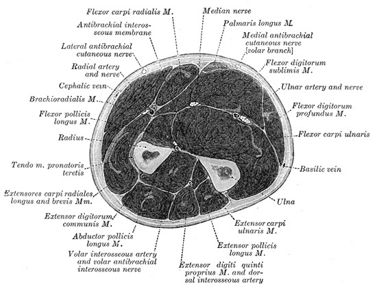
Figure 1. Transverse section of the mid-forearm showing the volar (flexor) and dorsal (extensor) compartments separated by the radius, ulna and interosseous membrane, with the radial, ulnar, median and anterior interosseous neurovascular bundles. Gray’s Anatomy plate 417 (public domain), via Wikimedia Commons.
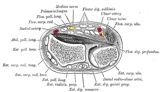
Figure 2. Transverse section of the distal forearm at the pronator quadratus and distal radio-ulnar joint, showing the volar and dorsal compartments and the radial, ulnar and median neurovascular relations. Gray’s Anatomy plate 421 (public domain), via Wikimedia Commons.
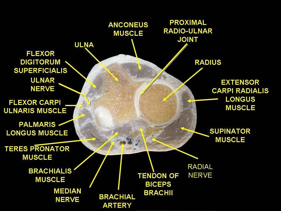
Figure 3. Cadaveric cross-section of the proximal forearm: the flexor and extensor muscle groups around the radius and ulna, with the median, ulnar and radial nerves and the brachial artery. Anatomist90, CC BY-SA 3.0, via Wikimedia Commons.
The bones and the interosseous membrane
The radius and ulna are bowed away from each other to allow pronation and supination, and the radial bow must be restored at fixation if rotation is to be preserved. The ulna is straight and subcutaneous along its whole posterior border, which is why the approach to it is the simplest in the forearm. The two bones are linked by the interosseous membrane, whose fibres run obliquely from the radius down to the ulna; the membrane both transmits load from the radius to the ulna and provides muscle attachment. The biceps inserts into the bicipital tuberosity of the radius, and the supinator wraps from the ulna and lateral epicondyle to insert onto the anterior aspect of the radius, the fact that determines how the proximal radius is exposed.[5]
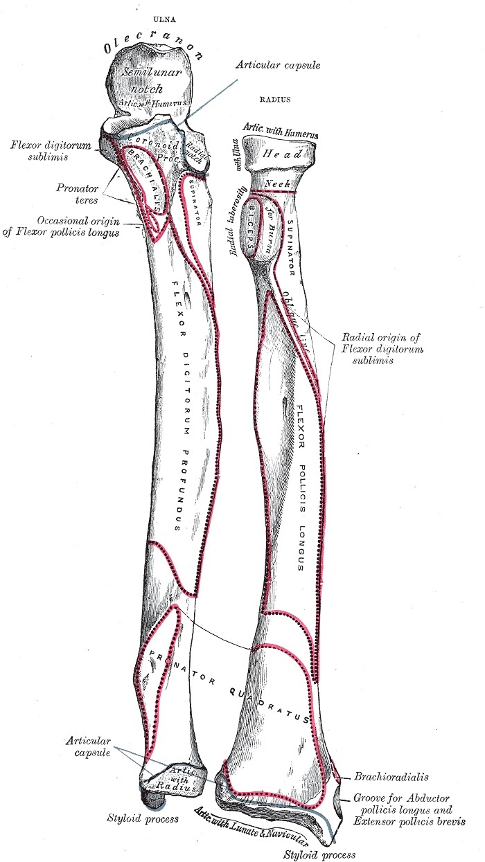
Figure 4. Bones of the left forearm, anterior aspect: radius and ulna with the radial head, neck and bicipital tuberosity, the subcutaneous border of the ulna, and the muscular attachments. Gray’s Anatomy plate 213 (public domain), via Wikimedia Commons.
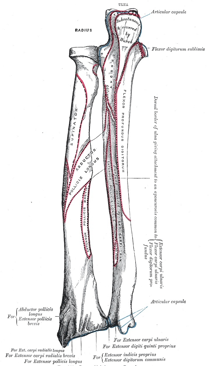
Figure 5. Bones of the left forearm, posterior surface: the subcutaneous dorsal border of the ulna (the landmark for the direct ulnar approach), the interosseous crest, and the dorsal muscle attachments. Gray’s Anatomy plate 214 (public domain), via Wikimedia Commons.
The median nerve and the anterior interosseous nerve
The median nerve enters the forearm by passing between the two heads of the pronator teres, then runs down the forearm adherent to the deep surface of the flexor digitorum superficialis. Just past pronator teres it gives off the anterior interosseous nerve, which runs down the interosseous membrane to supply the deep flexors (flexor pollicis longus, the radial half of flexor digitorum profundus, and pronator quadratus). The median nerve can be entrapped at the pronator teres (pronator syndrome), and the anterior interosseous nerve can be selectively palsied as a pure motor palsy with no sensory loss.[6]
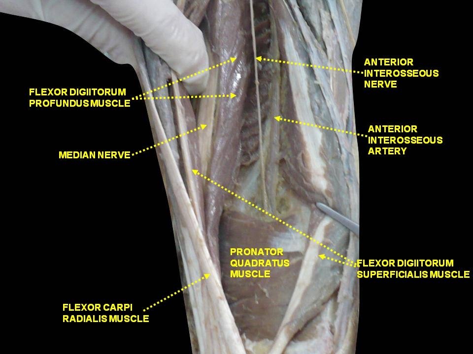
Figure 6. Median nerve in the forearm giving off the anterior interosseous nerve, which descends with the anterior interosseous artery on the interosseous membrane between flexor digitorum profundus and flexor pollicis longus. Anatomist90, CC BY-SA 4.0, via Wikimedia Commons.
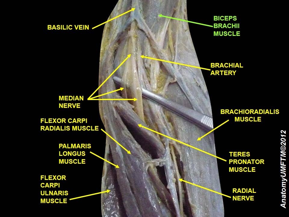
Figure 7. Anterior forearm / cubital fossa dissection: the median nerve and brachial artery entering the forearm between the heads of pronator teres, with the superficial flexors and brachioradialis. Anatomist90, CC BY-SA 3.0, via Wikimedia Commons.
The ulnar nerve and artery; the radial artery
The ulnar nerve runs down the medial forearm under cover of the flexor carpi ulnaris, lying on the flexor digitorum profundus; in the forearm it supplies the flexor carpi ulnaris and the ulnar half of the flexor digitorum profundus. The ulnar artery travels with it on the radial side of the nerve. On the radial side, the radial artery runs down the forearm beneath the brachioradialis, with the superficial (sensory) radial nerve on the undersurface of that muscle, and this nerve is the surgeon’s guide to the correct plane in the Henry approach. Near the elbow the radial artery gives a recurrent leash of vessels to the brachioradialis, the leash of Henry, which must be ligated to mobilise the muscle.[7]
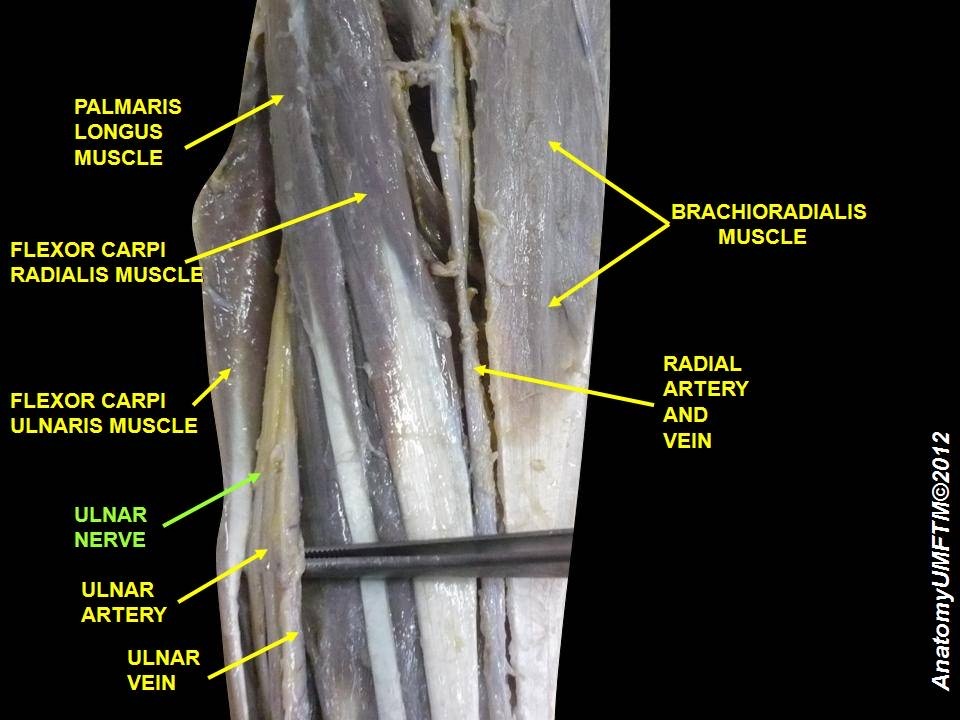
Figure 8. Ulnar neurovascular bundle in the forearm: the ulnar nerve and ulnar artery running deep to flexor carpi ulnaris (the interval between FCU and FDP), with the radial artery shown radially. Anatomist90, CC BY-SA 3.0, via Wikimedia Commons.
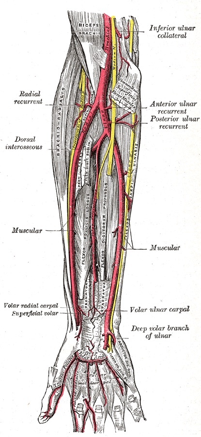
Figure 9. Arteries of the front of the forearm: the radial artery descending beneath the brachioradialis and the ulnar artery on the flexor digitorum profundus, with their recurrent and interosseous branches. Gray’s Anatomy plate 528 (public domain), via Wikimedia Commons.
The radial nerve, the superficial radial nerve, and the PIN
At the elbow the radial nerve divides into the superficial radial nerve, a purely sensory nerve that descends under the brachioradialis, and the posterior interosseous nerve, the motor nerve of the extensor compartment. The PIN enters the supinator beneath its proximal fibrous edge, the arcade of Frohse, runs between the muscle’s two heads, and emerges about 1 cm proximal to the distal edge of the supinator to branch to the wrist, finger, and thumb extensors. In about one patient in four the PIN lies in direct contact with the dorsal surface of the radius just below the bicipital tuberosity. That is why a plate placed too high on the dorsal radius can trap it, and why the nerve must be found and protected in every approach to the proximal radial shaft.[8]
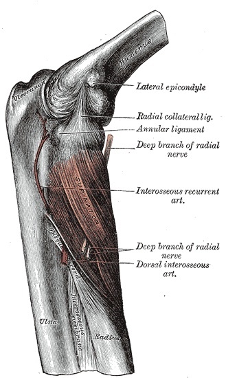
Figure 10. The deep branch of the radial nerve (posterior interosseous nerve) passing through the supinator at the arcade of Frohse to reach the posterior compartment, the structure at risk in approaches to the proximal radius. Gray’s Anatomy plate 420 (public domain), via Wikimedia Commons.
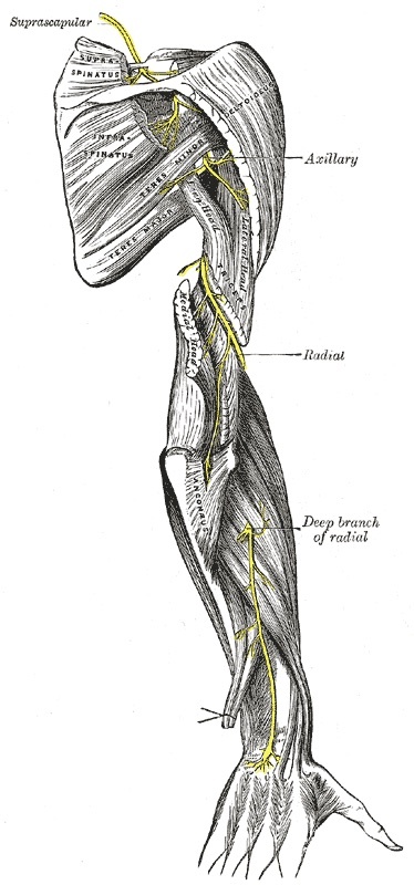
Figure 11. Course of the radial nerve and its deep branch continuing as the posterior interosseous nerve into the extensor compartment of the forearm. Gray’s Anatomy plate 818 (public domain), via Wikimedia Commons.
Part II - Anterior (Henry) Approach to the Radius
What it exposes and when to use it
The anterior approach, first described by Henry, gives a safe exposure of the entire length of the radius and is the classic extensile approach to the bone, relying on subperiosteal stripping of the supinator to protect the PIN. Its uses are ORIF of radial fractures, bone grafting of nonunions, radial osteotomy, biopsy and treatment of tumours, excision of sequestra in osteomyelitis, anterior exposure of the bicipital tuberosity, and treatment of compartment syndrome.[9]
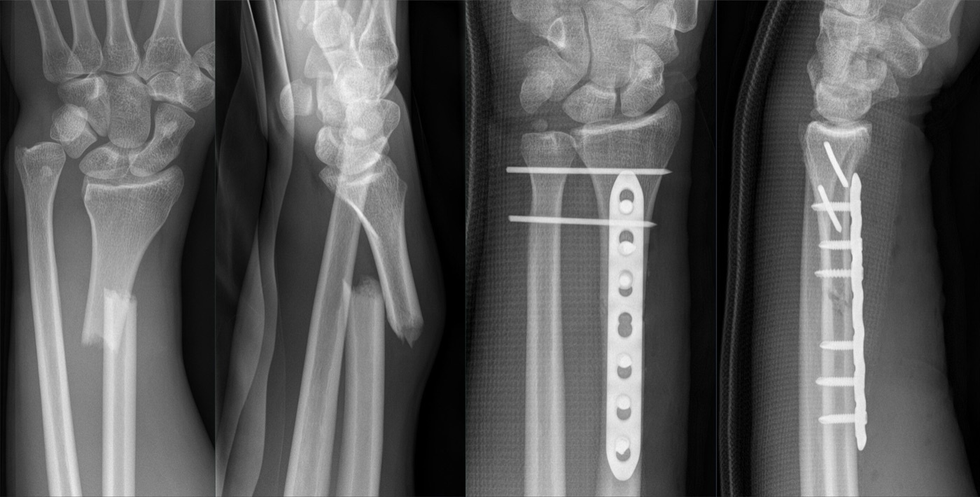
Figure 12. Galeazzi fracture (radial shaft fracture with distal radio-ulnar joint disruption) before and after open reduction and internal fixation of the radius with a volar plate. Hellerhoff, CC BY-SA 4.0, via Wikimedia Commons.
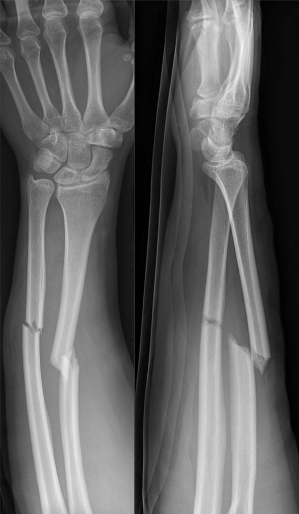
Figure 13. Displaced radial shaft fracture of the forearm from direct trauma, AP and lateral views. Hellerhoff, CC BY-SA 4.0, via Wikimedia Commons.
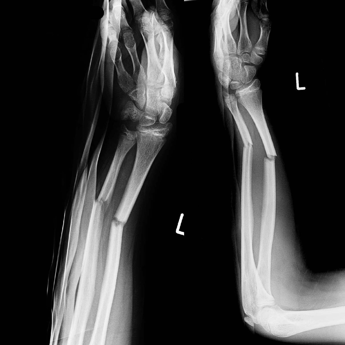
Figure 14. Both-bone (radius and ulna) diaphyseal forearm fracture with angulation, AP and lateral views. Thomas Zimmermann, CC BY-SA 3.0 DE, via Wikimedia Commons.
Position, landmarks, and incision
The patient is supine with the arm on an arm board and the forearm supinated. One useful pearl: the limb is not fully exsanguinated before the tourniquet is inflated, because the venous blood left behind keeps the venae comitantes of the radial artery prominent, and the artery itself is surprisingly small once a tourniquet is up. The incision is a straight line from the flexor crease of the elbow, just lateral to the biceps tendon, down to the radial styloid, taken as long as the lesion requires.[10]
Internervous plane
The internervous plane has the brachioradialis (radial nerve) as its constant lateral wall along the whole forearm; the medial wall changes with level, being the pronator teres (median nerve) proximally and the flexor carpi radialis (median nerve) distally. A classic pitfall is to mistake the plane between brachioradialis and extensor carpi radialis for the correct interval; the guide to the right plane is the superficial radial nerve, which runs on the undersurface of the brachioradialis.[11]
Superficial dissection
The brachioradialis is mobilised and retracted laterally, carrying the superficial radial nerve with it on its undersurface. The recurrent radial leash (the leash of Henry) is carefully ligated and divided, not avulsed, since avulsion causes a postoperative haematoma. The radial artery, lying beneath the brachioradialis near the medial edge of the wound with its two venae comitantes, is mobilised and retracted medially where necessary.[12]
Deep dissection: the three zones and forearm rotation
The deep dissection is governed by a single idea: rotate the forearm so that each muscle’s radial attachment is brought to the front, where it can be detached. The proximal third is the dangerous zone. The supinator cloaks it, with the PIN within its substance, so the forearm is fully supinated, which carries the PIN laterally and posteriorly out of the field and brings the supinator’s anterior insertion into view. The supinator is then detached by dividing its insertion (never by splitting the muscle belly) and stripped subperiosteally off the bone, with the nerve left inside the muscle. This is one of the rare places where subperiosteal stripping is justified, because protecting the nerve outweighs the vascular cost. No retractor is placed on the posterior radial neck, since in about a quarter of patients the PIN lies against the bone there, and excessive retraction causes a neurapraxia that recovers slowly, over six to nine months.[13]
The middle third is covered by the pronator teres and flexor digitorum superficialis; here the forearm is pronated (“turn the arm downward”) to bring the pronator teres insertion on the lateral radius into view, and the muscle is detached and stripped medially. The distal third gives origin to the flexor pollicis longus and pronator quadratus; the forearm is partially supinated and the periosteum incised lateral to these muscles, which are then lifted off medially. Distally the periosteum is left intact, unlike the deliberate subperiosteal stripping of the proximal third.[14]
Dangers
The PIN is the central danger, vulnerable as it winds round the radial neck in the supinator; it is protected by the correct subperiosteal detachment of the supinator with full supination and by avoiding posterior retractors. The superficial radial nerve is at risk when the mobile wad is retracted laterally, with a neuroma or temporary paraesthesia the price of careless handling. The radial artery is vulnerable twice: during mobilisation of the brachioradialis (its venae comitantes are the guide to the small vessel) and proximally as it passes to the medial side of the biceps tendon, where staying lateral to the tendon protects it.[15]
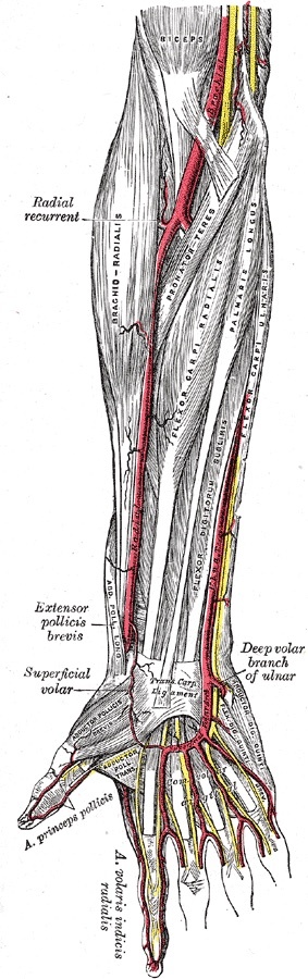
Figure 15. Superficial volar (flexor) muscles of the forearm, with the brachioradialis (mobile wad) radially: the brachioradialis-flexor carpi radialis and brachioradialis-pronator teres intervals are the internervous plane of the Henry approach. Gray’s Anatomy plate 527 (public domain), via Wikimedia Commons.
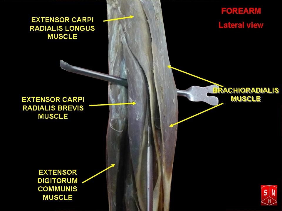
Figure 16. Lateral view of the forearm showing the mobile wad (brachioradialis, extensor carpi radialis longus and brevis), which is mobilised and retracted to develop the Henry plane. Anatomist90, CC BY-SA 3.0, via Wikimedia Commons.
Part III - Exposure of the Shaft of the Ulna
The ulnar approach is the simplest in the forearm, because the ulna is subcutaneous along its whole posterior border and can be reached directly. It exposes the entire length of the bone and is used for ORIF of ulnar fractures, nonunion, osteotomy, osteomyelitis, the fibrous anlage of ulnar clubhand, and for ulnar lengthening (in Kienböck disease) or shortening (for distal radial malunion). The patient is supine with the arm across the chest, or the elbow flexed 90° on the table, after exsanguination and a tourniquet. The incision is a straight line over the palpable subcutaneous border, centred on the fracture.[16]
The internervous plane lies between the flexor carpi ulnaris (ulnar nerve) and the extensor carpi ulnaris (posterior interosseous nerve); near the olecranon it becomes the flexor carpi ulnaris (ulnar nerve) against the anconeus (radial nerve), still a genuine internervous interval. Because the two muscles share a common aponeurosis at the subcutaneous border, the plane is hard to define, and some extensor carpi ulnaris fibres usually have to be detached to reach bone. Deeper dissection stays epiperiosteal, which is safe all the way around the bone because the flanking muscle masses protect the volar structures. The proximal fifth requires detaching part of the broad triceps insertion.[17]
The dangers are the ulnar nerve and ulnar artery, which lie together on the volar side under the flexor carpi ulnaris (the nerve on the flexor digitorum profundus, the artery on the radial side of the nerve). Both are safe as long as the flexor carpi ulnaris is stripped epiperiosteally, and both are endangered if the dissection strays into the muscle, especially proximally. The ulnar nerve is therefore identified as it passes between the two heads of the flexor carpi ulnaris before the proximal fifth is stripped. The approach cannot be extended distally, but it can be carried over the olecranon and up the back of the arm, either through an olecranon osteotomy to the elbow joint or onto the posterior distal humerus.[18]
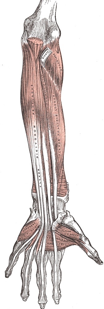
Figure 17. Deep volar (flexor) muscles of the forearm: flexor digitorum profundus, flexor pollicis longus and pronator quadratus; the ulnar nerve runs on the flexor digitorum profundus deep to flexor carpi ulnaris. Gray’s Anatomy plate 415 (public domain), via Wikimedia Commons.
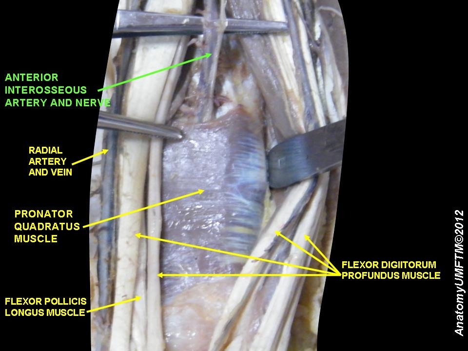
Figure 18. Deep flexor compartment, cadaveric dissection: flexor digitorum profundus, flexor pollicis longus and pronator quadratus, with the anterior interosseous neurovascular bundle on the interosseous membrane. Anatomist90, CC BY-SA 3.0, via Wikimedia Commons.
Part IV - Posterior (Thompson) Approach to the Radius
What it exposes and when to use it
The posterior approach, universally known as the Thompson approach, gives good access to the entire dorsal aspect of the radial shaft. Its principal aim, in Hoppenfeld’s words, is to isolate and retract the posterior interosseous nerve before the proximal radius is exposed, keeping the nerve under direct view throughout. Its uses are ORIF of radial fractures (the dorsal/extensor surface is the tension side, biomechanically the best place for a plate), nonunion, decompression of the PIN at the arcade of Frohse for nerve palsy or resistant tennis elbow, radial osteotomy, osteomyelitis, and tumours.[19]
Position, incision, and the internervous-plane caveat
The patient is supine with the forearm pronated to bring the extensor compartment into view (or the arm across the chest), after exsanguination and a tourniquet. The incision runs from just anterior to the lateral epicondyle to just distal to the ulnar side of Lister’s tubercle at the wrist. There is a caveat that examiners value: Hoppenfeld states plainly that no true internervous plane exists in this approach. Proximally the plane runs between the extensor carpi radialis brevis and the extensor digitorum communis, distally between the extensor carpi radialis brevis and the extensor pollicis longus. In this edition all of these are supplied by the PIN, so the interval is made safe not by different nerves but by the fact that the extensor carpi radialis brevis receives its branch well proximal to the dissection.[20]
Dissection and protecting the PIN
The superficial interval between the extensor carpi radialis brevis and the extensor digitorum communis is found distally, where the abductor pollicis longus and extensor pollicis brevis emerge between them, and then traced proximally where the two muscles share a common aponeurosis. The deep step is the whole point of the approach. The PIN is identified first in the proximal third, either proximal-to-distal (detaching the extensor carpi radialis brevis origin and palpating the nerve above the supinator) or distal-to-proximal (finding it where it emerges about 1 cm proximal to the supinator’s distal edge), preserving every muscular branch. Only then, with the nerve identified, is the forearm fully supinated to bring the supinator’s anterior insertion into view and move the PIN away, and the supinator detached subperiosteally from the anterior radius.[21]
A point of genuine importance for the viva: this fifth-edition deep step uses full supination, not the pronation of the classic “pronate-for-Thompson” mnemonic. The reason is anatomical. The supinator inserts on the anterior aspect of the radius, so supination, not pronation, delivers that insertion to the surgeon and at the same time rotates the nerve out of the dorsal field. The candidate who states the principle (rotate the forearm to bring the supinator insertion into view and carry the PIN away) is on safe ground; the candidate who recites the mnemonic without the anatomy may be caught out.[22]
Dangers and extensile measures
The PIN is the central danger throughout. In about a quarter of patients it touches the dorsal radius just below the bicipital tuberosity, where a high dorsal plate can trap it, and its position shifts with the type of trauma, so full dissection of the nerve is the only certain protection during proximal-third plating. The superficial radial nerve (sensory, supplying no forearm muscles) and the small posterior interosseous artery (which joins the nerve distal to the supinator and is protected by good collateral flow) are the other named structures. The approach extends distally to the dorsal wrist and proximally to the lateral epicondyle, though both extensions are rarely needed. For plate work, the dorsal surface is the tension side and biomechanically ideal, at the cost of possible extensor-tendon irritation.[23]
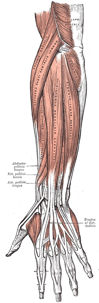
Figure 19. Superficial muscles of the posterior (extensor) compartment: brachioradialis, the extensors carpi radialis longus and brevis, extensor digitorum, extensor carpi ulnaris and anconeus. Gray’s Anatomy plate 418 (public domain), via Wikimedia Commons.
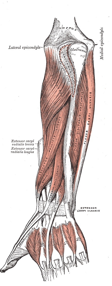
Figure 20. Deep muscles of the posterior compartment: the supinator proximally and the outcropping thumb extensors (abductor pollicis longus, extensor pollicis brevis and longus) and extensor indicis. Gray’s Anatomy plate 419 (public domain), via Wikimedia Commons.
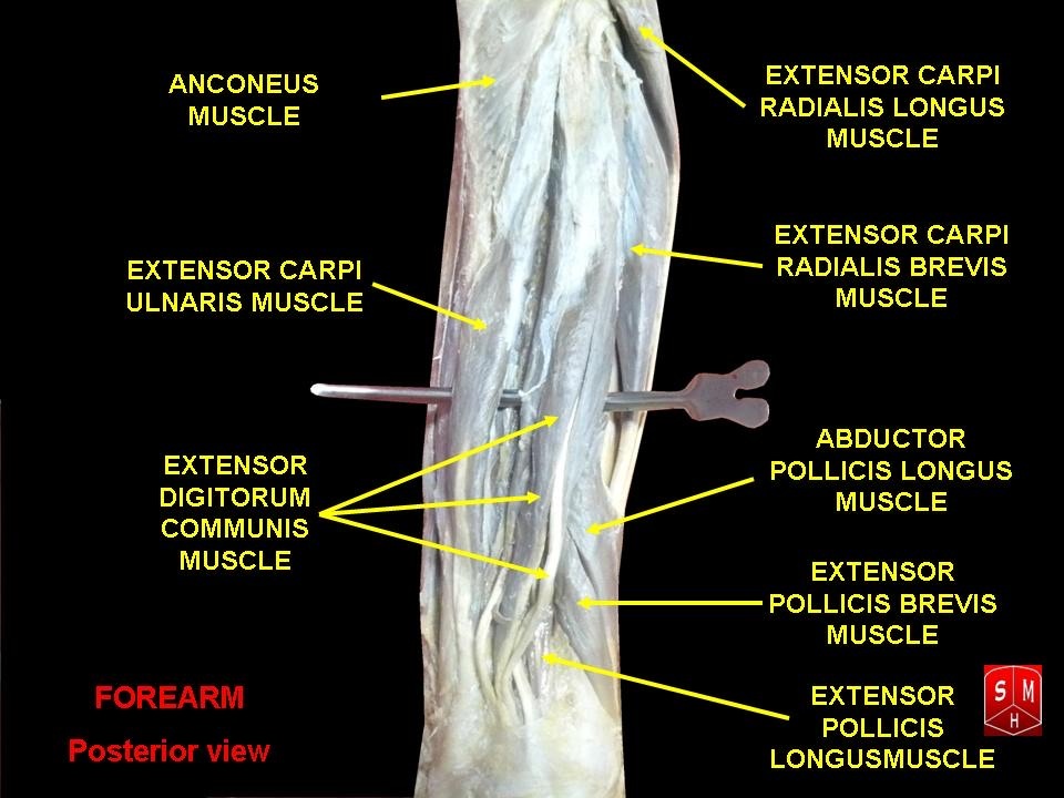
Figure 21. Posterior (extensor) compartment of the forearm, cadaveric dissection: the superficial and deep extensors including the extensor carpi radialis longus/brevis, extensor digitorum, extensor carpi ulnaris, anconeus and the outcropping thumb muscles. Anatomist90, CC BY-SA 3.0, via Wikimedia Commons.
Part V - Forearm Fasciotomy for Compartment Syndrome
Compartment syndrome is a surgical emergency in which rising pressure within the closed fascial compartments of the forearm reduces venous return and then arterial inflow, producing a self-perpetuating cycle of ischaemia and oedema that ends, if untreated, in the muscle necrosis and fibrosis of a Volkmann ischaemic contracture. It can occur without a fracture, classically after crush or roller injuries. The cardinal clinical sign is pain out of proportion to the injury, especially on passive stretch of the compartment muscles. The critical teaching point is that the distal pulses remain normal until very late, so the surgeon must never wait for pulse changes before decompressing.[24]
The volar (anterior) compartment is released through a long curved incision, with two described routes. The central route runs from the lateral epicondyle to the radial styloid, entering between the palmaris longus and flexor carpi radialis; the lacertus fibrosus is divided to free the median nerve, and the fascia of the flexor digitorum superficialis is incised to reach the deep flexors. The ulnar route runs from the medial epicondyle to a point just lateral to the ulnar styloid, entering between the flexor carpi ulnaris and the flexor digitorum superficialis; the median nerve is kept on the deep surface of the superficialis, the ulnar nerve is mobilised, and the three deep flexors (pronator quadratus, flexor pollicis longus, flexor digitorum profundus) are decompressed by epimysiotomy. The deep flexors are the muscles most at risk of ischaemic necrosis, and decompressing them is the purpose of the deep release.[25]
The dorsal (posterior) compartment and the mobile wad are released through a straight incision from the lateral epicondyle toward Lister’s tubercle, dividing the deep fascia only and preserving the extensor retinaculum. Often the volar release alone decompresses the dorsal side adequately, but the dorsal compartment is checked and released if still tight. The dangers are the median nerve and the ulnar neurovascular bundle on the volar side and the PIN in the supinator dorsally. The skin is left open, because closure under tension would recreate the very pressure the fasciotomy was meant to relieve, so the wound is managed by delayed closure or skin grafting once swelling settles.[26]
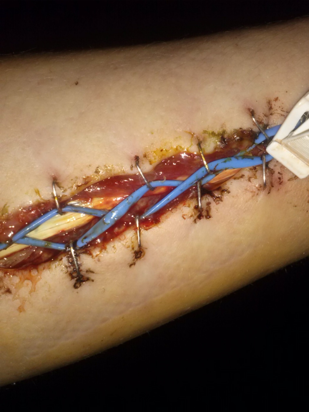
Figure 22. Forearm fasciotomy for compartment syndrome, managed with a vessel-loop “shoelace” closure to allow gradual delayed skin approximation. TPompert, CC BY-SA 4.0, via Wikimedia Commons.
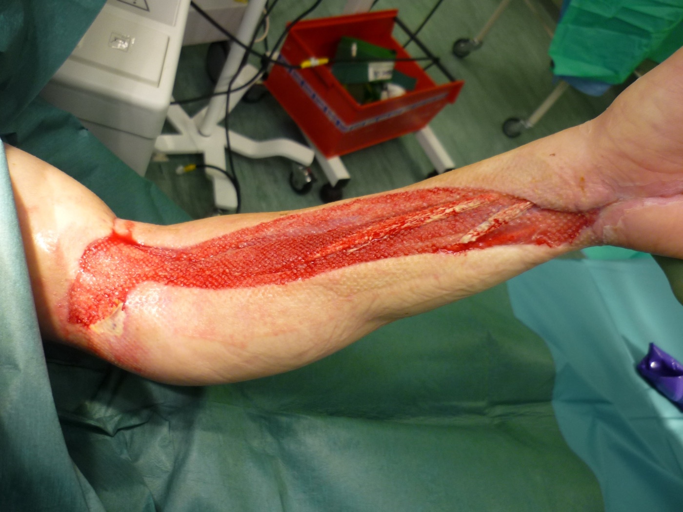
Figure 23. Open forearm fasciotomy wound (granulating) prior to split-skin grafting. Guyprocter, released under CC0, via Wikimedia Commons.
References
-
Hoppenfeld describes three approaches to the forearm bones, all giving complete bony exposure: the anterior (Henry) approach to the radius (the classic extensile approach), the posterior (Thompson) approach to the radius, and the direct approach to the subcutaneous border of the ulna; plus fasciotomy for compartment syndrome (Hoppenfeld, Surgical Exposures in Orthopaedics, 5th ed., ch.4, p.296, p.317, p.325, p.340). The PIN winds around the radial neck within the supinator, close to or touching the periosteum (p.296).
-
The forearm chapter presents two applied-anatomy sections (the anterior compartment, p.307-316, and the posterior approach, p.334-339) interleaved with the approaches; the highest-yield surgical items are the internervous planes and the named dangers around the PIN and the median/ulnar bundles (Hoppenfeld p.307, p.334).
-
The supinator inserts on the anterior aspect of the radius (Hoppenfeld p.339); in the Henry approach the proximal third is exposed by full supination, which moves the PIN laterally and posteriorly out of the field and brings the supinator insertion anterior (p.302); in the Thompson approach the deep step likewise uses full supination “to bring the insertion of the supinator into view and to move the posterior interosseous nerve away from the area of incision” (p.332, Fig. 4-30 caption), with pronation used only for initial positioning (p.326). The classic “supinate for Henry / pronate for Thompson” mnemonic is widely taught but does not match this edition’s Thompson deep step (standard teaching).
-
The volar/flexor compartment has three layers: superficial (pronator teres, flexor carpi radialis, palmaris longus, flexor carpi ulnaris), intermediate (flexor digitorum superficialis), and deep (flexor digitorum profundus, flexor pollicis longus, pronator quadratus); the dorsal/extensor side has twelve muscles in three groups: the mobile wad of three (brachioradialis, ECRL, ECRB), the four superficial extensors from the common extensor origin (anconeus, ECU, extensor digiti minimi, extensor digitorum communis), and five deep muscles (abductor pollicis longus, extensor pollicis brevis, extensor pollicis longus, supinator, extensor indicis) (Hoppenfeld p.307, p.334).
-
The radius and ulna are bowed to permit pronation/supination (the radial bow, standard teaching, must be restored at fixation); the ulna is subcutaneous along its posterior border (Hoppenfeld p.317); the interosseous membrane links the two bones; the supinator inserts on the anterior aspect of the radius (Hoppenfeld p.339), and the biceps on the bicipital tuberosity (p.302). The obliquity of the interosseous membrane fibres and the load-transfer function are standard teaching.
-
The median nerve passes between the two heads of pronator teres and runs deep to flexor digitorum superficialis; it gives the anterior interosseous nerve, which runs on the interosseous membrane to supply flexor pollicis longus, the radial half of flexor digitorum profundus, and pronator quadratus; entrapment occurs at the pronator teres (pronator syndrome) and the anterior interosseous nerve can be selectively affected (Hoppenfeld p.307-316). The AIN-syndrome “OK-sign” and the palmar cutaneous branch are standard teaching.
-
The ulnar nerve runs under flexor carpi ulnaris on the flexor digitorum profundus, supplying FCU and the ulnar half of FDP, with the ulnar artery on its radial side (Hoppenfeld p.324); the radial artery runs beneath the brachioradialis with the superficial radial nerve on the muscle’s undersurface, and gives a recurrent leash to the brachioradialis near the elbow (the “leash of Henry,” standard-teaching name) that must be ligated to mobilise the muscle (Hoppenfeld p.301).
-
The radial nerve divides into the superficial radial nerve (sensory, descending under brachioradialis) and the posterior interosseous nerve (motor to the extensor compartment); the PIN enters the supinator through the arcade of Frohse, runs between its two heads, and emerges about 1 cm proximal to the distal edge of the supinator; in about 25% of patients it touches the dorsal radius just below the bicipital tuberosity, so a high dorsal plate can trap it, and the nerve must be identified to protect it during proximal-radius plating (Hoppenfeld p.328, p.331, p.333, p.335, p.338).
-
The anterior (Henry) approach exposes the entire length of the radius and is the classic extensile approach, protecting the PIN by subperiosteal stripping of the supinator; uses = ORIF of fractures, grafting of nonunions, radial osteotomy, biopsy/treatment of tumours, excision of sequestra in osteomyelitis, anterior exposure of the bicipital tuberosity, and compartment-syndrome treatment (Hoppenfeld p.296-297). It was first described by Henry, whose name is associated with it (p.296).
-
Position supine, arm on an arm board, forearm supinated; do not fully exsanguinate before inflating the tourniquet, so the venae comitantes stay prominent as a guide to the small radial artery; incision a straight line from the elbow flexor crease just lateral to the biceps tendon to the radial styloid, scaled to the lesion (Hoppenfeld p.297).
-
Internervous plane: brachioradialis (radial nerve) laterally throughout, against pronator teres (median nerve) proximally and flexor carpi radialis (median nerve) distally; a common error is to mistake the brachioradialis-extensor carpi radialis plane for the correct interval, and the superficial radial nerve on the undersurface of brachioradialis confirms the right plane (Hoppenfeld p.298, p.300).
-
Mobilise and retract the brachioradialis laterally, carrying the superficial radial nerve on its undersurface; ligate and divide (do not avulse) the recurrent radial leash (leash of Henry) to avoid a postoperative haematoma; the radial artery with its venae comitantes lies beneath the brachioradialis near the medial wound edge and is retracted medially as needed (Hoppenfeld p.301).
-
Proximal third: the supinator cloaks it with the PIN inside; fully supinate the forearm to carry the PIN laterally/posteriorly out of the field and bring the supinator’s anterior insertion into view; detach the supinator by dividing its insertion (not by splitting the belly) and strip it subperiosteally, leaving the nerve in the muscle; subperiosteal stripping is justified here because nerve safety outweighs the vascular cost; place no retractor on the posterior radial neck (the PIN touches it in ~25%), and retract gently, since a neurapraxia recovers over 6-9 months (Hoppenfeld p.302, p.306).
-
Middle third: covered by pronator teres and flexor digitorum superficialis; pronate the forearm (“turn the arm downward”) to bring the pronator teres insertion on the lateral radius forward, then detach and strip it medially. Distal third: flexor pollicis longus and pronator quadratus arise here; partially supinate and incise the periosteum lateral to them, lifting them off medially, leaving the periosteum intact distally (Hoppenfeld p.304-305). The pronator quadratus detach-versus-divide debate (better repair and a soft-tissue pad over a volar plate) is unresolved in the text (p.304-305).
-
Dangers: the PIN winds round the radial neck in the supinator (protected by correct subperiosteal supinator detachment with full supination and no posterior retractors); the superficial radial nerve is at risk when the mobile wad is retracted laterally (neuroma/paraesthesia); the radial artery is vulnerable twice, during brachioradialis mobilisation (venae comitantes are the guide) and proximally as it passes medial to the biceps tendon (stay lateral to the tendon) (Hoppenfeld p.306). The recurrent radial vessels are ligated, not avulsed (p.301).
-
The ulnar approach is the simplest forearm approach, the ulna being subcutaneous along its posterior border, and exposes the whole bone; uses = ORIF of ulnar fractures, delayed/nonunion, osteotomy, chronic osteomyelitis, the fibrous anlage in ulnar clubhand, ulnar lengthening (Kienböck) and ulnar shortening (distal radial malunion); position supine, arm across the chest or elbow flexed 90°, exsanguinate and apply a tourniquet; incision a longitudinal line over the subcutaneous border, centred on the fracture (Hoppenfeld p.317-318).
-
Internervous plane: flexor carpi ulnaris (ulnar nerve) versus extensor carpi ulnaris (posterior interosseous nerve), becoming flexor carpi ulnaris (ulnar nerve) versus anconeus (radial nerve) near the olecranon; the shared aponeurosis makes the plane hard to define, so ECU fibres are usually detached; deep dissection stays epiperiosteal, which is safe circumferentially because the flanking muscles protect the volar structures, and the proximal fifth requires detaching part of the triceps insertion (Hoppenfeld p.317-323).
-
Dangers: the ulnar nerve and ulnar artery lie volar under the flexor carpi ulnaris (nerve on the flexor digitorum profundus, artery on the radial side of the nerve), safe if the FCU is stripped epiperiosteally and endangered if dissection strays into the muscle, most so proximally, so the ulnar nerve is identified between the two FCU heads before stripping the proximal fifth; the approach cannot be extended distally but can be carried over the olecranon up the back of the arm (olecranon osteotomy to the elbow, or onto the posterior distal humerus) (Hoppenfeld p.323-324). The dorsal cutaneous branch of the ulnar nerve at risk distally is standard teaching.
-
The posterior (Thompson) approach gives access to the entire dorsal radial shaft; its principal aim is to isolate and retract the PIN before exposing the proximal radius and keep it in view throughout; uses = ORIF of radial fractures (the dorsal surface is the tension side, the best place for a plate), nonunion, PIN decompression at the arcade of Frohse (nerve palsy or resistant tennis elbow), radial osteotomy, osteomyelitis, and tumours (Hoppenfeld p.325, p.332). The “Thompson” eponym is standard teaching.
-
Position supine, forearm pronated to bring the extensor compartment into view (or arm across the chest), exsanguinate and apply a tourniquet; incision from just anterior to the lateral epicondyle to just distal to the ulnar side of Lister’s tubercle (Hoppenfeld p.326-327). “No true internervous plane exists in this approach” (p.327): proximally extensor carpi radialis brevis/extensor digitorum communis, distally extensor carpi radialis brevis/extensor pollicis longus, all PIN-supplied in this edition, the interval being safe because the ECRB branch arises proximal to the dissection (p.327, p.334). ECRL is radial-nerve-supplied, ECRB is PIN-supplied per this edition’s muscle table (p.339); the “true internervous plane” framing with ECRB as radial-supplied is standard teaching from other texts.
-
Find the superficial interval between extensor carpi radialis brevis and extensor digitorum communis distally (where abductor pollicis longus and extensor pollicis brevis emerge) and trace it proximally through the common aponeurosis; identify the PIN first in the proximal third, proximal-to-distal (detach the ECRB origin, palpate the nerve above the supinator) or distal-to-proximal (it emerges ~1 cm proximal to the supinator’s distal edge), preserving all muscular branches; then fully supinate the forearm to bring the supinator’s anterior insertion into view and move the PIN away, and detach the supinator subperiosteally from the anterior radius (Hoppenfeld p.327-332). Fig. 4-30 caption confirms full supination “to move the posterior interosseous nerve away from the area of incision” (p.332).
-
Hoppenfeld 5e uses full supination for the Thompson deep step because the supinator inserts on the anterior radius, so supination delivers the insertion and carries the PIN out of the dorsal field (p.332, p.339); the classic “pronate for Thompson / supinate for Henry” mnemonic is a widely-taught simplification not followed for this edition’s deep step (standard teaching). Pronation here is only for initial positioning (p.326).
-
The PIN is the central danger: it touches the dorsal radius just below the bicipital tuberosity in ~25% of patients (where a high dorsal plate can trap it), its position varies with trauma, and full dissection is the only certain protection during proximal-third plating; the superficial radial nerve (sensory) and the small posterior interosseous artery (joining the nerve distal to the supinator, protected by collateral flow) are the other structures; the approach extends distally to the dorsal wrist and proximally to the lateral epicondyle, both rarely needed; the dorsal surface is the tension side, biomechanically best for a plate but prone to extensor-tendon irritation (Hoppenfeld p.333-338).
-
Compartment syndrome: rising compartment pressure reduces venous return then arterial inflow, a self-perpetuating ischaemia-oedema cycle ending in Volkmann ischaemic contracture; it can occur without a fracture (crush/roller injuries); the cardinal sign is pain out of proportion (especially on passive stretch), and distal pulses stay normal until very late, so do not wait for pulse changes before decompressing (Hoppenfeld p.340-345). Threshold pressures are deliberately not emphasised in the text.
-
The volar compartment is released through a long curved incision by one of two routes: the central route (lateral epicondyle to radial styloid, between palmaris longus and flexor carpi radialis, dividing the lacertus fibrosus to free the median nerve and incising the flexor digitorum superficialis fascia to reach the deep flexors), or the ulnar route (medial epicondyle to just lateral to the ulnar styloid, between flexor carpi ulnaris and flexor digitorum superficialis, keeping the median nerve on the deep surface of FDS, mobilising the ulnar nerve, and epimysiotomy of the deep flexors - pronator quadratus, flexor pollicis longus, flexor digitorum profundus, the muscles most at risk of necrosis) (Hoppenfeld p.340-348). The lazy-S/curved incision shape, the volar skin flap that protects the neurovascular bundle, and carpal tunnel release are standard teaching.
-
The dorsal compartment and mobile wad are released through a straight incision from the lateral epicondyle toward Lister’s tubercle, dividing the deep fascia only and preserving the extensor retinaculum, often unnecessary if the volar release decompresses the dorsal side; dangers are the median nerve and ulnar neurovascular bundle volarly and the PIN in the supinator dorsally; the skin is left open and managed by delayed closure or skin grafting, since closure under tension recreates the pressure (Hoppenfeld p.340-352). Detailed wound-closure and skin-graft guidance is largely standard teaching.
-
The PIN winds round the radial neck within the supinator and in ~25% of patients touches the dorsal radius below the bicipital tuberosity, so a high dorsal plate can trap it and proximal-third dissection can injure it; both radial approaches are built to protect it (Hoppenfeld p.296, p.302, p.333).
-
Henry internervous plane: brachioradialis (radial nerve) laterally throughout, against pronator teres (median nerve) proximally and flexor carpi radialis (median nerve) distally (Hoppenfeld p.298).
-
Fully supinate the forearm to move the PIN out of the field and bring the supinator insertion anterior; detach the supinator by its insertion (not by splitting the belly) and strip subperiosteally with the nerve in the muscle; no posterior radial-neck retractor; gentle retraction, as a neurapraxia recovers over 6-9 months (Hoppenfeld p.302).
-
Henry rotation by zone: full supination (proximal, supinator), pronation (middle, pronator teres), partial supination (distal, flexor pollicis longus and pronator quadratus) (Hoppenfeld p.302, p.304).
-
Ulnar approach plane: flexor carpi ulnaris (ulnar nerve) versus extensor carpi ulnaris (PIN), becoming FCU (ulnar) versus anconeus (radial) near the olecranon; the ulna is subcutaneous so the approach cuts directly onto bone, but the shared aponeurosis makes the plane hard to define (Hoppenfeld p.317-320).
-
Ulnar and ulnar-artery danger: both lie volar under the flexor carpi ulnaris (nerve on FDP, artery on the radial side of the nerve), safe if the FCU is stripped epiperiosteally, endangered if dissection enters the muscle, most so proximally; identify the ulnar nerve between the two FCU heads before stripping the proximal fifth (Hoppenfeld p.323-324).
-
Hoppenfeld states “no true internervous plane exists in this approach”; proximally ECRB/extensor digitorum communis, distally ECRB/extensor pollicis longus, all PIN-supplied, the interval safe because the ECRB branch arises proximal to the dissection (Hoppenfeld p.327, p.334).
-
Identify the PIN first (it emerges ~1 cm proximal to the supinator’s distal edge), preserving its branches, then fully supinate to deliver the supinator’s anterior insertion and move the nerve away, detaching the supinator subperiosteally; Hoppenfeld 5e specifies full supination for the deep step (not pronation), because the supinator inserts on the anterior radius (Hoppenfeld p.331-332, p.339). The “pronate for Thompson” mnemonic is standard teaching not followed here.
-
The dorsal/extensor surface is the tension side, biomechanically the best place for a plate, but extensor-tendon irritation over the plate may be a problem (Hoppenfeld p.325, p.332).
-
Henry = anterior/volar, whole radius, classic extensile, true internervous plane (brachioradialis vs pronator teres/flexor carpi radialis); Thompson = posterior/dorsal, whole dorsal shaft, no true internervous plane, favoured for a dorsal tension-side plate; both protect the PIN by identification and subperiosteal supinator detachment from the anterior radius with the forearm supinated (Hoppenfeld p.296, p.325, p.332). The regional preference is partly standard teaching.
-
Acute compartment syndrome can occur without a fracture (crush injury); the cardinal sign is pain out of proportion, especially on passive stretch, and distal pulses stay normal until very late, so do not wait for pulse changes before decompressing (Hoppenfeld p.340-345).
-
Volar fasciotomy: long curved incision, divide the lacertus fibrosus to free the median nerve, incise the FDS fascia to decompress the deep flexors (the muscles most at risk); release the dorsal compartment separately if tight; leave the skin open for delayed closure or grafting, since closure under tension recreates the pressure (Hoppenfeld p.340-352).