Contents
- Orientation
- Part I - Applied Surgical Anatomy of the Ankle and Foot
- The three tendon groups and three internervous planes
- The front of the ankle and the anterior bundle
- The medial ankle and the tarsal tunnel
- The lateral ankle and the superficial sensory triad
- The ankle joint, mortise, and ligaments
- The subtalar joint, tarsal canal, and sinus tarsi
- The dorsum and sole of the foot
- Part II - Approaches to the Ankle
- Part III - Approaches to the Hindfoot
- Part IV - Approaches to the Forefoot
- References
Orientation
A crowded traffic of tendons, vessels, and nerves crosses the ankle and foot, so almost every approach here comes down to a single decision: which structures to slip between, and which to protect. One organising idea does most of the work. Three groups of tendons cross the ankle, each with its own nerve: the flexors behind the medial malleolus (tibial nerve), the extensors in front (deep peroneal nerve), and the evertors behind the lateral malleolus (superficial peroneal nerve). Because their nerve supplies differ, these groups generate the three internervous planes of the ankle. The two neurovascular bundles they flank, the anterior bundle on the dorsum and the posterior bundle in the tarsal tunnel, are the structures every approach is built around.[1]
The other through-line of foot and ankle surgery is the soft-tissue envelope. The skin here is thin, frequently traumatised, and slow to heal, so poor wound healing is the commonest complication of the whole region. Three rules follow from this and recur in every section. Operate only once swelling has subsided and the circulation is assessed. Raise thick, full-thickness flaps rather than thin ones. And place incisions to spare the superficial sensory nerves, because a divided cutaneous nerve here means a painful neuroma in a shoe-bearing area. With those principles in hand, this summary covers the applied anatomy first and then works outward from the ankle, through the hindfoot, to the forefoot.[2]
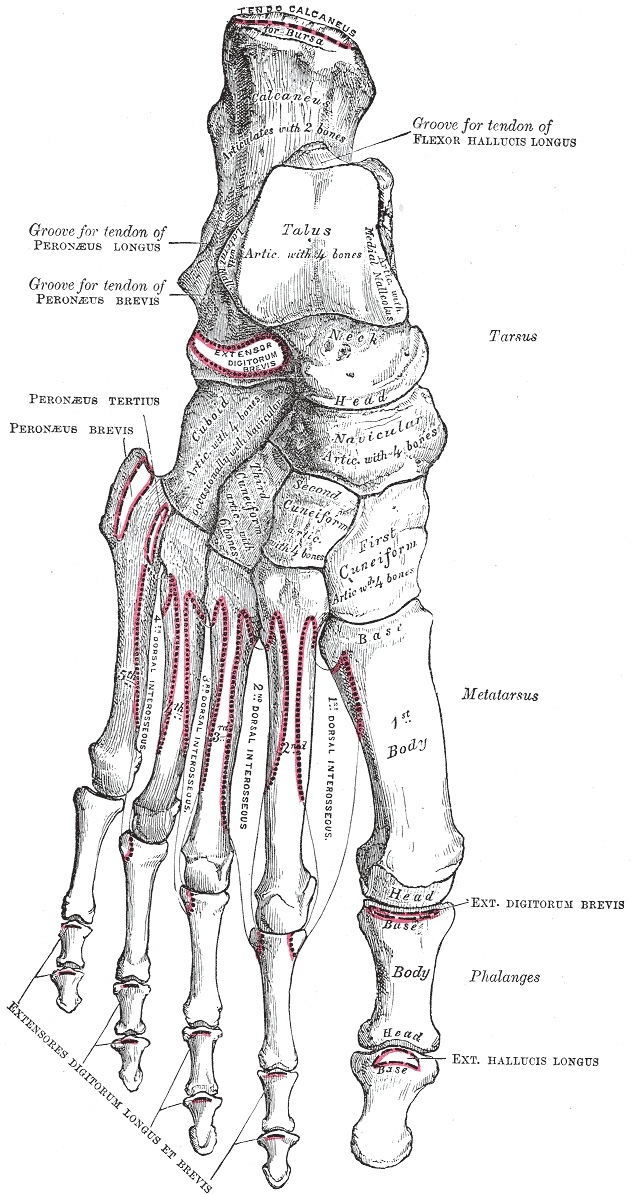
Figure 1. Bones of the right foot, dorsal surface: calcaneus, talus, navicular, cuboid, the three cuneiforms, metatarsals and phalanges. Gray’s Anatomy (1918), Plate 268, public domain, via Wikimedia Commons.
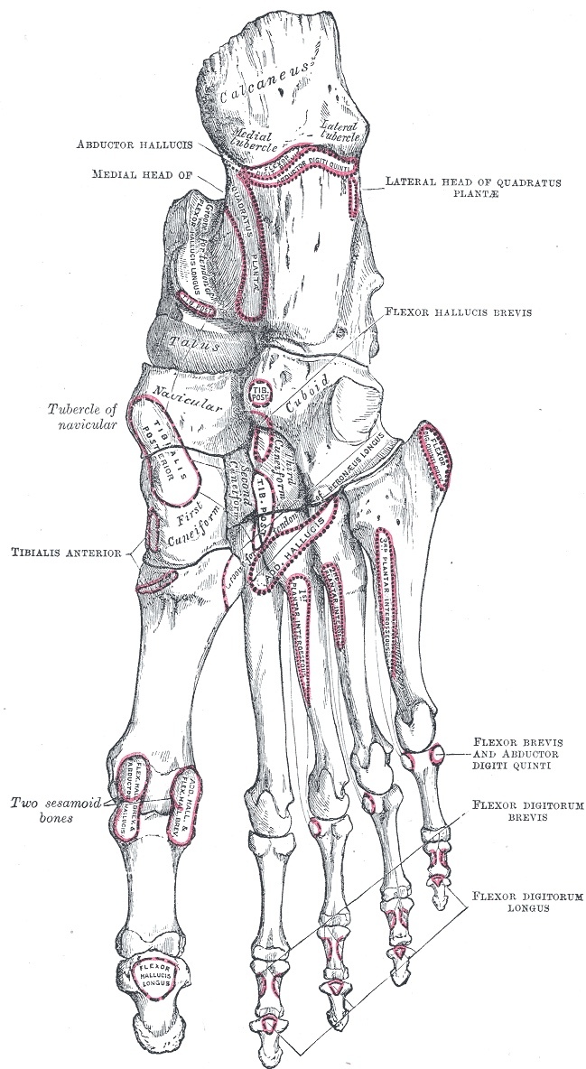
Figure 2. Bones of the right foot, plantar surface, with the plantar muscle attachments outlined. Gray’s Anatomy (1918), Plate 269, public domain, via Wikimedia Commons.
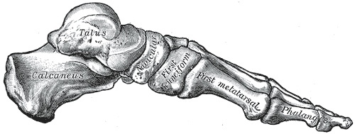
Figure 3. Skeleton of the foot, medial aspect, showing the medial longitudinal arch. Gray’s Anatomy (1918), Plate 290, public domain, via Wikimedia Commons.
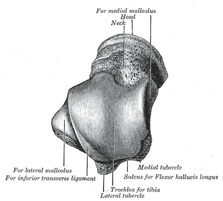
Figure 4. Left talus, superior surface: the trochlea and the facets for the medial and lateral malleoli. Gray’s Anatomy (1918), Plate 270, public domain, via Wikimedia Commons.
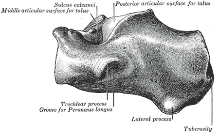
Figure 5. Left calcaneus, lateral surface, with the peroneal (trochlear) tubercle and the groove for peroneus longus. Gray’s Anatomy (1918), Plate 266, public domain, via Wikimedia Commons.
Part I - Applied Surgical Anatomy of the Ankle and Foot
The three tendon groups and three internervous planes
Apart from the Achilles and plantaris tendons, which lie posteriorly in the midline, three sets of tendons cross the ankle. The flexors (tibialis posterior, flexor digitorum longus, flexor hallucis longus) pass behind the medial malleolus and are supplied by the tibial nerve. The extensors (tibialis anterior, extensor hallucis longus, extensor digitorum longus, peroneus tertius) pass in front of the joint and are supplied by the deep peroneal nerve. The evertors (peroneus longus and brevis) pass behind the lateral malleolus and are supplied by the superficial peroneal nerve. All of them are held against the skeleton by thickened bands of deep fascia, the retinacula, which prevent bowstringing and which must be repaired whenever an approach divides them. The differing nerve supplies give three planes: medially between flexors and extensors, posterolaterally between flexors and evertors, and laterally between extensors and evertors.[3]
The front of the ankle and the anterior bundle
Four extensor tendons cross the front of the ankle. From medial to lateral they are tibialis anterior, extensor hallucis longus, extensor digitorum longus, and peroneus tertius. The anterior neurovascular bundle, the anterior tibial artery with the deep peroneal nerve, crosses roughly halfway between the malleoli. Above the joint it lies between tibialis anterior and extensor hallucis longus. The extensor hallucis longus tendon then crosses it from lateral to medial, so that at the level of the joint the bundle lies immediately lateral to the extensor hallucis longus tendon, and below the joint it lies between extensor hallucis longus and extensor digitorum longus. Beyond the ankle the anterior tibial artery becomes the dorsalis pedis, palpable on the dorsum, which dives into the first intermetatarsal space and anastomoses with the plantar circulation. A Lisfranc injury can disrupt that connection.[4]
The deep peroneal nerve accompanies the artery, supplies the two small dorsal muscles (extensor digitorum brevis and extensor hallucis brevis), and gives a sensory branch to the first web space. That web-space sensation matters clinically. The nerve becomes ischaemic before the muscles do, so numbness in the first web space is one of the earliest signs of an anterior compartment syndrome. The extensor tendons are kept down by the superior extensor retinaculum above the joint (split by the tibialis anterior tendon) and the inferior extensor retinaculum on the dorsum, and both should be repaired after any approach that cuts them.[5]
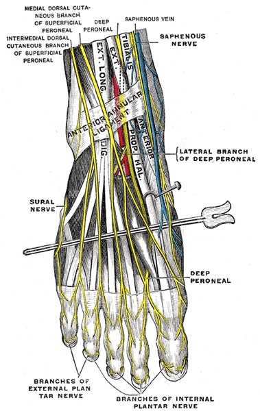
Figure 6. Nerves of the dorsum of the foot: the deep peroneal nerve, the dorsal cutaneous branches of the superficial peroneal nerve, and the extensor tendons beneath the extensor retinaculum. Gray’s Anatomy (1918), Plate 836, public domain, via Wikimedia Commons.
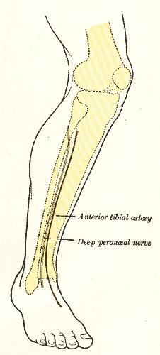
Figure 7. Surface markings of the anterior tibial artery continuing as the dorsalis pedis, and the deep peroneal nerve. Gray’s Anatomy (1918), Plate 1246, public domain, via Wikimedia Commons.
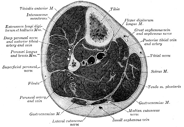
Figure 8. Cross-section through the middle of the leg, labelling the four compartments and the neurovascular bundles, the great and small saphenous veins, and the cutaneous nerves. Gray’s Anatomy (1918), Plate 440, public domain, via Wikimedia Commons.
The medial ankle and the tarsal tunnel
The tarsal tunnel runs behind the medial malleolus, deep to the flexor retinaculum, and its contents are a fixed examination favourite. From the malleolus backward they are the tibialis posterior tendon, the flexor digitorum longus tendon, the posterior tibial artery (with its veins), the tibial nerve, and the flexor hallucis longus tendon, captured by the mnemonic Tom, Dick, And Very Nervous Harry. The posterior tibial artery divides in the sole into medial and lateral plantar arteries. The tibial nerve gives a calcaneal branch to the heel before dividing into the medial and lateral plantar nerves that power the intrinsic muscles and supply sensation to the sole. Where the flexor retinaculum binds the nerve to the medial foot it may compress it, producing tarsal tunnel syndrome. Surgically, flexor hallucis longus is the deep landmark of the back of the ankle, since it is the only one of these muscles still muscular at this level.[6]
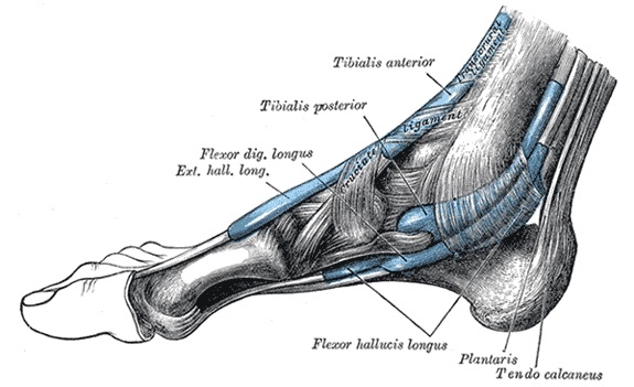
Figure 9. Synovial tendon sheaths around the medial ankle: tibialis posterior, flexor digitorum longus and flexor hallucis longus passing behind the medial malleolus deep to the flexor retinaculum. Gray’s Anatomy (1918), Plate 1242, public domain, via Wikimedia Commons.
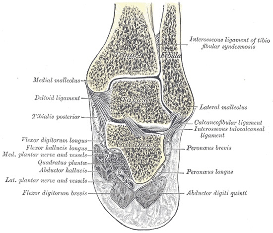
Figure 10. Coronal section through the ankle and subtalar joints, showing the mortise, the tibiofibular syndesmosis, and the medial neurovascular structures (medial and lateral plantar nerves and vessels). Gray’s Anatomy (1918), Plate 357, public domain, via Wikimedia Commons.
The lateral ankle and the superficial sensory triad
The two evertor tendons run behind the lateral malleolus, supplied by the superficial peroneal nerve and held by the superior and inferior peroneal retinacula. Peroneus brevis lies immediately behind the malleolus and stays muscular almost to the joint, while peroneus longus lies behind and lateral to it and is already tendinous in the distal leg. The two share a common synovial sheath around the malleolus and then gain separate sheaths at the peroneal tubercle of the calcaneus, the site of the jogger’s peroneal tendinitis. Whenever an approach divides the peroneal retinaculum it must be repaired, or the tendons will dislocate.[7]
Three superficial sensory nerves cross the ankle, all bound closely to a vein, and knowing their courses is what keeps a skin incision out of trouble. The saphenous nerve, the terminal branch of the femoral nerve, runs with the great saphenous vein in front of the medial malleolus. The superficial peroneal nerve crosses the anterior midline of the dorsum, where it is very superficial and easily cut. The sural nerve runs with the small saphenous vein behind the lateral malleolus and supplies the lateral foot. For both the saphenous and sural nerves, the accompanying vein is the surgical guide: preserve the vein and you preserve the nerve. Dividing the nerve leaves a painful neuroma in skin that, though it does not bear weight, meets the shoe.[8]
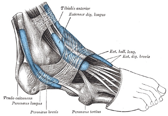
Figure 11. Synovial tendon sheaths around the lateral ankle: peroneus longus and brevis behind the lateral malleolus, with the superior and inferior peroneal retinacula. Gray’s Anatomy (1918), Plate 1241, public domain, via Wikimedia Commons.
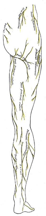
Figure 12. Cutaneous nerves of the lower limb, posterior view: the sural nerve laterally and the saphenous nerve medially, the superficial sensory nerves a foot incision must spare. Gray’s Anatomy (1918), Plate 830, public domain, via Wikimedia Commons.
The ankle joint, mortise, and ligaments
The weight-bearing joint is formed by the dome of the talus and the inferior surface of the tibia, stabilised by the two malleoli that grip the talus as a mortise. The medial malleolus is shorter and more anterior and stays in contact with the talus throughout motion, and the mortise as a whole points about 15 degrees laterally. The mortise is not rigid. The talus is wider in front than behind, so dorsiflexion drives the wide anterior talus in and forces the mortise to spread, while plantarflexion lets it narrow. Two practical consequences follow. An ankle that must be immobilised is held in dorsiflexion, the functional position. And a syndesmosis (diastasis) screw is inserted with the ankle in maximal dorsiflexion, so the mortise is not locked down too tightly around the narrow part of the talus.[9]
The ankle’s collateral ligaments are the medial deltoid ligament, a strong fan from the medial malleolus, and the lateral collateral complex. By standard teaching the lateral complex comprises the anterior talofibular ligament (weakest, the one torn in the common inversion sprain), the calcaneofibular ligament, and the posterior talofibular ligament (strongest). The calcaneofibular ligament is the one encountered surgically: it runs from the malleolus to the lateral calcaneus, is bound to the subtalar capsule, and is divided to expose the subtalar joint. The distal tibia and fibula are bound by the syndesmosis, whose anterior and posterior inferior tibiofibular ligaments and interosseous ligament resist the spreading force of weight-bearing.[10]
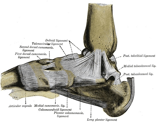
Figure 13. Ligaments of the medial aspect of the ankle and foot, showing the deltoid (medial) ligament and the spring ligament. Gray’s Anatomy (1918), Plate 354, public domain, via Wikimedia Commons.
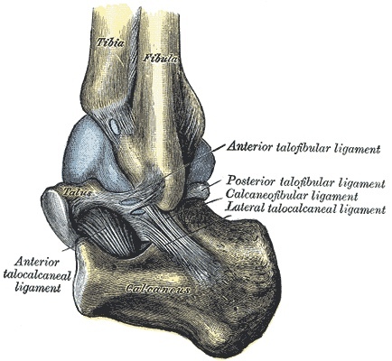
Figure 14. Lateral collateral ligaments of the ankle: the anterior talofibular, calcaneofibular and posterior talofibular ligaments. Gray’s Anatomy (1918), Plate 356, public domain, via Wikimedia Commons.
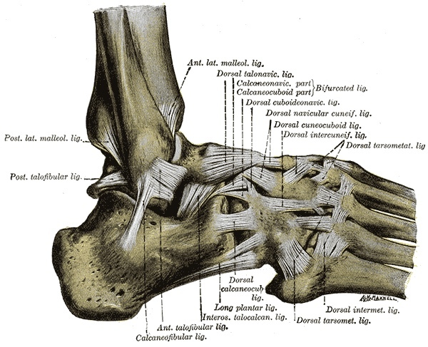
Figure 15. Ligaments of the lateral aspect of the ankle and foot, with the lateral collateral complex and the tarsal ligaments. Gray’s Anatomy (1918), Plate 355, public domain, via Wikimedia Commons.
The subtalar joint, tarsal canal, and sinus tarsi
Hindfoot surgery is almost entirely the surgery of three superficial joints: the posterior part of the subtalar (talocalcaneal) joint, the talocalcaneonavicular joint, and the calcaneocuboid joint. The key to their anatomy is the tarsal canal, an oblique tunnel between the grooves on the undersurface of the talus and the upper surface of the calcaneus. It separates the talocalcaneonavicular joint in front from the posterior talocalcaneal joint behind and serves as the landmark for reaching both. Laterally the canal widens into the sinus tarsi, which holds a tough interosseous (cervical) ligament and a fat pad, and from whose anterior wall the extensor digitorum brevis arises. One consequence matters surgically: the subtalar complex is locked. Forcible inversion will not open either the posterior subtalar or the talocalcaneonavicular joint until both have been incised.[11]
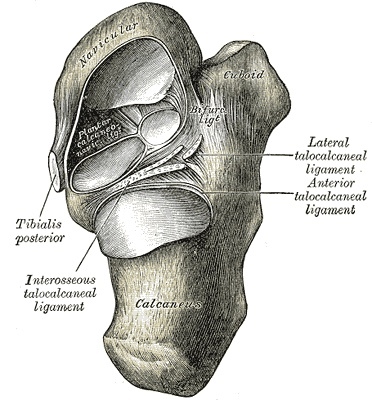
Figure 16. The subtalar (talocalcaneal and talocalcaneonavicular) joints exposed from above, showing the sinus tarsi and the interosseous talocalcaneal ligament. Gray’s Anatomy (1918), Plate 359, public domain, via Wikimedia Commons.
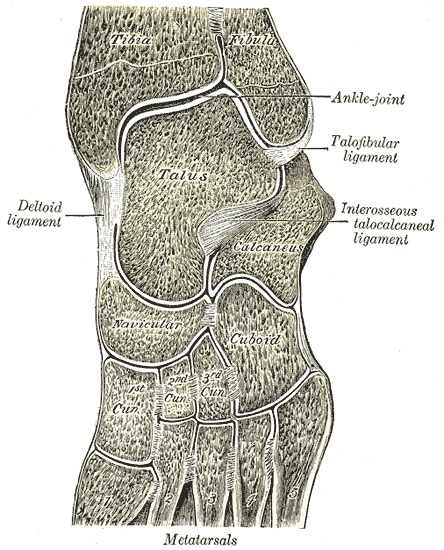
Figure 17. Sagittal/oblique section through the foot, showing the talus, calcaneus, the midtarsal joints and the arch. Gray’s Anatomy (1918), Plate 360, public domain, via Wikimedia Commons.
The dorsum and sole of the foot
Almost all foot bones are approached from the dorsum, for two reasons: the critical neurovascular structures lie on the plantar side of the metatarsals where they stay protected, and dorsal incisions spare the specialised weight-bearing skin of the sole. The dorsal skin is thin and loose, which allows the great swelling seen after foot trauma. Under it run the three dorsal cutaneous nerves (saphenous medially, superficial peroneal across most of the dorsum, sural laterally, with the deep peroneal nerve to the first web space) and the dorsal venous arch, which drains medially to the great and laterally to the small saphenous vein. The dorsalis pedis runs forward beneath the extensor hallucis brevis tendon before diving into the first intermetatarsal space.[12]
The sole is built for weight-bearing. Its tough skin hypertrophies into callosities under abnormal load, and the plantar aponeurosis, a thick central band from the medial calcaneal tubercle to the toes, both supports the arch and, when inflamed at its origin, produces plantar fasciitis. Beneath it the intrinsic muscles lie in four layers. The medial and lateral plantar nerves and arteries, the terminal branches of the tibial nerve and posterior tibial artery, run between the first and second layers, relatively superficial but protected by the tough fascia. The great toe carries two sesamoids in the flexor hallucis brevis insertion, and the adductor hallucis, inserting through the lateral sesamoid, is the most important deforming force in hallux valgus, while the abductor hallucis is the only muscle that opposes it. The deep transverse metatarsal ligament spans the metatarsal heads, with the plantar digital nerves and vessels running just beneath it. That is exactly why staying dorsal to it keeps the forefoot approaches safe.[13]
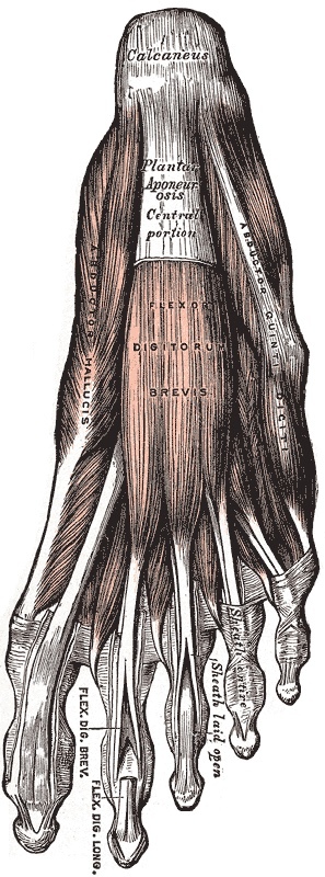
Figure 18. The plantar aponeurosis and the first layer of the sole, with abductor hallucis and abductor digiti minimi flanking flexor digitorum brevis. Gray’s Anatomy (1918), Plate 443, public domain, via Wikimedia Commons.
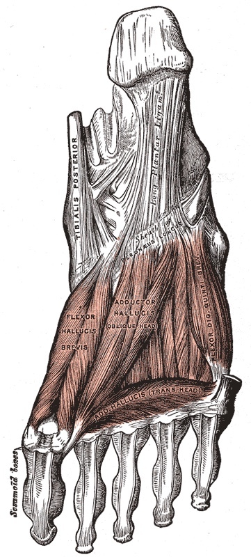
Figure 19. Deep muscles of the sole: adductor hallucis (oblique and transverse heads) and flexor hallucis brevis with the great-toe sesamoids. Gray’s Anatomy (1918), Plate 445, public domain, via Wikimedia Commons.
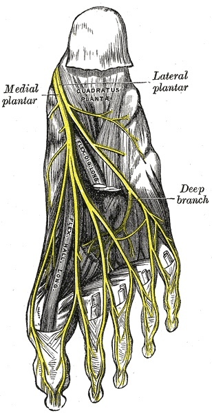
Figure 20. The medial and lateral plantar nerves, the terminal branches of the tibial nerve, distributed across the sole. Gray’s Anatomy (1918), Plate 833, public domain, via Wikimedia Commons.
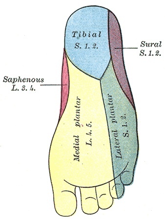
Figure 21. Cutaneous nerve distribution of the sole of the foot (medial plantar, lateral plantar, saphenous and sural territories). Gray’s Anatomy (1918), Plate 834, public domain, via Wikimedia Commons.
Part II - Approaches to the Ankle
The anterior approach to the ankle
The anterior approach is the standard wide exposure of the ankle joint, used for ankle arthrodesis and for open reduction of pilon (distal tibial plafond) fractures. The patient is supine, with only partial exsanguination so the dorsal veins remain visible, and a roughly 15 cm longitudinal incision is made midway between the malleoli. The plane is intermuscular rather than truly internervous, lying between extensor hallucis longus and extensor digitorum longus, both supplied by the deep peroneal nerve proximally. The cardinal danger is the anterior tibial artery and deep peroneal nerve running in the depth of the wound. Protect them by identifying and retracting them as a unit, and by remembering that at the joint line the bundle sits just lateral to the extensor hallucis longus tendon. The superficial peroneal nerve’s branches cross the field superficially and must be spared at the skin incision.[14]
The approaches to the medial malleolus and the medial side
Two short approaches address the medial malleolus for open reduction and internal fixation. Each cuts directly onto subcutaneous bone, so there is no internervous plane. The structures to respect are the great saphenous vein and saphenous nerve crossing in front of the malleolus and the posterior tibial tendon behind it. A separate, deliberate medial osteotomy approach is used to expose the talar dome, for example to address an osteochondral lesion. The malleolus is pre-drilled and tapped before the cut so the screw holes line up on reattachment, an oblique osteotomy is made, and the malleolar fragment is turned down on its intact deltoid ligament to swing the talus into view, the excursion limited by the intact fibula. An osteotomy lacks the rotational stability of interdigitating fracture fragments, so it is fixed with a screw supplemented by two anti-rotation K-wires.[15]
The posteromedial approach to the ankle
The posteromedial approach reaches the soft tissues around the back of the medial malleolus and the posterior ankle, used for medial soft-tissue release in clubfoot and for lengthening the medial flexor tendons. With the limb externally rotated (the figure-of-four position), an 8 to 10 cm incision is made midway between the medial malleolus and the Achilles tendon. There is no named internervous plane. The dissection is defined instead by flexor hallucis longus, the deep landmark, and the back of the joint is reached either lateral to it (between it and the peroneal tendons), medial to it (mobilising the neurovascular bundle and working between it and flexor digitorum longus), or directly when all three medial tendons are being lengthened. The danger throughout is the posterior tibial artery and tibial nerve, which must never be forcefully retracted lest a neurapraxia result. In children the tibial nerve is surprisingly large and the flexor digitorum longus tendon tiny, so every structure must be positively identified before anything is divided.[16]
The posterolateral approach to the ankle
The posterolateral approach gives the best visualisation of the posterior tibia among the posterior routes and is well suited to open reduction of posterior malleolar fractures. The patient is prone, which is its chief limitation: it cannot be combined with simultaneous fixation of the fibula and medial malleolus without repositioning. A 10 cm incision runs midway between the posterior border of the lateral malleolus and the Achilles tendon, and the plane is a true internervous one between peroneus brevis (superficial peroneal nerve) and flexor hallucis longus (tibial nerve). In the superficial dissection the sural nerve and small saphenous vein are kept anterior and out of harm’s way. The peroneal tendons are then released, and flexor hallucis longus is split off the fibula and retracted medially to expose the posterior tibia and the posterior capsule.[17]
The lateral approach to the lateral malleolus
The lateral approach is the workhorse for open reduction and internal fixation of lateral malleolar (distal fibular) fractures. It is a direct subperiosteal approach to subcutaneous bone, so there is no internervous plane at the malleolus, although for higher fibular fractures the plane runs between peroneus tertius (deep peroneal nerve) and peroneus brevis (superficial peroneal nerve). A 10 to 15 cm incision follows the posterior border of the fibula and curves below the tip. The sural nerve and small saphenous vein lie posteriorly and are preserved, and dissection is kept strictly subperiosteal because the terminal branches of the peroneal artery lie deep on the medial surface of the distal fibula. The tourniquet masks bleeding from those vessels, so it is deflated before closure to secure haemostasis, and the wound is drained.[18]
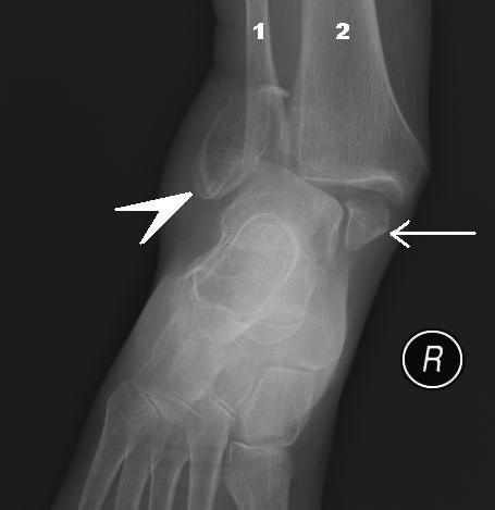
Figure 22. Bimalleolar ankle fracture with dislocation: fractures of both the fibula (1) and the tibial (medial) malleolus (2). Steven Fruitsmaak, CC BY-SA 3.0, via Wikimedia Commons.
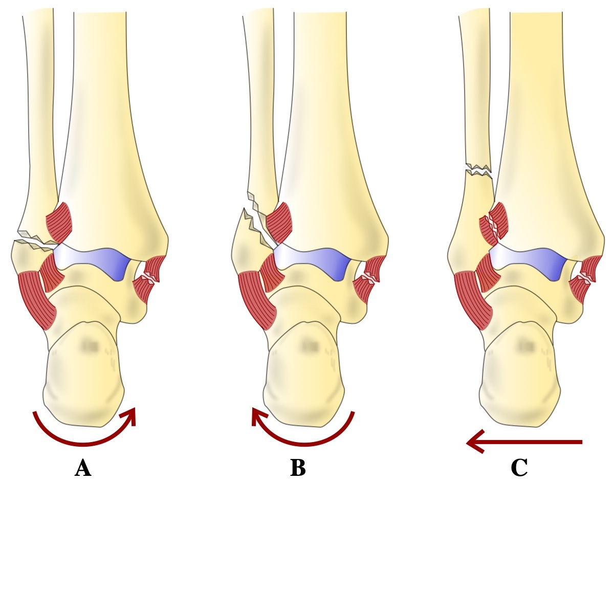
Figure 23. Danis-Weber classification of fibular fractures relative to the syndesmosis (type A below, B at, C above the syndesmosis, shown in blue). DrFO.Tn, CC BY 4.0, via Wikimedia Commons.
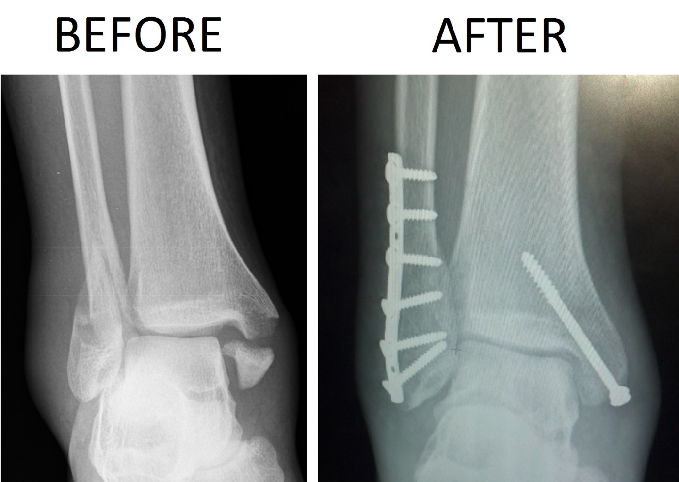
Figure 24. Ankle fracture before and after open reduction and internal fixation with a lateral fibular plate and a medial malleolar screw. Chaim Mintz, CC BY-SA 3.0, via Wikimedia Commons.
Part III - Approaches to the Hindfoot
The anterolateral approach to the ankle and hindfoot
This single anterolateral incision reaches a remarkable territory: the ankle joint together with the talonavicular, calcaneocuboid, and talocalcaneal joints. That makes it the common exposure for ankle fusion, triple arthrodesis, pantalar arthrodesis, and talectomy. With the limb internally rotated, a 15 cm curved incision crosses the front of the ankle just medial to the lateral malleolus and runs onto the foot toward the fourth metatarsal base. The plane is internervous, between the peroneal muscles (superficial peroneal nerve) and the extensor muscles (deep peroneal nerve). Skin flaps are not raised. The dorsal cutaneous branches of the superficial peroneal nerve are preserved, the deep peroneal nerve and anterior tibial artery are protected by staying close to bone, and the extensor digitorum brevis is detached from the calcaneus. Preserving the sinus tarsi fat pad both avoids an ugly dimple and helps the wound heal.[19]
The lateral approach to the hindfoot
A curved lateral incision over the sinus tarsi gives excellent exposure of the talocalcaneonavicular, posterior talocalcaneal, and calcaneocuboid joints for triple arthrodesis. Its plane lies between peroneus tertius (deep peroneal nerve) and the peroneal tendons (superficial peroneal nerve). The dominant warning is the soft tissue. Exposures here are notorious for skin-flap necrosis, so flaps are cut thick, stripping and retraction are minimised, and sharp curves in the incision are avoided. The extensor digitorum brevis origin is detached and the sinus tarsi fat pad left on the skin flap. The three joints are then opened by forced inversion, remembering that both the talocalcaneonavicular and the posterior subtalar joints must be incised before either will open. By standard teaching the sural nerve and small saphenous vein lie along this incision line and are at risk.[20]
The lateral approaches to the posterior talocalcaneal joint and the calcaneus
A dedicated lateral approach exposes the posterior facet of the subtalar joint more fully, for posterior subtalar fusion. It has no internervous plane (the peronei share the superficial peroneal nerve, supplied well proximal to the field), and its signature danger is the sural nerve with the small saphenous vein just behind the lateral malleolus. Dividing it leaves a neuroma and lateral-foot numbness. The peroneal tendons are released, the calcaneofibular ligament divided, and the joint opened by inversion. The separate lateral approach to the calcaneus is the extensile exposure for open reduction of calcaneal fractures: a two-limbed, L-shaped lateral incision is taken straight to bone and a single full-thickness flap of periosteum and all overlying tissue is raised in one piece, with the peroneal tendons carried up in it. This flap is perfused chiefly by the lateral calcaneal artery, which is why no layered dissection is permitted. These fractures also swell massively, so surgery is delayed until the swelling subsides, with diabetes and smoking as relative contraindications, since wound breakdown is the feared complication.[21]
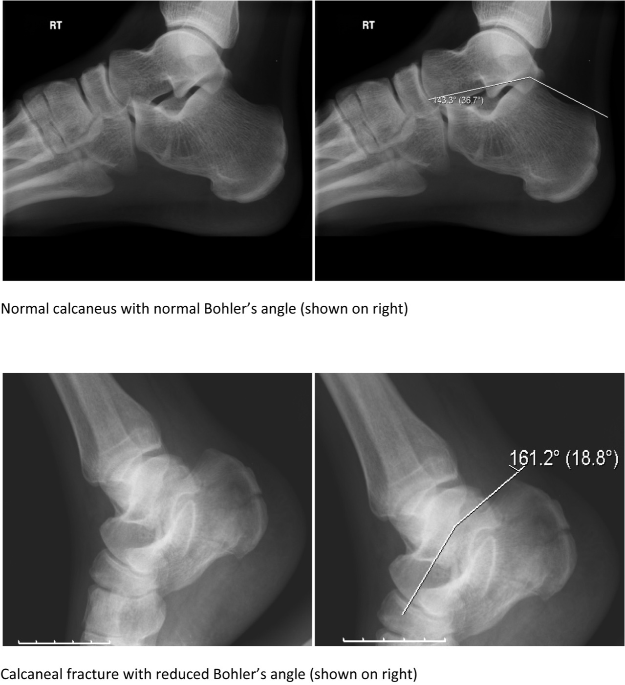
Figure 25. Boehler’s angle on the lateral calcaneal radiograph: normal (top) versus a calcaneal fracture with a reduced angle (bottom). Gilo1969, CC BY-SA 3.0, via Wikimedia Commons.
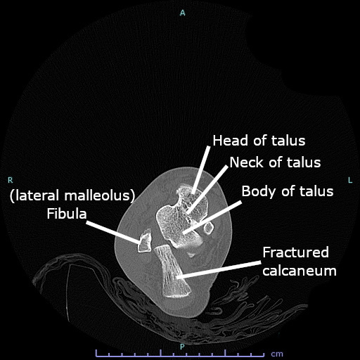
Figure 26. Axial CT of a calcaneal fracture through the hindfoot. Cerevisae, CC BY-SA 4.0, via Wikimedia Commons.
Part IV - Approaches to the Forefoot
The dorsal approaches to the midfoot
The bones and joints of the midfoot, including the tarsometatarsal (Lisfranc) joints, are reached through dorsal longitudinal incisions, which keep clear of the plantar neurovascular structures and the weight-bearing sole. There is no internervous plane. The structures to protect are the dorsalis pedis and deep peroneal nerve in the depth and the dorsal cutaneous nerves superficially. For a Lisfranc injury more than one incision is used, set apart so an adequate skin bridge remains between them. The midfoot incisions can be extended proximally behind either malleolus to reach the hindfoot tendons.[22]
The approaches to the great toe and bunion surgery
The metatarsophalangeal joint of the great toe is approached dorsally (through a straight incision just medial to the extensor hallucis longus tendon) or dorsomedially (through a curved medial incision, leaving a U-shaped flap of capsule on the phalangeal base for later reefing), for arthrodesis, the Keller excision, cheilectomy, and synovectomy. Bunion (hallux valgus) correction adds a dorsolateral first-web approach for the lateral soft-tissue release: the adductor hallucis is detached from the lateral sesamoid, the contracted lateral capsule is released, and the deep transverse metatarsal ligament is divided. Throughout, the dorsal digital nerves are spared, and the dorsal cutaneous nerves are protected by keeping to the midline of the web space. The digital nerve lies immediately beneath the transverse metatarsal ligament and is at risk when that ligament is cut.[23]
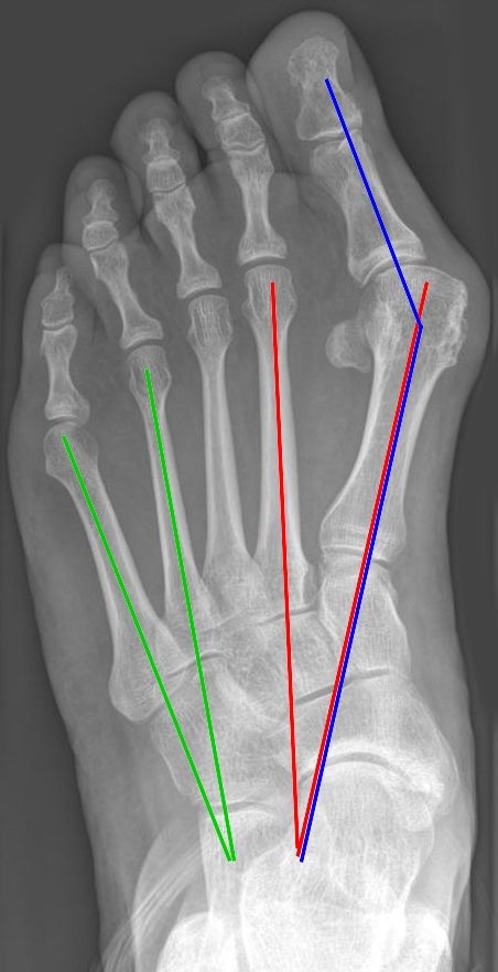
Figure 27. Weight-bearing radiograph of hallux valgus, with the hallux valgus angle (blue) and the intermetatarsal angles (red and green) measured. Jmarchn, CC BY-SA 3.0, via Wikimedia Commons.
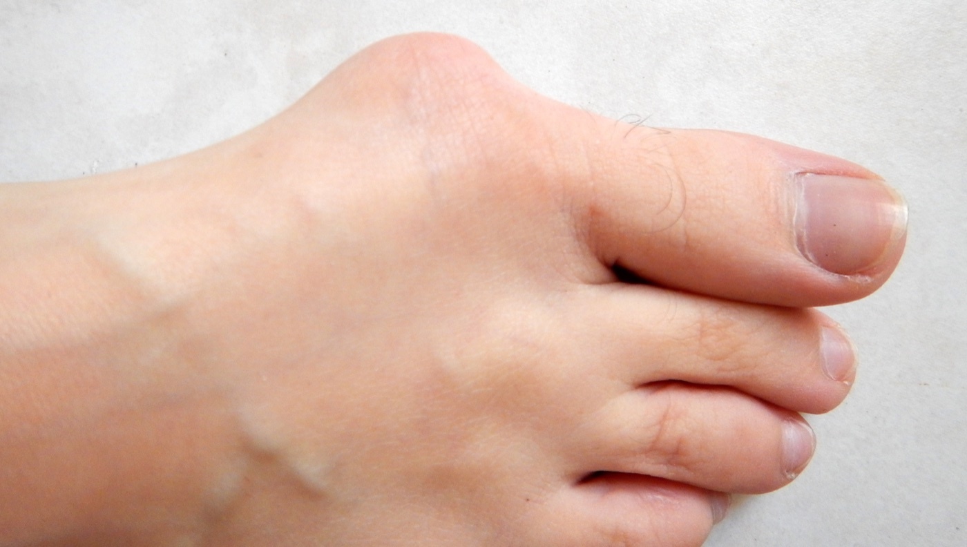
Figure 28. Clinical appearance of hallux valgus, with the prominent first metatarsophalangeal bunion and lateral deviation of the great toe. Lamiot, CC BY-SA 4.0, via Wikimedia Commons.
The lesser-toe metatarsophalangeal joints and the Morton neuroma
The metatarsophalangeal joints of the second to fifth toes are exposed through short dorsal longitudinal incisions just lateral to the long extensor tendon, sparing the plantar weight-bearing skin, for metatarsal-head excision, distal osteotomy, capsulotomy, and tenotomy. There is no internervous plane. The approaches stay well dorsal to the plantar nerves and vessels, which lie beneath the deep transverse metatarsal ligament. The Morton neuroma is reached through a dorsal longitudinal incision in the centre of the web space, most often the third web space: the deep transverse metatarsal ligament is divided to reveal the common plantar digital nerve and its vessel, and digital plantar pressure pushes the neuroma up into the wound. The target nerve and vessel are themselves the only structure at real risk. A single divided digital artery is tolerated, but cutting the second artery to the same toe in the adjacent web space can render it ischaemic, and excising the neuroma leaves the toe’s plantar surface partly numb without trophic change.[24]
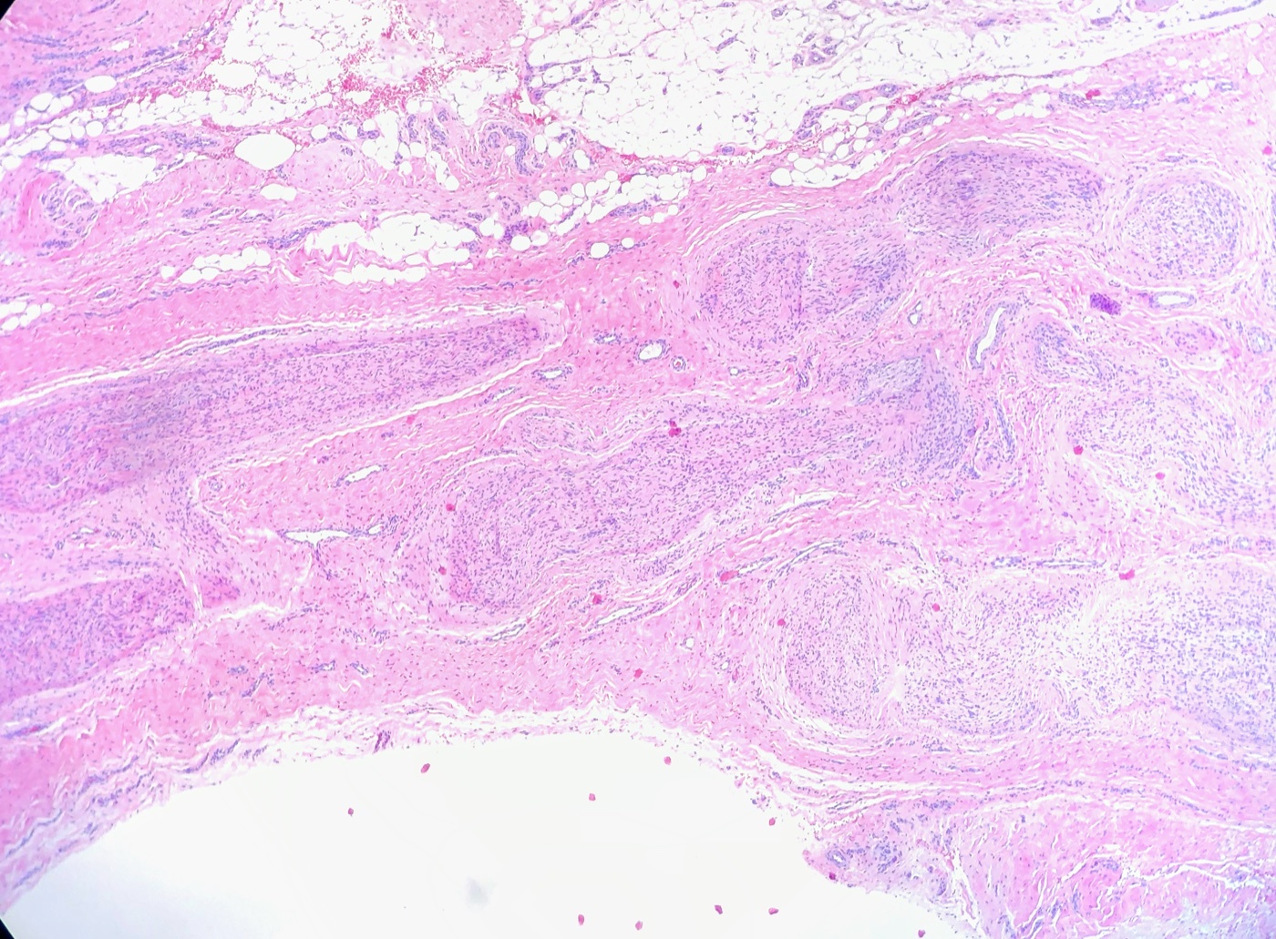
Figure 29. Histology of a Morton (plantar interdigital) neuroma, showing the perineural fibrosis of the interdigital nerve, H\&E stain. Ed Uthman, CC BY 2.0, via Wikimedia Commons.
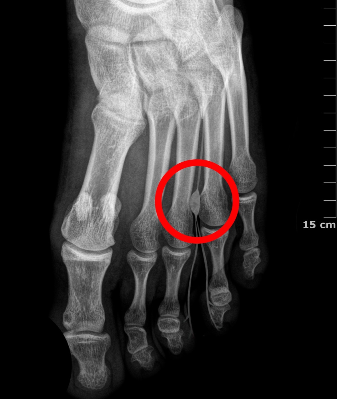
Figure 30. Forefoot radiograph indicating the third intermetatarsal web space, the typical site of a Morton neuroma. Nuñez Zapata Cascianini, CC BY-SA 4.0, via Wikimedia Commons.
References
-
The framing of the ankle by three tendon groups (flexors behind the medial malleolus, tibial nerve; extensors in front, deep peroneal nerve; evertors behind the lateral malleolus, superficial peroneal nerve), the three resulting internervous planes, and the two major neurovascular bundles as “the major surgical concerns for all approaches around the ankle” are from Hoppenfeld, Surgical Exposures in Orthopaedics: The Anatomic Approach, 5th ed. (2016), Chapter 12 (The Foot & Ankle), applied anatomy of the approaches to the ankle (Hoppenfeld p.1168). General teaching that supplements the source is flagged in-line as standard teaching and carries no page citation.
-
That poor wound healing is the most common complication of foot and ankle surgery, that the circulation and sensation must be assessed and surgery often delayed until swelling subsides, that flaps should be full thickness, and that incisions must spare the cutaneous nerves are from Hoppenfeld’s chapter introduction and applied anatomy (Hoppenfeld pp.1094-1096, p.1167). The aphorism that incisions in this region “heal side to side, not end to end” is standard teaching.
-
The three tendon groups crossing the ankle with their nerves (flexors behind the medial malleolus, tibial nerve; extensors in front, deep peroneal nerve; evertors behind the lateral malleolus, superficial peroneal nerve), the Achilles and plantaris lying posteriorly in the midline, the retinacula preventing bowstringing (and needing repair when divided), and the three internervous planes (medial flexors/extensors, posterolateral flexors/evertors, lateral extensors/evertors) are from Hoppenfeld’s applied anatomy of the ankle (Hoppenfeld p.1168).
-
The four extensor tendons crossing the front of the ankle from medial to lateral (tibialis anterior, extensor hallucis longus, extensor digitorum longus, peroneus tertius), the anterior bundle (anterior tibial artery + deep peroneal nerve) crossing halfway between the malleoli, lying between tibialis anterior and extensor hallucis longus proximally and between extensor hallucis longus and extensor digitorum longus distally, the extensor hallucis longus tendon crossing it lateral-to-medial so the bundle lies immediately lateral to that tendon at the joint, and the anterior tibial artery becoming the dorsalis pedis (communicating with the plantar circulation through the first intermetatarsal space, disrupted by Lisfranc injuries) are from Hoppenfeld (Hoppenfeld pp.1168-1169, p.1176).
-
The deep peroneal nerve accompanying the anterior tibial artery, supplying extensor digitorum brevis and extensor hallucis brevis and the first web space, with first-web-space numbness an early sign of anterior compartment syndrome (the nerve becoming ischaemic before the muscle), and the superior extensor retinaculum (split by the tibialis anterior tendon) and inferior extensor retinaculum holding the tendons and requiring repair when cut, are from Hoppenfeld (Hoppenfeld p.1169, pp.1176-1177).
-
The tarsal tunnel contents behind the medial malleolus from anterior to posterior (tibialis posterior, flexor digitorum longus, posterior tibial artery and veins, tibial nerve, flexor hallucis longus), the posterior tibial artery dividing into medial and lateral plantar arteries and the tibial nerve giving a calcaneal branch and dividing into medial and lateral plantar nerves, the flexor retinaculum binding and potentially compressing the nerve (tarsal tunnel syndrome), and flexor hallucis longus as the deep landmark (“the only muscle still muscular at this level”) are from Hoppenfeld (Hoppenfeld p.1169, p.1172, p.1175, p.1122). The mnemonic “Tom, Dick, And Very Nervous Harry” is standard teaching; Hoppenfeld names every structure but gives the older mnemonics (“Tom, Dick, and Harry”; “Timothy Doth Vex Nervous Housemaids,” p.1172).
-
The peroneal tendons behind the lateral malleolus (supplied by the superficial peroneal nerve, held by the superior and inferior peroneal retinacula), peroneus brevis lying immediately behind the malleolus and muscular almost to the joint while peroneus longus lies posterior and is tendinous distally, the common synovial sheath splitting into two at the peroneal tubercle (a site of jogger’s peroneal tendinitis), and the rule that the divided peroneal retinaculum must be repaired to prevent tendon dislocation are from Hoppenfeld (Hoppenfeld p.1178, p.1182, p.1159).
-
The three superficial sensory nerves crossing the ankle (saphenous nerve with the great saphenous vein anteromedially, terminal branch of the femoral nerve; superficial peroneal nerve along the anterior midline of the dorsum, very superficial; sural nerve with the small saphenous vein posterolaterally, supplying the lateral foot), and the principle that preserving the vein preserves the nerve while division leaves a painful neuroma in shoe-bearing skin, are from Hoppenfeld (Hoppenfeld pp.1170-1171, p.1159). Hoppenfeld describes the sural nerve simply as a terminal branch of the tibial nerve; that it is formed by a medial sural cutaneous branch plus a peroneal communicating branch is standard teaching.
-
The ankle joint formed by the talar dome and inferior tibial surface stabilised by the malleoli, the medial malleolus being shorter and more anterior and staying in contact with the talus, the mortise pointing 15 degrees laterally, the talus being wider anteriorly so dorsiflexion spreads the mortise and plantarflexion narrows it, and the resulting rules that an immobilised ankle is held in dorsiflexion (functional position) and a syndesmosis/diastasis screw is inserted in maximal dorsiflexion, are from Hoppenfeld (Hoppenfeld p.1171).
-
The deltoid (medial) ligament and the calcaneofibular ligament are named and depicted in Hoppenfeld (deltoid, Fig. 12-56, p.1174; calcaneofibular ligament divided to expose the subtalar joint and visible deep to the superior peroneal retinaculum, p.1167, p.1180), and the syndesmosis is treated functionally via the diastasis-screw rule (Hoppenfeld p.1171). The three named components of the lateral collateral complex (anterior talofibular, calcaneofibular, posterior talofibular) with the anterior talofibular the weakest/most commonly sprained and the posterior talofibular the strongest, and the named syndesmotic ligaments (anterior and posterior inferior tibiofibular and interosseous), are standard teaching; Hoppenfeld names only the deltoid and calcaneofibular ligaments in the text.
-
Hindfoot surgery being confined to the posterior subtalar, talocalcaneonavicular, and calcaneocuboid joints (all superficial), the tarsal canal as the key landmark (between grooves on the talus and calcaneus, separating the talocalcaneonavicular from the talocalcaneal joint and widening laterally into the sinus tarsi), the sinus tarsi containing the cervical ligament and a fat pad with extensor digitorum brevis arising from its anterior wall, and the “locking” principle that forcible inversion opens neither subtalar joint until both are incised, are from Hoppenfeld’s applied anatomy of the hindfoot (Hoppenfeld p.1181, p.1153, p.1160). The talus’s tenuous retrograde blood supply and vulnerability to avascular necrosis after talar neck fracture are standard teaching and are not discussed in this extract.
-
That all foot bones can be approached dorsally because the critical neurovascular structures lie plantar to the metatarsals and dorsal incisions spare the weight-bearing sole, the thin loose dorsal skin accounting for marked post-traumatic swelling, the dorsal cutaneous nerves (saphenous medially, superficial peroneal over most of the dorsum, sural laterally, deep peroneal to the first web space with first-web numbness the earliest sign of a deep peroneal lesion), the dorsal venous arch draining to the great and small saphenous veins, and the dorsalis pedis running beneath the extensor hallucis brevis tendon into the first intermetatarsal space are from Hoppenfeld’s applied anatomy of the foot (Hoppenfeld pp.1214-1216).
-
The specialised weight-bearing sole skin forming callosities under load, the plantar aponeurosis from the medial calcaneal tubercle to the toes supporting the arch and causing plantar fasciitis when inflamed, the four muscle layers with the medial and lateral plantar nerves and arteries running between the first and second layers (protected by the tough fascia), the great-toe sesamoids in the flexor hallucis brevis insertion, the adductor hallucis (through the lateral sesamoid) being “the most important deforming force in hallux valgus” while abductor hallucis is the only muscle opposing it, and the deep transverse metatarsal ligament with the plantar digital nerves and vessels beneath it (so dorsal approaches stay safe), are from Hoppenfeld’s applied anatomy of the foot (Hoppenfeld pp.1216-1218, p.1209). That the medial and lateral plantar nerves and arteries are terminal branches of the tibial nerve and posterior tibial artery is standard teaching.
-
The anterior approach to the ankle as the exposure for ankle arthrodesis and pilon fracture fixation, supine with partial exsanguination to keep veins visible, the ~15 cm incision midway between the malleoli, the intermuscular plane between extensor hallucis longus and extensor digitorum longus (both deep peroneal nerve, supplied proximally), the anterior tibial artery and deep peroneal nerve as the chief deep danger (the bundle lying just lateral to the extensor hallucis longus tendon at the joint), and the superficial peroneal nerve branches crossing the field, are from Hoppenfeld’s anterior approach to the ankle (Hoppenfeld pp.1097-1102).
-
The anterior and posterior approaches to the medial malleolus (for ORIF, cutting onto subcutaneous bone with no internervous plane, protecting the great saphenous vein and saphenous nerve), the medial osteotomy approach to the talar dome (pre-drill and tap before the cut, oblique osteotomy, malleolus turned down on the intact deltoid ligament, excursion limited by the intact fibula), and the rule that an osteotomy needs a screw plus two anti-rotation K-wires (because it lacks the interdigitation of a fracture) are from Hoppenfeld’s approaches to the medial malleolus and medial side of the ankle (Hoppenfeld pp.1103-1120).
-
The posteromedial approach exposing the soft tissues behind the medial malleolus and the posterior ankle (for clubfoot release and medial flexor tendon lengthening), the figure-of-four position, the 8-10 cm incision midway between the medial malleolus and Achilles tendon, the absence of a named internervous plane with flexor hallucis longus as the deep landmark and three deep routes (lateral to flexor hallucis longus, medial to it between the neurovascular bundle and flexor digitorum longus, or direct during triple lengthening), and the dangers (posterior tibial artery and tibial nerve, no forceful retraction lest neurapraxia; the large tibial nerve and tiny flexor digitorum longus tendon in children) are from Hoppenfeld’s posteromedial approach to the ankle (Hoppenfeld pp.1121-1128).
-
The posterolateral approach giving the best visualisation of the posterior tibia and being well suited to posterior malleolar fracture fixation, the prone position limiting simultaneous fibular/medial fixation, the 10 cm incision midway between the posterior border of the lateral malleolus and the Achilles tendon, the internervous plane between peroneus brevis (superficial peroneal nerve) and flexor hallucis longus (tibial nerve), and the superficial danger to the sural nerve and small saphenous vein (kept anterior) with flexor hallucis longus split off the fibula and retracted medially, are from Hoppenfeld’s posterolateral approach to the ankle (Hoppenfeld pp.1128-1136).
-
The lateral approach to the lateral malleolus as the workhorse for ORIF of lateral malleolar fractures, being a direct subperiosteal approach to subcutaneous bone with no internervous plane (the higher-fracture plane between peroneus tertius/deep peroneal nerve and peroneus brevis/superficial peroneal nerve), the 10-15 cm incision along the posterior fibular border curving below the tip, the sural nerve and small saphenous vein preserved posteriorly, the strictly subperiosteal dissection (terminal branches of the peroneal artery deep on the medial distal fibula), and deflating the tourniquet before closure with a drain, are from Hoppenfeld’s lateral approach to the lateral malleolus (Hoppenfeld pp.1136-1140).
-
The anterolateral approach exposing the ankle plus the talonavicular, calcaneocuboid, and talocalcaneal joints (for ankle fusion, triple and pantalar arthrodesis, and talectomy), the internally rotated limb and 15 cm curved incision crossing 2 cm medial to the lateral malleolus toward the fourth metatarsal base, the internervous plane between the peroneal muscles (superficial peroneal nerve) and extensor muscles (deep peroneal nerve), not raising skin flaps, preserving the dorsal cutaneous branches of the superficial peroneal nerve and protecting the deep peroneal nerve and anterior tibial artery by staying on bone, detaching extensor digitorum brevis, and preserving the sinus tarsi fat pad to avoid a dimple and aid healing, are from Hoppenfeld’s anterolateral approach to the ankle and hindfoot (Hoppenfeld pp.1141-1147).
-
The lateral approach to the hindfoot exposing the talocalcaneonavicular, posterior talocalcaneal, and calcaneocuboid joints for triple arthrodesis, its plane between peroneus tertius (deep peroneal nerve) and the peroneal tendons (superficial peroneal nerve), the notorious skin-flap necrosis risk (thick flaps, minimal stripping/retraction, no sharp curves), detaching extensor digitorum brevis and leaving the sinus tarsi fat pad on the flap, and the locking principle (both the talocalcaneonavicular and posterior subtalar joints incised before inversion opens either), are from Hoppenfeld’s lateral approach to the hindfoot (Hoppenfeld pp.1147-1156). The sural nerve and small saphenous vein along this incision line are standard teaching here (named explicitly in the adjacent posterior talocalcaneal approach, p.1158).
-
The lateral approach to the posterior talocalcaneal joint (for posterior subtalar fusion, no internervous plane because the peronei share the superficial peroneal nerve supplied proximally, the sural nerve with the small saphenous vein behind the lateral malleolus the signature danger, dividing the calcaneofibular ligament and opening the joint by inversion) and the lateral approach to the calcaneus (the L-shaped extensile incision for calcaneal fracture ORIF, a single full-thickness flap raised to bone with the peroneal tendons carried in it, no layered dissection, surgery delayed until swelling subsides with diabetes/smoking relative contraindications and wound breakdown the feared complication) are from Hoppenfeld (Hoppenfeld pp.1156-1166). The lateral calcaneal artery as the flap’s blood supply is standard teaching; the extract prescribes the full-thickness no-layer flap that protects it.
-
The dorsal approaches to the midfoot (including the tarsometatarsal/Lisfranc joints) through dorsal longitudinal incisions that avoid the plantar neurovascular structures and weight-bearing sole, with no internervous plane and the dorsalis pedis/deep peroneal nerve and dorsal cutaneous nerves to protect, and the use of separate incisions for Lisfranc fixation, are from Hoppenfeld’s dorsal approaches to the middle part of the foot (Hoppenfeld pp.1184-1191). The two-incision adequate-skin-bridge rule is standard teaching.
-
The dorsal approach to the great-toe metatarsophalangeal joint (straight incision just medial to extensor hallucis longus) and the dorsomedial approach (curved medial incision, U-shaped capsular flap left on the phalangeal base), their uses (arthrodesis, Keller excision, cheilectomy, synovectomy), and the dorsolateral first-web approach for bunion surgery (detaching adductor hallucis from the lateral sesamoid, releasing the lateral capsule, dividing the deep transverse metatarsal ligament), with the dorsal digital and cutaneous nerves protected by staying in the web-space midline and the digital nerve immediately beneath the transverse metatarsal ligament, are from Hoppenfeld’s great-toe and bunion approaches (Hoppenfeld pp.1191-1205).
-
The dorsal approach to the lesser-toe (2nd-5th) metatarsophalangeal joints (short dorsal longitudinal incision just lateral to the long extensor tendon, sparing plantar skin, for metatarsal-head excision, distal osteotomy, capsulotomy, tenotomy; no internervous plane, staying dorsal to the plantar nerves and vessels beneath the deep transverse metatarsal ligament) and the dorsal approach for Morton neuroma (a dorsal longitudinal web-space incision, most often the third web space, dividing the deep transverse metatarsal ligament to expose the common plantar digital nerve and vessel, with plantar pressure delivering the neuroma; the target nerve/vessel the only real danger, one digital artery tolerated but the second to the same toe risking ischaemia, and excision leaving partial plantar numbness without trophic change) are from Hoppenfeld’s lesser-toe and Morton neuroma approaches (Hoppenfeld pp.1205-1214).
-
The three tendon groups, their nerves, and locations, and the resulting internervous planes, are from Hoppenfeld’s applied anatomy of the ankle (Hoppenfeld p.1168).
-
The medial-to-lateral order at the front of the ankle and the bundle lying immediately lateral to the extensor hallucis longus tendon at the joint are from Hoppenfeld (Hoppenfeld pp.1168-1169, p.1176).
-
The tarsal tunnel contents in order, the tibial nerve dividing into medial and lateral plantar nerves, and tarsal tunnel syndrome are from Hoppenfeld (Hoppenfeld p.1169, p.1172, p.1175); the “Tom, Dick, And Very Nervous Harry” mnemonic is standard teaching.
-
The talus being wider anteriorly, the functional (dorsiflexion) position for immobilisation, and the rule to insert a syndesmosis screw in maximal dorsiflexion are from Hoppenfeld (Hoppenfeld p.1171).
-
The posterolateral approach plane (peroneus brevis/superficial peroneal nerve vs flexor hallucis longus/tibial nerve), the prone position, and the sural nerve and small saphenous vein danger are from Hoppenfeld (Hoppenfeld pp.1128-1136).
-
The delay until swelling subsides, the diabetes/smoking contraindications, and the single full-thickness no-layer L-flap for the lateral calcaneal approach are from Hoppenfeld (Hoppenfeld p.1163, p.1166); the lateral calcaneal artery as the flap’s supply is standard teaching.
-
The joints exposed by the anterolateral approach, its uses, and its internervous plane (peroneal/superficial peroneal vs extensor/deep peroneal) are from Hoppenfeld (Hoppenfeld pp.1141-1142).
-
That the calcaneofibular ligament is divided to expose the subtalar joint is from Hoppenfeld (Hoppenfeld p.1167, p.1180); the three named components of the lateral complex and the anterior talofibular ligament being the weakest/most commonly sprained are standard teaching.
-
The two reasons for dorsal approaches (plantar neurovascular structures stay protected; the weight-bearing sole skin is spared) and the exception for abnormal plantar skin are from Hoppenfeld’s applied anatomy of the foot (Hoppenfeld p.1214).
-
The Morton neuroma approach (dorsal web-space incision, usually the third, dividing the deep transverse metatarsal ligament to expose the common plantar digital nerve and vessel), the digital-artery ischaemia rule, and the expected partial numbness without trophic change are from Hoppenfeld (Hoppenfeld pp.1211-1213).
-
The adductor hallucis as the most important deforming force in hallux valgus (and its reattachment in bunion surgery) and abductor hallucis as the only opposing muscle are from Hoppenfeld’s applied anatomy of the foot (Hoppenfeld pp.1217-1218).
-
The three superficial sensory nerves, their courses and accompanying veins, and the principle that preserving the vein preserves the nerve are from Hoppenfeld (Hoppenfeld pp.1170-1171).